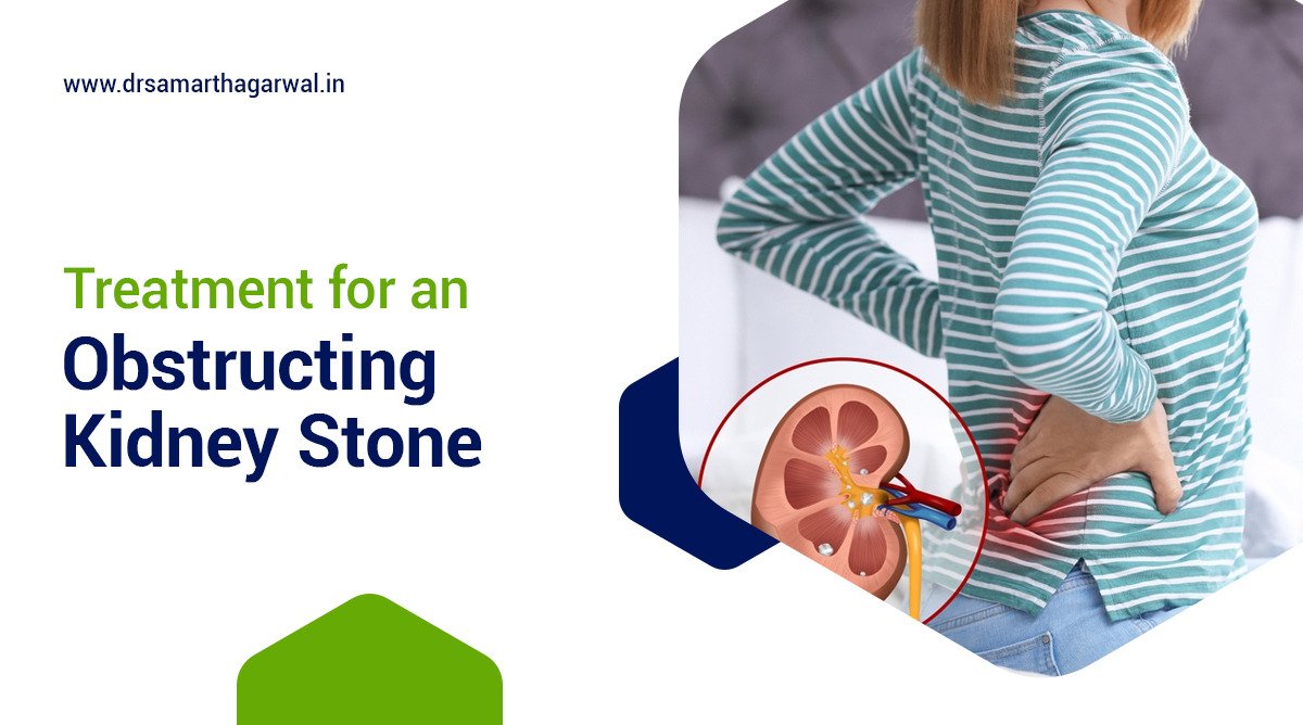
Kidney stones are a prevalent condition that affects a significant number of individuals worldwide, leading to severe pain and discomfort. The formation of kidney stones can severely obstruct the flow of urine from the kidney to the bladder, causing a range of symptoms and increasing the risk of complications. This article explores the triggers for the formation of kidney stones, their symptoms, various treatment options, and effective prevention strategies. Furthermore, it probes into understanding the different types of kidney stones and how they influence treatment choices, the diagnosis process, and managing emergency situations in which kidney stones might precipitate.
What Triggers the Formation of Kidney Stones?
The formation of kidney stones is primarily influenced by dehydration, dietary choices, and genetic predisposition. Dehydration, by reducing the volume of urine, leads to higher concentrations of minerals which can precipitate and form stones.
Diets high in sodium, oxalate, and protein can increase the risk of stone formation by elevating the levels of stone-forming substances in the urine. Moreover, individuals with a family history of kidney stones are at a heightened risk, indicating a strong genetic component to stone susceptibility.
Other factors contributing to kidney stone development include certain medical conditions like hyperparathyroidism, which alters calcium metabolism, leading directly to the formation of calcium stones.
Several factors can trigger the formation of kidney stones, significantly impacting an individual’s risk profile. Chronic dehydration, dietary habits laden with high salt or protein intake, and a genetic predisposition are principal contributors. Dehydration decreases urine output, which results in highly concentrated urine where minerals can crystallize and form stones.
Excessive salt in the diet can increase calcium in the urine, while high protein intake can increase uric acid levels, both of which are known to contribute to kidney stone formation.
Additionally, obesity, certain medications, and medical conditions such as inflammatory bowel disease (IBD) and renal tubular acidosis can increase the likelihood of developing kidney stones, underlining the multifactorial origins of this condition.

If you have any Common Symptoms of Urinary Problems and want a professional evaluation then consult with Urologist Dr. Samarth Agawarwal in Siliguri
Symptoms of kidney stones
Kidney stones often manifest through various symptoms, the most notable being severe pain or renal colic. This pain typically starts in the flank or lower abdomen and can radiate to the groin area, varying in intensity. Other symptoms include hematuria (blood in the urine), frequent urination, urination in small amounts, nausea, vomiting, and fever if an infection is present. These symptoms occur as the stone moves from the kidney to the ureter, obstructing urine flow and causing inflammation and irritation in the urinary tract. The size of the stone and its exact location significantly influence the severity and type of symptoms experienced by the individual.
Symptoms associated with kidney stones are varied and can significantly impact an individual’s quality of life. Renal colic, characterized by intense, sharp pain in the back, belly, or groin, is a hallmark symptom. Additionally, sufferers may experience blood in the urine (hematuria), which can be visible or microscopic. Frequent urges to urinate, painful urination, urine that is cloudy or foul-smelling, and episodes of nausea and vomiting are other common manifestations. If the stone leads to a blockage causing urinary tract infection, symptoms might escalate to include fever and chills, highlighting the importance of prompt treatment to prevent further complications.
Obstructing Kidney Stone Treatment Options
When it comes to obstructing kidney stone treatment, the options vary depending on the size, type of stone, and severity of the obstruction. Small kidney stones may often pass through the urinary tract without the need for medical intervention, supported by increased water intake to facilitate stone passage.
- Shockwave lithotripsy (SWL): A non-invasive procedure that uses ultrasound to pinpoint the location of the kidney stone and sends shock waves to break it into smaller pieces, allowing it to pass through the urinary tract.
- Ureteroscopy: A procedure that involves passing a thin, flexible telescope called a ureteroscope through the urethra, bladder, and ureter to locate the stone. The stone is then either removed or broken into smaller pieces using laser energy.
- Percutaneous nephrolithotomy (PCNL): A procedure that involves making a small incision in the back and using a thin telescope called a nephroscope to locate and remove the kidney stone directly from the kidney.
- Medical therapy: This includes the use of alpha blockers, such as tamsulosin, to help pass the stone by relaxing the muscles in the ureter. However, this is an off-label use of the drug and its effectiveness remains controversial.
- Extracorporeal shock wave lithotripsy (ESWL): A non-invasive procedure that uses shock waves to break up the kidney stone into smaller pieces, which can then pass through the urinary tract.
- Percutaneous nephrolithotripsy (PCNL): A procedure that involves gaining access to the kidney stones through a small incision in the lower back and breaking them into fragments using ultrasound or laser.
- Pyelolithotomy: A procedure that involves the removal of a stone from within the renal pelvis or from the ureter, and can be done as an open or laparoscopic procedure.
- Medications: Oral alkalinization can be used to increase urine pH for uric stones, and hypercalciuria for calcium stones.
- Hydration: Drinking plenty of fluids, particularly water, can help flush out smaller kidney stones and prevent new stones from forming.
- Dietary changes: Limiting salt intake, avoiding fizzy drinks, and adding fresh lemon juice to water can help prevent kidney stones.
- Ureteral stents: A thin, flexible tube that may be left in the urinary tract to help urine flow or a stone to pass.
However, for larger stones causing significant obstruction or pain, more active treatment options are considered. These can include medications to relax the ureter and facilitate stone passage, extracorporeal shock wave lithotripsy (ESWL) to break up stones into smaller pieces, and ureteroscopy, where a small scope is used to remove the stone directly. In more severe cases, percutaneous nephrolithotomy, a surgical procedure to remove kidney stones, may be necessary.
Treatments for obstructing kidney stones are determined by factors like the stone’s size, composition, and location, as well as the patient’s health. Small stones may be treated with enhanced fluid intake and pain management. Alpha-blockers can help with larger stones. Extracorporeal shock wave lithotripsy (ESWL) uses shock waves to break up stones. Ureteroscopy involves inserting a scope to fragment or remove the stone. In severe cases, percutaneous nephrolithotomy, a surgical procedure, may be used to remove the stone directly.
Overactive Bladder treatment options
Preventing Recurrent Kidney Stones: Strategies That Work
Preventing the recurrence of kidney stones is crucial for individuals with a history of this condition. Dietary modifications, including increasing fluid intake to maintain dilute urine, reducing salt intake, and limiting foods high in oxalates (such as spinach and almonds) and animal proteins, can significantly decrease the risk of stone formation.
Regular exercise is also beneficial in managing body weight and reducing the risk of kidney stones. In some cases, doctors may prescribe medications that alter the composition of urine to make it less conducive to stone formation, particularly for those with a history of recurrent stones. Monitoring and adjusting calcium intake, while once advocated, is now approached with caution, as calcium plays a critical role in binding oxalates in the gut, potentially reducing stone risk.
To safeguard against the recurrence of kidney stones, adopting lifestyle and dietary changes is essential alongside monitoring by healthcare professionals.
Staying well-hydrated is paramount; individuals are encouraged to drink at least 8 glasses of water daily, as adequate hydration dilutes the substances in urine that lead to stone formation. A balanced diet low in salt and animal proteins and rich in fruits and vegetables helps in reducing the risk factors associated with kidney stones. For individuals with specific types of stones, such as uric acid stones, a reduction in purine-rich foods (like red meat and shellfish) may be recommended.
Furthermore, certain medications that adjust urinary pH levels or decrease calcium or oxalate levels in the urine can be effective in preventing stone recurrence, tailored to the individual’s unique medical history and stone composition. Engaging in regular physical activity and maintaining a healthy weight also contribute to lowering the likelihood of developing additional kidney stones, emphasizing a holistic approach to prevention.
When Should You Consult a Urologist in Siliguri ?
Understanding the Different Types of Kidney Stones
Calcium stones: The common culprit
To summarize, calcium stones are the most common type of kidney stones, consisting of calcium oxalate and calcium phosphate. Dietary factors, such as high oxalate intake and metabolic disorders, contribute to their formation. High sodium intake can exacerbate the risk by increasing calcium levels in urine. Prevention strategies focus on dietary adjustments, including reduced oxalate and salt intake, and maintaining adequate hydration to dilute urine concentration.
Uric acid stones and dietary influences
Uric acid stones are a common type of kidney stone, often resulting from a high-protein diet rich in purines, such as meat and fish. Gout and genetic factors can also increase the risk. Prevention and treatment include a low-purine diet, proper hydration, and in some cases, medications to reduce uric acid levels or adjust urine pH. Dietary habits, especially the consumption of meat, poultry, and fish, play a significant role in stone formation, emphasizing the importance of dietary measures in prevention.
Struvite and cystine stones: Causes and treatment nuances
Struvite stones are formed due to bacteria in the urinary tract elevating the pH of urine, leading to stone formation. These stones can grow large and cause significant obstruction. Treatment involves managing the underlying infection and may require surgical intervention. Cystine stones result from a genetic disorder causing excessive cystine excretion in the urine. Treatment includes high fluid intake, dietary adjustments, and medications to decrease cystine concentration or alter urinary pH.
Diagnosing a Kidney Stone: What You Need to Know
Diagnosing a kidney stone typically involves a combination of physical examination, review of symptoms, and diagnostic imaging tests. The intense pain associated with kidney stones often prompts individuals to seek medical attention, at which point a healthcare provider will assess symptoms such as pain location, urinary habits, and the presence of blood in the urine.
Imaging tests play a crucial role in diagnosis, with non-invasive options like ultrasound and CT scans being preferred for their accuracy in detecting the size, location, and number of stones present. In some cases, urinalysis, blood tests, and a detailed medical history are utilized to identify underlying conditions that may contribute to stone formation, guiding the approach to treatment and prevention.
The process of diagnosing kidney stones is comprehensive, aiming to accurately identify the presence and characteristics of stones for effective treatment planning.
Severe pain typically leads individuals to consult with a healthcare provider, who will inquire about specific symptoms, including the nature and duration of the pain, any changes in urinary patterns, and the presence of hematuria.
Diagnostic imaging is pivotal in confirming the diagnosis and mapping out the stones; ultrasound and computed tomography (CT) scans are among the most reliable methods for this purpose. These imaging techniques can ascertain the stone’s size and location, crucial for determining the appropriate treatment path.
Supplemental diagnostic tools such as urinalysis can detect signs of infection or other abnormalities in the urine, while blood tests help uncover any biochemical imbalances that may indicate the stone’s composition or underlying metabolic causes, thereby informing targeted preventative and therapeutic strategies.
Emergency Situations: Kidney Stones Leading to Complications
Identifying a kidney infection:
Kidney stones can lead to significant complications, with kidney infection being among the most urgent. If a stone obstructs the flow of urine, it creates an environment conducive to bacterial growth, potentially resulting in an infection. Symptoms of a kidney infection include severe pain, fever, chills, nausea, and vomiting, which necessitate prompt medical intervention.
The diagnosis is typically confirmed through urinalysis to identify bacteria or pus in the urine and may require antibiotic treatment to address the infection and measures to remove or bypass the obstructing stone to restore urine flow and prevent further damage to the kidney.
A kidney infection, or pyelonephritis, is a serious complication of kidney stones and warrants immediate medical attention. When a stone causes a blockage, urine becomes trapped, creating an ideal environment for bacteria to multiply, leading to infection. Signs of a kidney infection are distinct and can rapidly escalate, including high fever, intense back or side pain, nausea, vomiting, and cloudy or foul-smelling urine.
Diagnosing a kidney infection involves a combination of clinical evaluation, urinalysis to detect the presence of bacteria or white blood cells indicating infection, and sometimes imaging tests to assess the extent of the obstruction. Treatment typically comprises antibiotics to combat the infection and, depending on the stone’s size and location, procedures to remove the stone or relieve the obstruction, emphasizing the critical need for addressing both the cause and the complications of the condition.
When a kidney stone causes urinary tract obstruction
A urinary tract obstruction caused by a kidney stone can lead to severe pain and potential kidney damage if not promptly addressed. The obstruction prevents urine from flowing freely from the kidney to the bladder, causing the urine to back up, leading to pain, swelling, and increased pressure within the kidney.
Symptoms may include intense pain, visible blood in the urine, and reduced urine output. Treatment focuses on relieving the obstruction, either through medical management to enable the stone to pass on its own or through procedural interventions such as ureteroscopy or shock wave lithotripsy to remove or break up the stone, ensuring the restoration of normal urine flow and preventing long-term damage to the kidney.
Urinary tract obstructions are acute complications arising from kidney stones, which, if unaddressed, can cause significant discomfort and escalate to renal damage. The obstruction hampers the normal flow of urine, resulting in accumulation and backflow towards the kidney, exacerbating pressure and swelling, and manifesting as severe pain, hematuria, and decreased urine volume. Immediate intervention aimed at removing the blockage is crucial to alleviate symptoms and avert permanent kidney damage.
Depending on the size and location of the stone, treatment may involve pharmacological methods to facilitate the stone’s passage or surgical techniques like ureteroscopy, which involves using a scope to extract or disintegrate the stone, and shock wave lithotripsy, which employs sound waves to break the stone into smaller, more manageable pieces. These measures are essential to resuming normal urine flow and safeguarding renal health.
The critical importance of acute management in preventing renal damage
Acute management of kidney stones is pivotal in preventing renal damage, which can result from prolonged obstruction or infection. Immediate treatment aims to alleviate the obstruction and address any infection to restore urine flow and reduce pressure on the kidney. Failure to promptly treat kidney stones can lead to complications like hydronephrosis (swelling of the kidney due to urine buildup), kidney infection, and in severe cases, permanent kidney damage.
Interventions may include pain management, medical therapy to facilitate stone passage, or surgical removal of the stone, underscoring the importance of early diagnosis and treatment to prevent the progression to irreversible kidney damage.
Effective acute management of kidney stones is crucial to mitigate the risk of renal damage, emphasizing the necessity of swift and appropriate treatment strategies. Prolonged urinary tract obstruction caused by stones can lead to increased pressure on the kidneys, impaired kidney function, and heightened risk of infection, each posing a serious threat to renal health.
Prompt intervention, tailored to the stone’s characteristics and the patient’s clinical profile, is essential to relieve obstruction, treat any concurrent infections, and prevent complications such as hydronephrosis, which if left unmanaged, can progress to irreversible kidney damage. Treatment modalities span from conservative management for smaller stones to more aggressive surgical options for larger or more stubborn stones, highlighting the importance of a proactive approach to care in preserving kidney function and preventing long-term damage.
FAQ
Can you pass an obstructing kidney stone?
Passing a kidney stone is possible, especially for small stones. Healthcare providers recommend increasing fluid intake, engaging in physical activity, and, in some cases, taking medication to relax the ureter. The ability to pass a stone depends on its size, location, and individual’s pain tolerance and kidney health. For larger stones causing obstruction or pain, medical or surgical intervention may be needed.


