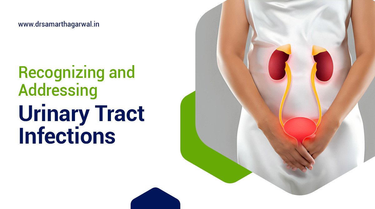
How Long Does Painful Urination Last With Gonorrhea?

How Long Does Painful Urination Last With Gonorrhea?

The duration of painful urination caused by gonorrhea varies based on factors such as the timing of diagnosis and treatment. Without proper antibiotic intervention, symptoms can last for weeks or months. However, most individuals experience significant relief from gonorrhea symptoms within days to a week after starting treatment. Painful urination results from urethral inflammation due to the infection, with severity differing among individuals. Untreated gonorrhea can lead to complications like pelvic inflammatory disease in women and epididymitis in men, potentially causing chronic pain. Early diagnosis and adherence to prescribed antibiotic regimens are crucial to prevent long-term damage and alleviate symptoms. Regular STI testing is essential, as many individuals may be asymptomatic for sexually transmitted infections like gonorrhea and chlamydia.

How long does painful urination last with gonorrhea

The duration of painful urination caused by gonorrhea can vary depending on several factors, including the timeliness of diagnosis and treatment. Without proper antibiotic treatment, the symptoms of gonorrhea, including painful urination, can persist for weeks or even months. However, once appropriate treatment is initiated, most people experience significant relief from symptoms within a few days to a week. It’s important to note that even if symptoms subside, the infection may still be present, emphasizing the need to complete the full course of antibiotics as prescribed by a healthcare provider.

The Centers for Disease Control and Prevention (CDC) recommend specific antibiotic regimens for the treatment of gonorrhea, a common sexually transmitted infection. These treatments are highly effective in eliminating the infection and resolving symptoms when taken as directed. It’s crucial for individuals diagnosed with gonorrhea to abstain from sexual activity until they and their partner(s) have completed treatment and are confirmed to be free of the infection. This helps prevent reinfection and reduces the risk of developing antibiotic-resistant strains of the bacteria that cause gonorrhea.

Does gonorrhea hurt when you pee?

Gonorrhea often causes painful urination, a symptom that can be distressing for those infected. This discomfort is typically due to the inflammation of the urethra, the tube that carries urine from the bladder out of the body. The bacteria that cause gonorrhea can infect the urethra and rectum, leading to irritation and pain during urination. It’s important to note that while painful urination is a common symptom, gonorrhea can also cause no symptoms in some individuals, making regular STI testing crucial for sexually active people.

The severity of pain during urination can vary from person to person. Some may experience mild discomfort, while others may find it extremely painful to urinate. This variation in symptoms underscores the importance of getting tested for gonorrhea and other STIs, even if symptoms include mild or non-existent discomfort. Early detection and treatment are key to preventing complications and reducing the risk of transmission to sexual partners.

How bad does it hurt to pee with gonorrhea?

The intensity of pain during urination with gonorrhea can vary significantly among individuals. Some people may experience a mild burning sensation, while others might endure severe discomfort or sharp pain. The level of pain often depends on the extent of the infection and how long it has been present. In addition to pain, individuals may notice a pus-like discharge from the penis or vagina, which can further exacerbate discomfort during urination. It’s crucial to understand that the severity of symptoms does not necessarily indicate the seriousness of the infection.

Factors such as the individual’s overall health, immune system response, and the strain of the gonorrhea bacteria can influence the intensity of symptoms. Some people may find the pain so severe that it affects their daily activities, while others might only notice mild discomfort. Regardless of the pain level, it’s essential to seek medical attention promptly if any symptoms of gonorrhea are present. Early diagnosis and treatment can help alleviate symptoms and prevent potential complications associated with untreated gonorrhea.

Can gonorrhea cause permanent pain?

While gonorrhea itself does not typically cause permanent pain, untreated or inadequately treated gonorrhea can lead to complications that may result in chronic pain or discomfort. One of the most serious potential complications is pelvic inflammatory disease (PID) in women, which can cause long-term pelvic pain and increase the risk of infertility. In men, untreated gonorrhea can lead to epididymitis, an inflammation of the epididymis that can cause chronic testicular pain if not properly treated.

Additionally, gonorrhea can spread to other parts of the body, potentially causing systemic infections that may result in joint pain or other long-lasting symptoms. It’s crucial to emphasize that these long-term consequences are largely preventable with timely diagnosis and appropriate antibiotic treatment. Regular STI screening, practicing safe sex, and seeking prompt medical attention for any symptoms are key strategies to prevent the potential for permanent pain or other complications associated with gonorrheal infections.

Can gonorrhea damage urethra?

Gonorrhea can indeed cause damage to the urethra if left untreated. The bacteria that cause gonorrhea can lead to inflammation and scarring of the urethral tissues, a condition known as urethral stricture. This narrowing of the urethra can result in long-term complications such as difficulty urinating, increased risk of urinary tract infections, and chronic pain or discomfort. In severe cases, urethral strictures may require surgical intervention to correct. The risk of urethral damage underscores the importance of early detection and treatment of gonorrheal infections.

Furthermore, untreated gonorrhea can also lead to more extensive damage to the reproductive system. In men, it can cause epididymitis, which may result in infertility if not promptly treated. In women, the infection can ascend to the upper reproductive tract, potentially causing pelvic inflammatory disease (PID), which can lead to scarring of the fallopian tubes and increased risk of ectopic pregnancy. These potential complications highlight the critical need for regular STI testing, especially for sexually active individuals, and immediate treatment upon diagnosis to prevent long-term urethral and reproductive system damage.

Does gonorrhea cause bladder pain?

While gonorrhea primarily affects the urethra, it can also cause discomfort and pain in the bladder area. As the infection progresses, it may spread to nearby organs, including the bladder, leading to symptoms such as frequent urination, urgency, and pain or pressure in the lower abdomen. These symptoms can be similar to those of a urinary tract infection (UTI), which is why proper diagnosis by a healthcare provider is crucial. It’s important to note that gonorrhea can coexist with other STIs or urinary tract infections, further complicating the symptom profile.

The presence of bladder pain or discomfort in cases of gonorrhea underscores the importance of comprehensive STI testing. Many people with gonorrhea don’t have any symptoms, or their symptoms may be mild and easily mistaken for other conditions. This asymptomatic nature of many gonorrheal infections highlights the need for regular screening, especially for individuals with multiple sexual partners or those engaging in high-risk sexual behaviors. Early detection and treatment not only alleviate symptoms like bladder pain but also prevent the spread of the infection to sexual partners and reduce the risk of complications associated with untreated gonorrhea.

2

Painful urination associated with gonorrhea can vary in duration depending on the individual and whether treatment has been received. Typically, symptoms may persist until appropriate antibiotics are administered. If you have questions or concerns about painful urination or gonorrhea, contact Dr. Samarth Agarwal for professional guidance regarding your urinary health!

Can Urinary Tract Infection Cause Back Pain?

Urinary tract infections (UTIs) are common bacterial infections that can affect various parts of the urinary system, including the bladder, urethra, and kidneys. While UTIs are typically associated with symptoms such as frequent urination and burning sensations, many people wonder if these infections can also cause back pain. This article will explore the relationship between UTIs and back pain, as well as discuss treatment options and preventive measures.

Can back pain be a symptom of a UTI?

Yes, UTI can cause back pain. Back pain can indeed be a symptom of a urinary tract infection, particularly when the disease has spread to the kidneys. Lower back pain is often associated with UTIs that have progressed beyond the bladder. Bacteria entering the urinary system and traveling upwards can infect the ureters and kidneys, causing inflammation and discomfort. This type of infection, known as pyelonephritis or a kidney infection, can result in pain in the lower back, sides, or groin area.

According to Rosen, J. M., & Klumpp, D. J. (2014). Mechanisms of pain from urinary tract infection. International journal of urology, UTIs can cause transient pelvic pain, but certain E. coli strains lacking O-antigen can lead to chronic pelvic pain, which may persist long after the infection clears. This chronic pain is associated with central sensitization and can mimic symptoms of interstitial cystitis, suggesting a potential link between UTIs and chronic back pain.

It’s important to note that not all UTIs cause back pain, and not all back pain is indicative of a UTI. However, suppose you experience back pain along with other common UTI symptoms such as frequent urination, burning sensation while urinating, cloudy or bloody urine, and a strong urge to urinate. In that case, it may be a sign that the infection has spread to your kidneys. In such cases, it’s crucial to seek medical attention promptly, as kidney infections can lead to more severe complications if left untreated.

How can a person treat back pain caused by a UTI?

Treating back pain caused by a urinary tract infection primarily involves addressing the underlying infection. The most common and effective treatment for UTIs is a course of antibiotics prescribed by a healthcare professional. The type and duration of antibiotic treatment will depend on the severity of the infection and the specific bacteria causing it. It’s essential to complete the entire course of antibiotics as prescribed, even if symptoms improve, to ensure the infection is fully eradicated.

In addition to antibiotics, there are several measures that can help alleviate back pain and other UTI symptoms. Drinking plenty of water can help flush out bacteria from the urinary system and reduce discomfort. Some people find relief by using a heating pad on the affected area or taking over-the-counter pain relievers. While cranberry juice is often touted as a natural remedy for UTIs, its effectiveness is debated. However, maintaining good hygiene practices, such as wiping from front to back after using the bathroom, can help prevent future infections.

Where does a UTI hurt in your back?

When a urinary tract infection causes back pain, it typically manifests in the lower back region. This pain is often described as a dull ache or a more intense, sharp sensation in the area just below the rib cage, on one or both sides of the spine. The discomfort may extend to the sides of the body, particularly in the area where the kidneys are located. Some individuals may also experience pain in the upper back, although this is less common.

The location of the pain can provide clues about the extent of the infection. Lower back pain is often associated with infections that have spread to the kidneys, while pain in the lower abdomen or pelvic area may indicate a bladder infection. It’s important to note that the intensity and exact location of the pain can vary from person to person, and some individuals may experience discomfort in multiple areas simultaneously.

What does it feel like when a UTI spreads to your kidneys?

When a urinary tract infection spreads to the kidneys, the symptoms can become more severe and may include a range of uncomfortable sensations. In addition to lower back pain, individuals may experience a high fever, often accompanied by chills and shaking. Nausea and vomiting are also common symptoms of a kidney infection, as well as fatigue and general malaise. The pain associated with a kidney infection is typically more intense than that of a lower UTI and may be constant or come in waves.

Other signs that a UTI has spread to the kidneys include increased urinary frequency and urgency, often with little urine output. The urine may appear cloudy, dark, or bloody, and there may be a strong, unpleasant odor. Some people also report experiencing confusion or mental fogginess when the infection reaches the kidneys. If you suspect that your UTI has progressed to a kidney infection, it’s crucial to seek immediate medical attention, as this condition can lead to serious complications if left untreated.

1

If you’re experiencing back pain along with symptoms of a urinary tract infection (UTI), it’s important to consult with a healthcare professional. Contact Dr. Samarth Agarwal if you have any questions or concerns about your urinary health and how it may relate to back pain. Your well-being is a priority!

What Can Cause Pressure in Lower Abdomen and Frequent Urination for Female?

Experiencing pressure in the lower abdomen accompanied by frequent urination can be a source of concern for many women. Gaining a comprehensive understanding of the potential causes can offer valuable insights into one’s health. Various factors may contribute to these symptoms, ranging from urinary tract infections to conditions such as endometriosis and ovarian cysts. This article will examine these common causes, outline when it is advisable to seek medical consultation, and discuss possible treatment options, with insights from Dr. Samarth Agarwal, a urologist committed to women’s health.

What Are The Common Causes of Pressure in Lower Abdomen and Frequent Urination for Female?

Pressure in the lower abdomen and frequent urination in females can arise from various health issues, ranging from urinary tract infections (UTIs) to chronic conditions such as interstitial cystitis and hormonal changes associated with menstrual cycles. Understanding these causes is crucial to managing symptoms effectively and maintaining good pelvic health.

Factors like emotional stress and pelvic health also contribute significantly to this discomfort, thereby affecting the quality of life for many women. Additionally, issues such as bladder dysfunction and bladder control problems can exacerbate these symptoms.

Being aware of these conditions can guide individuals towards seeking appropriate medical attention to alleviate discomfort and ensure a healthier lifestyle.

Urinary Tract Infection (UTI)

A urinary tract infection (UTI) is a common cause of pressure in the lower abdomen and frequent urination, often presenting symptoms like increased urinary urgency and discomfort.

This discomfort is primarily due to the inflammation of the bladder wall, which can lead to an intense sensation of needing to void, even when the bladder is not full. This inflammation can also cause bladder pain and bladder irritation.

Individuals may experience burning sensations during urination, cloudy urine, or even a particular odor. As the infection progresses, symptoms may worsen, indicating a crucial need for prompt intervention.

  • Causes: Common culprits include bacteria such as Escherichia coli, which may enter the urinary tract through improper hygiene practices or sexual activity.
  • Symptoms: Alongside urgency, individuals may notice fever, chills, or back pain, suggesting a potential kidney involvement.

Many treatment options are available, ranging from prescribed antibiotics to increased fluid intake to flush the system. Additionally, practicing bladder retraining and maintaining a bladder diary can help manage symptoms effectively.

Importantly, consulting a healthcare provider is essential not only to confirm the diagnosis but to tailor an effective treatment plan, ensuring recovery is swift and complete.

Bladder Infection

Bladder infections can lead to significant bladder pain and increased urinary frequency, often requiring prompt medical intervention to avoid further complications.

When these infections occur, individuals frequently experience a burning sensation during urination, urgency to urinate, and sometimes even blood in the urine. These symptoms may also be accompanied by urinary frequency and night urination.

It’s crucial to recognize these urinary symptoms as signs that should not be ignored, as they may indicate a more severe underlying issue like bladder stones or chronic bladder pain.

Treatment options typically involve antibiotics, which can effectively eliminate the bacterial infection. Incorporating adequate hydration and possibly using pain relief medications can also alleviate discomfort.

  • Patients are advised to consult healthcare providers to establish tailored treatment plans.
  • Understanding personal risk factors and preventive measures is equally important.

Seeking timely medical attention give the power tos individuals to manage this condition effectively and enhances overall urinary health.

Overactive Bladder

An overactive bladder is characterized by an uncontrollable urge to urinate frequently, which can drastically affect a woman’s quality of life and overall bladder control, leading to interruptions in daily activities, social engagements, and even sleep patterns. This condition may result in bladder spasms and bladder signaling issues.

Many individuals experience discomfort and embarrassment due to unexpected leaks or constant restroom trips, contributing to not only physical distress but emotional struggles as well.

This persistent condition often results in anxiety and frustration, making it difficult for individuals to engage fully in their lives. The psychological impact can amplify feelings of isolation and depression, creating a negative cycle that is hard to escape.

To manage symptoms effectively, individuals are encouraged to explore a variety of treatment options. These may include:

  • Behavioral therapies: Strategies such as bladder training and pelvic floor muscle exercises can enhance control.
  • Medications: Certain prescriptions may help relax the bladder muscles, providing relief from urgency.
  • Lifestyle changes: Adjustments in diet, such as reducing caffeine and alcohol intake, along with maintaining a healthy weight, can significantly alleviate symptoms.

Considering these approaches can help restore balance and improve overall emotional well-being, enabling individuals to regain a sense of normalcy in their lives.

Interstitial Cystitis

Interstitial cystitis, often referred to as painful bladder syndrome, is a chronic condition that can manifest as persistent pelvic pain and pressure in the bladder area. This condition can involve chronic pain and significantly affect quality of life.

This complex ailment can significantly disrupt daily life, leading not only to physical discomfort but also emotional distress. Individuals may experience a range of symptoms that vary in intensity, including:

  • Frequent urination and urinary urgency
  • Urgency to urinate
  • Pelvic pressure or discomfort
  • Pain during sexual intercourse

The exact causes of interstitial cystitis remain uncertain; however, several factors are believed to contribute to its onset. These may include:

  • Protective lining of the bladder
  • Autoimmune reaction
  • Chronic infections
  • Pelvic floor dysfunction and weakened bladder capacity

Given the significant impact on emotional well-being, it is crucial to recognize the importance of bladder health and to implement effective management strategies, such as pelvic floor exercises and pain management techniques.

Treatment options like bladder instillations, dietary modifications, physical therapy, and medications can prove beneficial in alleviating symptoms and improving quality of life.

Pelvic Inflammatory Disease (PID)

Pelvic inflammatory disease (PID) is an infection of the female reproductive organs that can result in significant pelvic pain and pressure in the lower abdomen, often influenced by sexual activity. This condition can cause severe health issues if not promptly treated.

This condition is primarily a result of untreated sexually transmitted infections (STIs) such as chlamydia and gonorrhea, as these bacteria can ascend from the vagina into the uterus, fallopian tubes, and surrounding tissues, leading to inflammation.

As PID progresses, symptoms may intensify and can include:

  • Chronic pelvic pain: persistent pain that can drastically affect daily activities and sexual intimacy.
  • Abnormal vaginal discharge: which may have an unusual odor.
  • Fever: often indicating a more severe infection.
  • Painful intercourse: discomfort during or after sexual activity.

The impact on pelvic health can be considerable, potentially resulting in long-term complications such as infertility, ectopic pregnancy, or continuous pelvic pain. Additionally, untreated PID can lead to more severe infections and chronic pain.

Due to these risks, it is essential for individuals experiencing symptoms to seek medical attention promptly. Treatment often involves antibiotics to eradicate the infection, and in severe cases, surgery may be necessary. Regular follow-ups with a healthcare provider are also crucial to monitor and manage the condition effectively.

Early intervention is crucial for managing this condition effectively and safeguarding reproductive health.

Endometriosis

Endometriosis is a chronic condition where tissue similar to the lining of the uterus grows outside it, often leading to significant pelvic pain and discomfort, especially during menstruation, affecting both physical well-being and emotional health. This condition frequently involves chronic pain and may require specialized treatment options.

Women experiencing this condition may note various symptoms such as heavy periods, pain during intercourse, and even complications with fertility. The hormonal changes associated with endometriosis can exacerbate these symptoms, resulting in inflammation and scarring in the pelvic area. Recognizing these signs is crucial because prompt diagnosis can help prevent further complications.

Medical intervention is essential for managing endometriosis effectively, offering various treatment options including:

  • Hormonal therapies that regulate the menstrual cycle
  • Pain management strategies
  • Surgical options to remove endometrial tissue
  • Fertility treatments for those affected by infertility

With proper care and treatment, individuals can significantly improve their quality of life and tackle the challenges posed by this condition.

Ovarian Cysts

Ovarian cysts are fluid-filled sacs on the ovaries that can lead to pelvic pain and pressure, often exacerbated by hormonal fluctuations during the menstrual cycle.

These cysts can develop at various stages of a woman’s life, particularly during reproductive years. They may form due to normal physiological processes, such as ovulation, or can arise from other underlying issues like hormone fluctuations and autoimmune reactions.

While many women experience minimal symptoms, some may notice discomfort, bloating, or irregular menstrual cycles as their signs.

As these cysts grow, they can exert increased pressure on surrounding organs, including the bladder, leading to frequent urination or difficulty in maintaining bladder control. Some women may also experience symptom-free periods wherein the cysts shrink temporarily.

  • Common symptoms include:
  • Pelvic pain
  • Nausea
  • Difficulty emptying the bladder

In most cases, ovarian cysts resolve independently, but persistent or severe symptoms vary and warrant medical evaluation.

Effective treatment strategies range from lifestyle modifications to hormonal therapies, and, in certain instances, surgical intervention may be necessary. It’s crucial to consult a healthcare provider if unusual symptoms arise or exacerbate over time, as they can lead to significant health consequences.

Uterine Fibroids

Uterine fibroids are noncancerous growths in the uterus that can cause pelvic pain, increased pressure in the lower abdomen, and heavy menstrual bleeding. These are important considerations in women’s health.

These growths, which vary greatly in size and can be found on the uterine wall or inside the uterus, can contribute to a wide range of symptoms that may affect an individual’s quality of life significantly. Women experiencing uterine fibroids might notice:

  • Frequent urination due to pressure on the bladder
  • A sense of fullness in the lower abdomen
  • Pain during intercourse

In some cases, fibroids might lead to complications such as infertility or pregnancy-related issues. It’s crucial for those facing these symptoms to seek guidance from a healthcare provider, who can offer insights on management options such as medication, lifestyle changes, or surgical procedures. Consulting a health care provider ensures appropriate treatment.

Understanding the available treatment options can give the power to individuals to make informed decisions about their health.

Pregnancy

During pregnancy, women often experience increased pelvic pressure and urinary frequency due to hormonal changes and the growing uterus pressing on the bladder, sometimes exacerbated by diuretic beverages.

This pressure can lead to the frequent urge to urinate, particularly in the later stages as the fetus grows larger. These physiological changes can cause discomfort and sometimes feelings of anxiety about being too far from a restroom.

To alleviate these symptoms, it’s beneficial to stay hydrated but also to manage fluid intake, particularly before bedtime. Kegel exercises can help strengthen pelvic floor muscles, offering some relief from pressure.

If urinary issues are accompanied by pain, burning sensations, or changes in the appearance of urine, it is advisable to consult a healthcare provider to rule out potential urinary tract infections, Interstitial cystitis, or other complications.

Menopause

Menopause brings about hormonal fluctuations that can significantly impact bladder function, leading to symptoms such as frequent urination and discomfort. These changes are a crucial part of the urinary network.

During this transitional phase, many women may find that these alterations not only disrupt daily activities but also detract from overall quality of life. The decline in estrogen levels can exacerbate urinary tract issues, making it crucial to understand the relationship between hormonal changes and urinary health. Proper education on this matter can give the power to individuals to seek effective solutions, ensuring informed decisions regarding health consequences.

  • Potential Treatments: Various treatments exist, including hormone replacement therapy (HRT), which can alleviate symptoms for some.
  • Lifestyle Adjustments: Adopting a balanced diet, maintaining hydration, and incorporating pelvic floor exercises can also significantly improve symptoms.
  • Consultation: Regular consultations with healthcare providers can facilitate personalized treatment plans tailored to specific needs.

Being proactive and informed can lead to better management of urinary health challenges associated with menopause.

When Should You See a Doctor?

It is crucial to seek medical attention when experiencing persistent urinary symptoms or severe pelvic pain, as these may indicate underlying health issues such as prostate inflammation or other conditions that require professional evaluation.

Ignoring such discomfort can lead to complications that may significantly impact quality of life. Common warning signs include:

Recognizing these symptoms early can facilitate a more effective treatment plan, making it vital for individuals to prioritize their health. Addressing these issues promptly not only allows for early diagnosis and intervention but also helps in managing potential chronic conditions that could escalate without proper care.

How is Pressure in Lower Abdomen and Frequent Urination Diagnosed?

Diagnosing the causes of pressure in the lower abdomen and frequent urination typically involves consulting a healthcare provider, who may recommend tests to evaluate bladder function and pelvic health.

During this diagnostic process, the healthcare provider may suggest various assessments to gain a comprehensive understanding of the underlying issues. Initially, keeping a bladder diary can help track urinary habits and symptoms over several days. This documentation aids in identifying patterns and potential triggers that may contribute to the discomfort.

  • A thorough physical examination might be conducted to locate any anatomical abnormalities or tenderness in the pelvic region.

For further investigation, imaging studies such as:

  • ultrasounds
  • CT scans

may be utilized to visualize the bladder and surrounding organs. Depending on the findings, specialized tests to assess bladder function, including urodynamic studies, can provide critical insights into how effectively the bladder is performing its role.

Through these methods, a tailored treatment plan can be developed, addressing the unique needs of the individual.

What are the Treatment Options for Pressure in Lower Abdomen and Frequent Urination?

Discover effective treatment options to alleviate pressure in the lower abdomen and frequent urination. Explore physical therapy, medication, behavioral changes, antibiotics, electrical nerve stimulation, surgery, chemotherapy, and biofeedback therapy for potential solutions.

Treatment Options for Pressure in Lower Abdomen and Frequent Urination

  1. Physical Therapy: Strengthening the pelvic floor muscles through exercises like Kegels can improve bladder control.
  2. Pentosan Polysulfate Sodium: This medication may be used to block irritants in urine, reducing bladder pressure.
  3. Behavioral Changes: Modifying diet and fluid intake, especially reducing caffeine and alcohol, can help alleviate symptoms.
  4. Antibiotics: Infections are typically treated with antibiotics, if they are the cause of the symptoms.
  5. Electrical Nerve Stimulation (TENS): This treatment may help relieve symptoms by increasing blood flow to the bladder and strengthening pelvic muscles.
  6. Surgery: For severe cases, surgery may be considered to correct any physical issues causing the pressure and frequent urination.
  7. Chemotherapy, Radiation, and Other Treatments: These are common treatments for cancer, which can cause pressure in the lower abdomen and frequent urination. Other options include targeted therapy, immunotherapy, and hormonal therapy.
  8. Biofeedback Therapy: Combined with Kegel exercises, this treatment can help a person become more aware of their body’s functions to better manage symptoms.
4

Contact Dr. Samarth Agarwal if you have any questions or concerns about your Urinary health!

Can Urine Infection Cause Stomach Pain?

Urinary tract infections (UTIs) are common bacterial infections that can indeed cause stomach pain, particularly in the lower abdominal region. These infections typically affect the bladder and urethra, but can sometimes spread to the kidneys, leading to more severe symptoms. Understanding the connection between UTIs and abdominal discomfort is crucial for proper diagnosis and treatment.

Can urinating cause abdominal pain?

Urinating can cause abdominal pain, especially in individuals suffering from a urinary tract infection or bladder infection. When bacteria enter the urinary system, they can irritate the bladder and urethra, leading to discomfort during urination and potential infection symptoms. This pain may radiate to the lower abdomen, causing a sensation of pressure or cramping. In some cases, the infection may spread to the kidneys, resulting in more intense abdominal pain and other serious symptoms.

According to Rosen, J. M., & Klumpp, D. J. (2014). Mechanisms of pain from urinary tract infection. International journal of urology : official journal of the Japanese Urological Association, Urinary tract infections (UTIs) can cause abdominal pain due to the response of the body to uropathogenic E. coli (UPEC). Unlike asymptomatic bacteriuria, UPEC induces transient pelvic pain. This pain response is linked to the bacterial lipopolysaccharide (LPS) and its interaction with the TLR4 receptor, rather than bladder inflammation. Repeated infections can lead to chronic pelvic pain, especially when certain bacterial components, like O-antigen, are absent. Chronic pain can persist even after the bacteria are cleared and is associated with voiding dysfunction and anxiety or depression. Overall, a UTI can trigger both acute and chronic pain, resembling symptoms of interstitial cystitis.

The abdominal pain associated with UTIs can vary in intensity and location. It may be felt as a dull ache or sharp, stabbing sensations in the lower belly. Additionally, frequent urination and a persistent urge to urinate are common symptoms that can exacerbate abdominal discomfort. If left untreated, the infection can spread and potentially lead to more severe complications, increasing the risk of kidney damage or sepsis, which may cause serious health concerns.

How do you get rid of a stomach ache from a urine infection?

To alleviate stomach aches caused by a urine infection, it’s essential to treat the underlying UTI. The most effective treatment for UTIs is typically a course of antibiotics prescribed by a healthcare provider. These medications help eliminate the bacterial infection and reduce symptoms, including abdominal pain. It’s crucial to complete the entire antibiotic course as prescribed to prevent the infection from recurring or becoming antibiotic-resistant.

In addition to antibiotics, there are several self-care measures that can help relieve stomach aches associated with UTIs. Drinking plenty of water helps flush out bacteria from the urinary system and dilutes urine, which can help prevent irritation. Applying a warm compress to the lower abdomen can also provide relief from pain and discomfort. Over-the-counter pain relievers, such as ibuprofen or acetaminophen, may be used to manage pain and reduce inflammation. However, it’s important to consult a healthcare provider before taking any medications.

What does stomach pain from UTI feel like?

Stomach pain from a UTI can manifest in various ways, depending on the severity and location of the infection. Typically, individuals experience a dull ache or pressure in the lower abdomen, which may intensify during urination. The pain can range from mild discomfort to sharp, stabbing sensations. In some cases, the abdominal pain may be accompanied by a feeling of fullness or bloating in the bladder area.

As the infection progresses, the abdominal pain may become more pronounced and constant. Some people describe it as a burning sensation or a feeling of heaviness in the lower belly. If the infection spreads to the kidneys, the pain may extend to the sides or lower back, indicating a possible kidney infection. It’s important to note that UTI symptoms can vary from person to person, and some individuals may experience more severe abdominal pain than others.

Do UTI cramps feel like period cramps?

UTI cramps can indeed feel similar to period cramps, which can sometimes lead to confusion in distinguishing between the two. Both types of cramps typically occur in the lower abdominal area and can cause discomfort or pain. However, there are some key differences to consider. UTI cramps are often accompanied by other urinary symptoms, such as frequent urination, burning sensation during urination, and cloudy or strong-smelling urine.

Unlike period cramps, which usually follow a predictable pattern and timing related to the menstrual cycle, UTI cramps can occur at any time and may persist until the infection is treated. Additionally, UTI cramps may be more focused in the bladder area, while period cramps tend to be more diffuse across the lower abdomen. It’s important to pay attention to other accompanying symptoms and consult a healthcare provider if there’s uncertainty about the cause of the cramps, as they may cause further complications.

What is the cause of abdominal pain after urinating?

Abdominal pain after urinating can have several causes, with urinary tract infections being one of the most common. In the case of a UTI, the pain is typically due to inflammation and irritation of the bladder and urethra caused by bacterial infection. As urine passes through the infected urinary system, it can trigger pain and discomfort that may persist even after urination is complete.

Other potential causes of abdominal pain after urinating include interstitial cystitis, bladder stones, or prostate problems in men, so it’s advisable to see a doctor. In some cases, the pain may be related to underlying conditions such as pelvic inflammatory disease or endometriosis, which may cause similar symptoms. If the abdominal pain after urinating is severe, persistent, or accompanied by other concerning symptoms, it’s crucial to seek medical attention for proper diagnosis and treatment.

4

Contact Dr. Samarth Agarwal if you feel any discomfort while urinating or feel any abdominal pain!

Why Do I Have Lower Back Pain and Frequent Urination at Night?

Experiencing lower back pain accompanied by frequent nighttime urination can be both concerning and disruptive to daily life. These symptoms may stem from a range of underlying conditions, including urinary tract infections, kidney issues, or prostate-related problems. This article aims to explore the potential causes of these discomforts, assess their impact on overall health, and discuss the available treatment options, including effective treatments and lifestyle changes. Gaining a comprehensive understanding of these factors can empower individuals to take proactive measures toward relief and ascertain when it is necessary to seek guidance from a healthcare professional or health provider.

Why Can One Have Lower Back Pain and Frequent Urination at night?

Experiencing lower back pain and frequent urination at night, known medically as nocturia, can be distressing and may indicate underlying health issues that require attention.

Nocturia is characterized by the need to wake at night to urinate, which can disrupt sleep and lead to health consequences, including sleep disturbances, over time. Understanding the potential causes—ranging from urinary tract infections, prostate problems, to diabetes and obstructive sleep apnea—can assist in determining the appropriate medical interventions and lifestyle adjustments needed for relief and management.

What Are the Causes of Lower Back Pain and Frequent Urination at Night?

Understanding the causes of lower back pain and frequent urination at night is crucial for effective diagnosis and treatment. These symptoms can arise from various medical conditions and lifestyle factors, including urinary tract infections (UTIs), prostate problems such as benign prostatic hyperplasia, diabetes, obstructive sleep apnea, and lifestyle factors like caffeine consumption and alcohol intake.

Each of these conditions may have overlapping symptoms and varying degrees of severity, necessitating a thorough evaluation by a medical professional to identify the root cause.

Urinary Tract Infection (UTI)

A urinary tract infection (UTI) is a common condition that can lead to symptoms like lower back pain and frequent urination at night. UTIs occur when bacteria infect the urinary system, often causing discomfort such as a burning sensation during urination, abdominal pain, and an increased urgency to urinate. If left untreated, a UTI can lead to more severe complications, including chronic kidney problems or pyelonephritis.

This condition can arise from various causes, including poor hydration, sexual activity, and existing urinary tract abnormalities. Risk factors often involve age, gender, and personal hygiene practices.

Diagnosis typically begins with a urinalysis, which screens for signs of infection, followed by a urine culture to identify the specific bacteria responsible. Blood tests may also be conducted to rule out other conditions. Early detection is crucial, as timely intervention can prevent complications.

  • Symptoms to watch for include:
  • Increased urgency and frequency of urination
  • Cloudy or strong-smelling urine
  • Pelvic pain or pressure

Treatment primarily involves antibiotics, which effectively target the bacterial infection. Home care strategies such as increasing fluid intake, consuming cranberry products, practicing good hygiene, and maintaining proper sleep hygiene can support recovery and help reduce the likelihood of future infections.

Kidney Infection

Kidney infection, medically referred to as pyelonephritis, is a serious condition that can cause lower back pain and frequent urination at night.

This type of urinary tract infection affects the kidneys and may lead to severe abdominal pain, fever, and symptoms of systemic illness. Aside from the discomfort of lower back pain, individuals may experience chills, nausea, a burning sensation during urination, and potentially chronic kidney failure if left untreated. Early detection is vital to mitigate potential complications like chronic kidney failure and sepsis.

  • Causes: Bacteria from the bladder or urethra often ascend to the kidneys, which may trigger an infection. Other risk factors include urinary blockages and weakened immune systems.
  • Symptoms: Common signs include fever, flank pain, increased frequency of urination, and malaise.
  • Diagnosis: Physicians frequently rely on blood tests and imaging techniques to ascertain the presence of infection and assess kidney function.
  • Treatment: Management typically involves antibiotics to combat the infection alongside medications to relieve symptoms. Staying well-hydrated is crucial for flushing out bacteria and supporting overall kidney health.

Prostate Problems

Prostate problems, particularly benign prostatic hyperplasia (BPH), can significantly contribute to lower back pain and frequent urination at night. The prostate gland plays a crucial role in these symptoms. As the prostate gland enlarges, it may obstruct the urethra, leading to urinary symptoms such as increased urination frequency, dysuria, and discomfort in the pelvic area. Recognizing obstruction as a key factor is important. Understanding this condition is vital for appropriate management and treatment options.

Additionally, BPH and other prostate issues such as chronic nonbacterial prostatitis can also impact men’s health significantly. Symptoms often include nagging pelvic pain, difficulties in urination, and, sometimes, lower back pain, complicating the individual’s quality of life.

  • Risk factors for these prostate problems may encompass age, family history, and lifestyle choices, such as diet and exercise.
  • Diagnostic methods typically involve a combination of physical examinations, blood tests, and imaging studies to assess prostate health accurately.
  • Effective treatment strategies vary depending on the specific diagnosis; options can include medication adjustments, dietary changes, and surgical interventions for more severe cases.

Recognizing the multifaceted nature of prostate health is essential for proactive management.

Bladder Problems

Bladder problems can lead to symptoms like lower back pain and frequent urination at night, frequently presenting as nocturnal polyuria or incontinence. Conditions such as bladder stones or infections may cause discomfort, including a burning sensation during urination, frequent urges to void, and pain during urination. Understanding the various bladder conditions is essential for determining effective treatments.

These issues can arise from diverse underlying causes, such as bacterial infections, dietary factors, or dehydration. Symptoms frequently associated with these conditions often include:

  • Fluctuating urinary patterns
  • Pain during voiding
  • Unexplained abdominal discomfort

It is crucial to address bladder health proactively, as early intervention can lead to more effective management strategies. Integrating adequate hydration and balanced fluid intake into one’s lifestyle not only aids in flushing out harmful bacteria but also can prevent the formation of bladder stones. Making conscious lifestyle adjustments, such as limiting caffeine and alcohol, can further enhance bladder function and overall well-being.

Diabetes

Diabetes can significantly affect urinary health, often resulting in elevated thirst and frequent urination at night, known as nocturia. High blood sugar levels can lead to osmotic diuresis, prompting increased urination frequency and potential health consequences if not managed properly. Understanding the relationship between diabetes and urinary symptoms is crucial for effective diabetes management.

Diabetes introduces complex challenges that extend beyond glucose control, impacting various bodily functions and potentially affecting bladder capacity. When blood sugar levels soar, the kidneys work harder to filter and absorb excess glucose, occasionally resulting in increased urine production. This can lead to various urinary symptoms, including:

  • Increased frequency of urination during the day
  • Urgency to urinate
  • Nighttime urination, disrupting sleep
  • Possibility of urinary tract infections

Diagnosis encompasses blood glucose testing and monitoring urinary patterns, often performed by a medical professional. Effective management strategies require a holistic approach that includes:

  • Regular blood sugar monitoring
  • Adopting a balanced diet and exercising
  • Staying hydrated, but managing fluid intake wisely to avoid excessive urination

Consultation with a healthcare provider is essential for personalized guidance and regular check-ups to prevent complications associated with urinary health, considering the patient’s medical history and urination habits.

Pregnancy

Pregnancy can lead to lower back pain and frequent urination due to hormonal changes and pressure on the bladder as the fetus grows, affecting bladder capacity and urination frequency. These changes can reduce bladder capacity and increase the frequency of nighttime urination, known as nocturnal polyuria, which can disrupt sleep patterns.

Understanding these changes is vital for managing discomfort and maintaining overall health during pregnancy.

As the body adapts to nurture new life, the shift in hormonal balance affects various systems, resulting in physical symptoms that can be quite bothersome, sometimes necessitating a physical exam. Commonly, the increase in progesterone relaxes pelvic muscles, contributing to urinary incontinence and pain in the lower back.

To alleviate these discomforts, expectant mothers can adopt a range of coping strategies:

  • Regular Exercise: Gentle activities like prenatal yoga can strengthen the back and pelvic floor muscles, benefiting those with conditions like a herniated disc.
  • Pillows for Support: Using supportive pillows while sleeping or sitting can improve posture and reduce strain on the lower back.
  • Staying Hydrated: Drinking adequate water throughout the day can maintain normal bladder function and minimize nighttime disruptions.
  • Heat Therapy: Applying a warm compress to the lower back can relieve muscle tension.

Implementing these adjustments not only eases the symptoms but also contributes to a more comfortable pregnancy experience, helping manage back pain and Nocturia.

How Does Lower Back Pain and Frequent Urination at Night Affect Your Health?

Lower back pain and frequent urination at night can have profound effects on overall health and quality of life. These conditions can lead to significant sleep disturbances, increased fatigue, dysuria, and emotional stress, which may ultimately affect daily functioning and overall well-being.

Seeking guidance from a health provider, especially for a urologic patient, can aid in understanding the implications and establishing effective management strategies.

What Are the Treatment Options for Lower Back Pain and Frequent Urination at Night?

There are various treatment options available for managing lower back pain and frequent urination at night, tailored to the underlying causes of these symptoms, such as diabetes or benign prostatic hyperplasia.

Effective treatments may include medication adjustments, lifestyle changes, and home care strategies that promote overall health and wellness, including managing fluid deprivation and avoiding caffeine and alcohol.

Collaborating with a healthcare provider is essential for developing a personalized treatment plan that addresses specific needs.

When Should You See a Doctor for Lower Back Pain and Frequent Urination at Night?

Knowing when to see a doctor for lower back pain and frequent urination at night is essential for maintaining health and well-being. If symptoms are persistent, severe, or accompanied by other concerning symptoms such as fever, blood in urine, or significant pain, prompt medical attention is necessary.

Keeping a symptom log can help communicate effectively with your health provider about any changes or concerns.

1

Contact Dr. Samarth Agarwal if you have any questions or concerns about your Urinary health!

Are Kidney Cancer and Breast Cancer Related?

Kidney and breast cancer represent two of the most prevalent malignancies affecting millions globally. An in-depth understanding of these cancers, including their associated risk factors, symptoms, and treatment options, is essential for facilitating early detection and effective management. This article examines the relationship between kidney and breast cancer, including the potential for dual malignancies, as articulated by Dr. Samarth Agarwal, a urologist based in Siliguri. It addresses the impact of these diseases on individuals, highlights the commonalities in their risk factors, such as genetic predispositions and lifestyle choices, and discusses potential lifestyle modifications that may help reduce the risk. We invite readers to engage with us as we explore the complexities surrounding these two significant health challenges.

What is Kidney Cancer?

Kidney cancer primarily refers to renal cell carcinoma, which originates in the lining of the kidney’s tubules, comprising about 90% of kidney cancer cases. It often requires comprehensive diagnostic imaging and clinical evaluation for effective oncological care.

Clear cell carcinoma, a subtype of renal cell carcinoma, is characterized by distinct histopathological features and is known for its aggressive nature. This type of malignancy is often diagnosed during routine medical examinations or imaging tests, such as ultrasound examination or CT scans, making early detection crucial for successful treatment outcomes.

What is Breast Cancer?

Breast cancer is a multifaceted disease characterized by the uncontrolled growth of breast cells, often manifesting as breast masses, bilateral breast symptoms, or changes in breast parenchyma. It can present with atypical features that may be detected through mammogram findings, emphasizing the need for regular breast imaging and clinical evaluation.

Understanding the risk factors associated with breast cancer, including genetic predispositions, mammographic density, and lifestyle choices, plays a pivotal role in early detection and effective oncological care.

Are Kidney Cancer and Breast Cancer Related?

Understanding the relationship between kidney cancer and breast cancer is critical, as research indicates that patients can present with dual malignancy or synchronous cancers, where both cancer types are diagnosed concurrently or sequentially.

This phenomenon, known as synchronous cancers, highlights the necessity of comprehensive diagnostic workups, including histopathological assessment, and interdisciplinary patient management strategies. Furthermore, cases of breast cancer metastasizing to the kidneys, although rare, underscore the importance of thorough surveillance and multi-cancer detection methods in oncology.

What are the Risk Factors for Developing Both Cancers?

Identifying the risk factors for developing both kidney cancer and breast cancer is crucial for early intervention and prevention strategies.

Factors such as age, family history, and lifestyle choices, including diet and exercise, can significantly influence an individual’s likelihood of developing these cancer types. Understanding the tumor characteristics can further aid in assessing risk.

Understanding the cancer stages, radiological characteristics, and their implications in the context of patient history can help in tailoring effective treatment and management plans.

Several lifestyle changes can play a pivotal role in mitigating these risks. For instance, maintaining a healthy weight, engaging in regular physical activity, and adopting a balanced diet rich in fruits, vegetables, and whole grains are essential.

Reducing the intake of processed foods and red meats has also been linked to lower cancer risk. Regular screenings, such as mammography, and self-examinations are critical for early detection, especially in individuals with a family history of these diseases.

The stage of cancer significantly impacts prognosis and treatment decisions; for example, early-stage kidney and breast cancers typically have a higher survival rate and more treatment options available.

Patient education about these stages, including understanding treatment pathways and the importance of adhering to recommended interventions, give the power tos individuals to take an active role in their health journey.

Fostering awareness about both the risk factors and the importance of continuous education can aid in better health outcomes.

How Can Kidney Cancer and Breast Cancer be Diagnosed?

The diagnosis of kidney cancer and breast cancer relies heavily on advanced diagnostic imaging techniques, such as the Breast Imaging Reporting and Data System, and clinical evaluation, which play a pivotal role in identifying malignancies at an early stage.

Methods such as ultrasound examination, MRI, and mammography system are essential for accurate imaging assessment, while histopathological diagnosis through biopsy provides definitive confirmation of neoplastic growth in tissues.

A thorough clinical examination, along with a detailed patient history and immunohistochemistry analysis, further enhances the effectiveness of the diagnostic workup.

What are the Common Symptoms of Kidney Cancer and Breast Cancer?

Common symptoms of kidney cancer and breast cancer can vary, but vigilance for atypical features is essential for timely diagnosis. Early recognition of these signs plays a crucial role in initiating appropriate preventive measures and treatments that can significantly influence patient outcomes. Kidney cancer may present with symptoms such as hematuria, pain in the flank area, and potential lymph node involvement, while breast cancer can manifest as a noticeable lump, bilateral breast changes, alterations in breast discharge, or other alterations in breast appearance. Recognizing these symptoms can lead to prompt medical evaluation, malignancy diagnosis, and effective oncological care.

Understanding the overlapping symptoms is vital, as both cancers may present vague symptoms like fatigue and unexplained weight loss. Some patients may be unaware that these subtle indicators could signify more serious conditions.

The significance of early symptom recognition cannot be overstated, as it directly impacts the effectiveness of treatment plans and patient survival rates.

  • For kidney cancer: Pay attention to persistent abdominal pain, swelling, or unexplained fever.
  • For breast cancer: Look out for skin changes such as dimpling or puckering, which might be easily dismissed.

By prioritizing awareness of these diverse symptoms, individuals can facilitate quicker diagnoses, leading to earlier interventions and comprehensive care strategies in their treatment journeys.

What are the Screening Tests for Kidney Cancer and Breast Cancer?

Screening tests play a vital role in the early detection of kidney cancer and breast cancer, allowing for timely intervention that can significantly improve patient outcomes. For breast cancer, mammography is the gold standard screening tool, while ultrasound examination is often utilized for further assessment in high-risk individuals. In the case of kidney cancer, imaging assessments such as CT scans and MRIs are critical for identifying renal masses and determining their characteristics.

These essential tests not only help in spotting abnormalities at an early stage but also facilitate a deeper understanding of the disease’s progression and aid in creating an effective treatment plan.

For breast cancer, health professionals recommend women aged 40 and over to undergo a mammogram annually, while those with a family history may need to start earlier. Similarly, kidney cancer screening mostly involves imaging tests for those at higher risk, such as individuals with a history of kidney disease or smoking, with evaluations often recommended every few years.

  • The effectiveness of these screening tests, including multi-cancer early detection methods is well-documented, demonstrating a reduction in mortality rates when cancer is detected early.
  • These tests provide more options for treatment, allowing healthcare providers to tailor a patient’s care effectively.

Ultimately, the importance of early detection cannot be overstated, as it is directly linked to increased survival rates and better overall prognoses.

What are the Treatment Options for Kidney Cancer and Breast Cancer?

The treatment options for kidney cancer and breast cancer encompass a wide array of medical, surgical, and supportive therapies, tailored to the individual needs of patients.

Surgical oncology is often the first line of treatment, with nephrectomy and partial nephrectomy being prevalent for kidney cancer, while lumpectomy or mastectomy may be employed for breast cancer.

Additionally, advancements in immunotherapy, such as GATA3 and PAX9 targeting, and chemotherapy have transformed the landscape of cancer treatment, offering promising avenues for managing both malignancies.

What are the Surgical Procedures for Treating Kidney Cancer and Breast Cancer?

Surgical procedures are fundamental components in the treatment of kidney cancer and breast cancer, with nephrectomy and partial nephrectomy being common for renal tumors. For breast cancer, surgical options include mastectomy and lumpectomy, which are determined by the tumor’s size, location, and patient preferences. These surgical techniques necessitate careful consideration of oncological principles, case presentations, and patient management to ensure optimal outcomes.

The choice of surgical intervention, guided by histopathology reports and tumor characteristics, plays a crucial role in shaping the overall treatment strategy. Each option comes with distinct considerations, including:

  • indications: The selection often depends on tumor staging and patient health.
  • benefits: Effective surgery can lead to significant tumor reduction.
  • risks: Potential complications should be thoroughly discussed.

These procedures may be complemented by adjuvant therapies, such as chemotherapy or radiation, often assessed through Breast Imaging Reporting and Data System (BI-RADS) guidelines, emphasizing the need for a multidisciplinary approach. By integrating surgery with other treatment modalities, healthcare providers aim to enhance prognosis and improve the quality of life for those affected.

What are the Chemotherapy and Radiation Therapy Options for Kidney Cancer and Breast Cancer?

Chemotherapy and radiation therapy are critical adjuncts in the treatment of kidney and breast cancers, particularly for patients with advanced disease, bilateral breast metastasis, or high-risk features. While chemotherapy can be employed to target systemic disease, radiation therapy may be utilized for palliation or as an adjunct to surgical procedures, depending on the treatment plan. Effective patient management and oncological care are essential to navigate the complexities of these therapies.

These approaches play a pivotal role in the overall treatment strategy, particularly when the goal is to achieve maximal therapeutic benefit while minimizing potential side effects.

Understanding the specific protocols for each therapy type is crucial—chemotherapy regimens often involve cycles of medication tailored to the individual’s needs, such as the consideration of PAX9 and GATA3 markers, while radiation therapy can range from localized treatment to whole-body applications based on cancer staging.

Potential side effects such as nausea and fatigue from chemotherapy or skin irritation from radiation should be carefully monitored and managed to enhance:

  • patient comfort
  • adherence to treatment plans

Integrating these therapies effectively, as often highlighted in a case report, not only helps in targeting the malignancy but also aims to preserve or improve the patient’s quality of life during challenging times.

Can Kidney Cancer, Including Renal Cell Carcinoma, and Breast Cancer be Prevented?

While not all kidney and breast cancer cases can be prevented, adopting certain lifestyle changes can significantly reduce the risk factors associated with these malignancies.

Emphasizing early detection through regular screenings, including mammographic density assessments and awareness of personal and family health history, is essential for effective cancer prevention.

Additionally, patient education about modifiable risk factors, such as diet and exercise, along with information from the study population, can give the power to individuals to take proactive measures in their cancer prevention strategies.

What are the Lifestyle Changes that can Help Lower the Risk of Developing Both Cancers?

Implementing lifestyle changes is a key strategy in lowering the risk of developing kidney cancer and breast cancer, as numerous studies, including those from the Egyptian Journal of Radiology, support the connection between healthy living and cancer prevention. Eating a balanced diet rich in fruits and vegetables, maintaining a healthy weight, and engaging in regular physical activity are essential components that can positively influence overall health. Reducing alcohol consumption and avoiding tobacco products also play vital roles in minimizing risk factors associated with these malignancies.

Making informed choices can further enhance protective measures against cancer. For instance, adopting a plant-based diet not only aids in achieving optimal weight but also ensures exposure to beneficial antioxidants. Likewise, incorporating activities such as brisk walking, cycling, or even yoga into a daily routine helps maintain both physical and mental well-being.

  • Focus on whole foods: Select natural, minimally processed options over refined products to improve nutrient intake.
  • Stay hydrated: Drinking plenty of water can help flush out potential toxins from the body.
  • Avoid sun exposure: Utilizing sunscreen and protective clothing can significantly reduce skin cancer risks.

Insights from Egypt and other regions further emphasize these preventive measures.

Comprehensive patient education is paramount in guiding individuals to understand these factors, fostering an environment ripe for healthier choices and ultimately reducing cancer risk.

Who is at Risk for Developing Both Kidney Cancer and Breast Cancer, Including Multiple Malignancies?

While anyone can develop kidney cancer or breast cancer, women with a family history of breast cancer, as well as those who have a genetic mutation called BRCA1 or BRCA2, are at a higher risk for both types of cancer.

Can Breast Cancer Treatment, Such as Radiation Therapy, Increase the Risk of Kidney Cancer?

Yes, certain breast cancer treatments, such as radiation therapy, can increase the risk of developing kidney cancer. This is why it is important for breast cancer patients to undergo regular screenings for kidney cancer.

Is There Anything I Can Do to Lower My Risk of Developing Kidney Cancer and Breast Cancer, Including Metastatic Disease?

Maintaining a healthy lifestyle, including regular exercise, a balanced diet, and avoiding tobacco and excessive alcohol consumption, can help lower your risk of developing both kidney cancer and breast cancer.

What Should I Do if I Have Been Diagnosed with Both Kidney Cancer and Breast Cancer, According to Experts like Crystal Fancher and Ramkishen Narayanan?

In this case, it is important to work closely with your healthcare team to develop a treatment plan that addresses both types of cancer. This may involve multiple specialists and a personalized approach to your care.

5

Contact Dr. Samarth Agarwal if you have any questions or concerns about your Urinary health!

Jaboulay Procedure

The Jaboulay Procedure is a specialized surgical technique designed to address specific medical conditions, particularly those related to the gastrointestinal tract.

Understanding this procedure is essential for patients considering it, as well as for healthcare professionals looking to broaden their surgical expertise.

This guide explores the purpose of the Jaboulay Procedure, the detailed steps involved in its execution, potential risks, recovery expectations, and its overall success rate.

Whether seeking information for yourself or a loved one, this comprehensive guide will provide the knowledge needed about this intricate surgical approach.

What is the Jaboulay Procedure?

The Jaboulay Procedure, a specialized surgical intervention, is primarily utilized for the management of adult hydroceles, which are fluid-filled sacs within the scrotum. This technique, developed to address hydroceles, involves sac excision to prevent recurrence and improve overall patient outcomes. Through this process, surgeons ensure minimal trauma to the surrounding tissues, leading to effective resolution of symptoms associated with hydrocele. The Jaboulay technique has gained recognition for its clinical efficacy in treating large hydroceles and enhancing patient satisfaction post-surgery.

Why is the Jaboulay Procedure Performed?

The Jaboulay Procedure is performed primarily to effectively manage adult hydrocele, which can cause discomfort, swelling, and potential complications such as infections or hernias.

This surgical intervention addresses not only the visible symptoms but also the underlying issues associated with the condition. Hydroceles can lead to significant patient distress, affecting daily activities and overall quality of life. The procedure is particularly indicated in cases where patients experience:

  • Persistent discomfort
  • Increased size of the hydrocele
  • Signs of inflammation

Untreated hydroceles may escalate into complications such as chronic pain and even testicular atrophy. By opting for the Jaboulay Procedure, patients can expect not just relief from these symptoms but also a substantial improvement in satisfaction levels post-surgery. The benefits of this approach significantly outweigh the risks, making it a pivotal choice for effective management.

How is the Jaboulay Procedure Performed?

The Jaboulay Procedure is a meticulously structured surgical approach designed for hydrocelectomy, involving several key steps that ensure effective treatment for adult hydroceles. This technique emphasizes the use of advanced tools such as the LigaSure device and harmonic scalpel, which optimize vessel sealing and minimize blood loss during the operation. Surgeons typically start by making an incision in the scrotum to access the hydrocele sac, followed by sac excision and proper closure to prevent recurrence. The precision of this hydrocelectomy technique significantly enhances patient outcomes and satisfaction.

What is the Surgical Technique for the Jaboulay Procedure?

The surgical technique for the Jaboulay Procedure involves a series of precise maneuvers, focusing on achieving optimal results in hydrocelectomy while minimizing complications, ensuring a thorough understanding of the anatomy is paramount for the surgeon. This meticulous approach not only enhances the efficacy of the procedure but also significantly lowers the risk of postoperative issues that could arise from inadequate excision or improper tissue handling.

During the procedure, the first step is to create an incision, followed by careful dissection to expose the hydrocele sac. The specific surgical steps involve excising the sac completely, with attention given to preserving surrounding structures.

A vessel sealer is then employed, playing a crucial role in achieving effective hemostasis by sealing blood vessels as they are encountered, which minimizes bleeding and enhances visibility.

After the sac excision, the cavity is then closed with precise suturing, which helps reduce recurrence rates significantly. This meticulous closure is vital since any residual sac can lead to the reformation of the hydrocele. The use of the vessel sealer not only streamlines the procedure but also contributes to a smoother recovery for the patient.

  • Importance of sac excision
  • Utilization of vessel sealer for effective hemostasis
  • Reduction of recurrence rates through thorough closure

What are the Pre-operative and Post-operative Instructions for the Jaboulay Procedure?

Pre-operative and post-operative instructions for the Jaboulay Procedure are crucial in ensuring a smooth recovery and minimizing postoperative complications, including edemas, as well as enhancing the overall success of the surgical intervention. Proper adherence to these guidelines not only prepares individuals physically and mentally for the procedure but also significantly influences the healing trajectory following surgery. Patients should be keenly aware of the requirements and recommendations set forth by healthcare professionals.

Before undergoing the procedure, a series of essential preparations need to be taken into account:

  • Medical Assessments: Comprehensive evaluations, including blood tests and imaging studies, should be performed to identify any underlying conditions that could affect the surgery.
  • Lifestyle Adjustments: Patients are advised to quit smoking, limit alcohol intake, and maintain a balanced diet rich in nutrients to promote healing.
  • Medication Review: A thorough discussion regarding the current medications must occur to avoid any potential interactions during surgery.

After the Jaboulay Procedure, following specific post-operative care instructions is vital:

  • Wound Care: Keep the surgical site clean and dry to prevent infections. Look out for any signs of complications.
  • Pain Management: Appropriate pain relief should be administered as prescribed to keep discomfort in check.
  • Monitoring Recovery: Regular follow-ups with the healthcare provider are essential to evaluate the healing process and address any complications swiftly.

By following these recommendations, patients can greatly enhance their recovery experience and satisfaction with the surgical outcomes.

What are the Risks and Complications of the Jaboulay Procedure?

While the Jaboulay Procedure aims for clinical efficacy in managing adult hydrocele, it is essential to consider the potential risks and complications that may arise during or after surgery.

In particular, complications such as infections, hematomas, and recurrence of hydrocele can pose significant challenges for both the patient and healthcare providers. The likelihood of these issues can be influenced by various factors, including:

  • Age: Older patients may have a higher risk due to decreased healing capacity.
  • Pre-existing conditions: Individuals with diabetes or vascular disease might face an increased risk of infection.
  • Post-operative care: Poor adherence to aftercare instructions can lead to complications.

A thorough understanding of these risk factors allows for better patient management and tailored post-operative follow-up, ultimately enhancing the success rate of the procedure.

What is the Recovery Process for the Jaboulay Procedure?

The recovery process for the Jaboulay Procedure plays a critical role in determining patient satisfaction and overall surgical success, often featuring a relatively short hospital stay.

What is the Expected Recovery Time for the Jaboulay Procedure?

The expected recovery time for the Jaboulay Procedure typically ranges from a few days to a couple of weeks, depending on individual patient factors and the presence of any postoperative complications.

Numerous aspects play a crucial role in determining the timeline for recovery. For instance, age significantly influences healing, as younger patients often experience a quicker recovery. The overall health status of the individual, including pre-existing medical conditions, can impact the healing process.

Adherence to post-surgery instructions is another vital factor, as those who follow their doctor’s guidelines meticulously are likely to recover more swiftly.

  • Age: Younger patients tend to heal faster.
  • Health Status: Pre-existing conditions may impede recovery.
  • Post-Surgery Instructions: Following guidelines aids in quicker recovery.

Ultimately, understanding these variables can help set realistic expectations for those undergoing the procedure.

What are the Post-operative Care Instructions for the Jaboulay Procedure?

Post-operative care instructions following the Jaboulay Procedure are vital for minimizing complications and ensuring a high level of patient satisfaction.

These guidelines not only enable a smoother recovery process but also contribute significantly to reducing the length of hospital stays.

Effective medication management is crucial; patients must adhere to prescribed dosages to alleviate pain and prevent infections. Regular wound care is another essential aspect, including keeping the surgical site clean and dry as instructed.

Additionally,

  • Activity restrictions should be followed diligently, as overexertion can lead to unwanted complications.
  • It’s also important for patients to monitor their condition and recognize signs of complications, such as increased swelling, redness, or unusual discharge.

By rigorously following these post-operative care instructions, individuals can foster better recovery outcomes and enhance their overall well-being.

Dr.Samarth Agarwal

Contact Dr. Samarth Agarwal if you have any questions or concerns about your Urinary health!
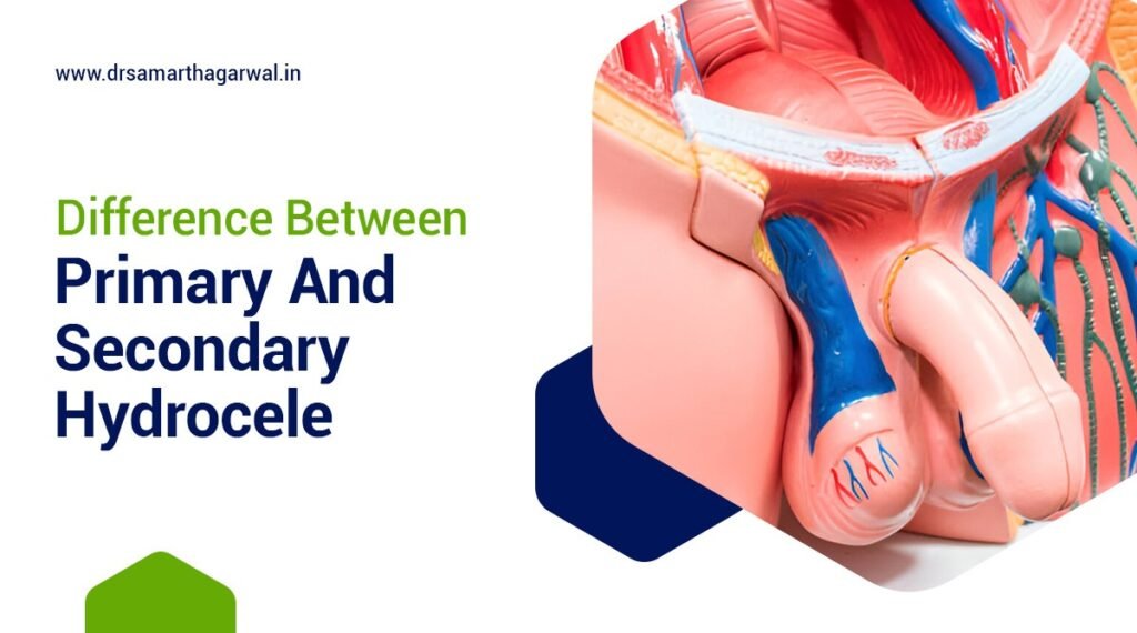
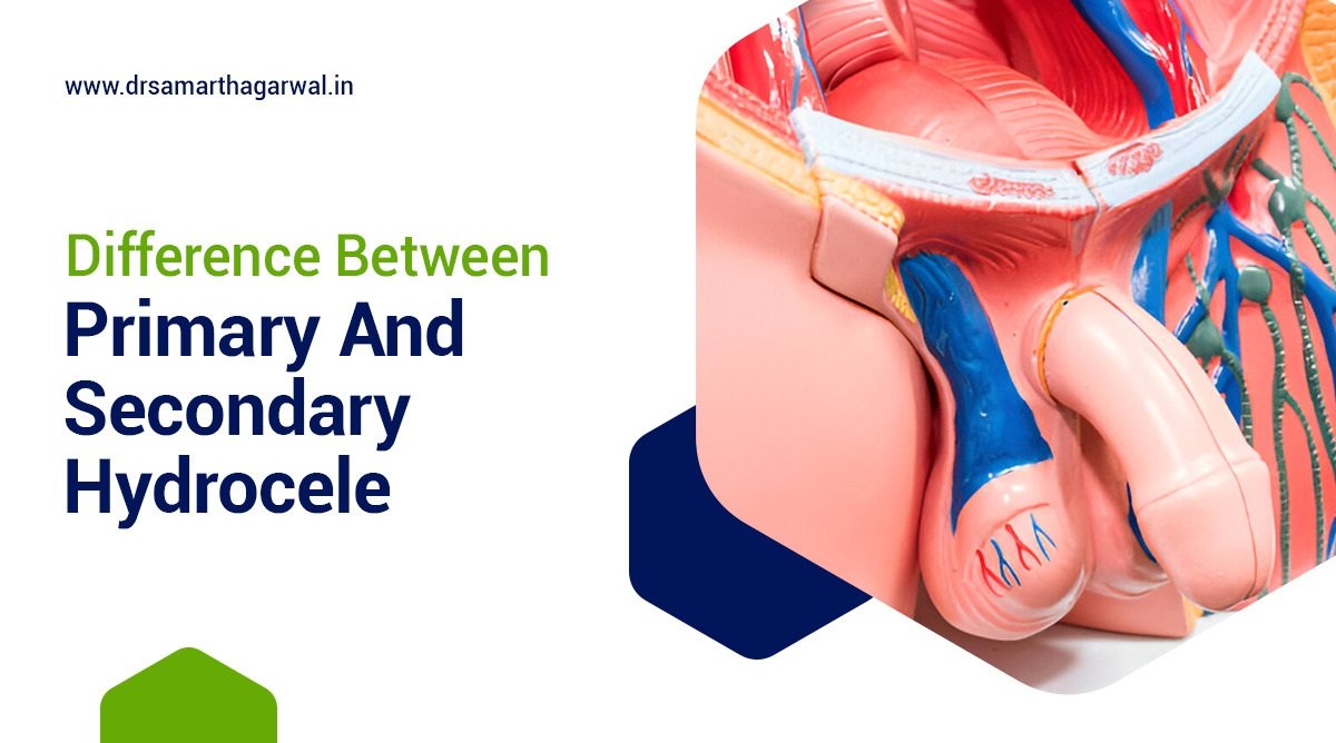
Difference Between Primary and Secondary Hydrocele

Hydrocele is a medical condition characterized by the accumulation of serous fluid within the tunica vaginalis, the protective sheath surrounding the testis in the scrotum. This fluid buildup can cause noticeable scrotal swelling and discomfort. While the general definition remains consistent, hydroceles are categorized into two main types: primary and secondary.

The key differences between primary and secondary hydrocele lie in their underlying causes, onset, and associated symptoms. Primary hydrocele typically occurs due to developmental issues, often presenting in infancy or childhood, and is generally painless. In contrast, secondary hydrocele results from underlying pathological conditions such as testicular tumors or infections, can occur at any age, and is frequently accompanied by pain.

Treatment options for these two types of hydrocele vary based on their distinct characteristics. Primary hydrocele may be managed through observation for asymptomatic cases or surgical intervention for more severe instances. Secondary hydrocele treatment focuses primarily on addressing the underlying cause, which may involve infection treatment or surgical procedures to alleviate symptoms and prevent complications.

Understanding these differences is crucial for accurate diagnosis and effective management of hydrocele. The following detailed comparison provides a comprehensive overview of primary and secondary hydrocele, their distinguishing features, and appropriate treatment approaches.

What Is a Hydrocele?

A hydrocele is a medical condition characterized by the accumulation of serous fluid within the tunica vaginalis, the protective sheath surrounding the testis in the scrotum. This fluid buildup can lead to noticeable scrotal swelling, often causing discomfort or anxiety for those affected. Hydroceles can occur in various forms and may be congenital, appearing at birth, or acquired later in life. 

Understanding hydrocele is essential for identifying its types, causes, symptoms, diagnosis, and treatment options, particularly as the condition may affect both pediatric and adult patients differently.

What Are the Types of Hydrocele?

Hydroceles can be categorized into two main types: primary hydrocele and secondary hydrocele, each with distinct characteristics and underlying causes that influence treatment options and patient outcomes.

Primary Hydrocele

Primary hydrocele refers to a type of hydrocele that arises without any underlying pathological conditions, often occurring due to developmental issues in the tunica vaginalis, leading to fluid accumulation that causes noticeable scrotal swelling.

This condition usually manifests in infancy or early childhood and can be linked to a variety of congenital conditions that may affect the normal anatomy of the scrotum. Typically, affected individuals experience symptoms such as:

  • Swelling in one or both testicles
  • Discomfort or heaviness in the scrotum
  • Changes in the size of the swelling depending on activity level and time of day

Diagnosis is often straightforward, as a physical examination usually suffices to identify the distinctive scrotal swelling. In some cases, a scrotal ultrasound may be conducted to confirm the presence of fluid. Individuals with primary hydrocele generally lead normal lives; however, they may need to adjust certain physical activities to manage discomfort. Timely intervention, including prenatal diagnosis for congenital hydrocele, can alleviate symptoms and prevent complications, emphasizing the importance of understanding this condition.

Furthermore, early diagnosis and proper management strategies can significantly enhance the quality of life for those affected.

Secondary Hydrocele

Secondary hydrocele is related to underlying conditions such as testicular tumors, epididymo-orchitis, or trauma to the testis, resulting in fluid accumulation due to inflammation or disruption in the lymphatic system.

Understanding the causes and risk factors for this condition is crucial for effective management. While primary hydrocele may occur without any identifiable reason, secondary hydrocele often stems from significant medical issues that require attention. For instance, testicular tumors can obstruct lymphatic pathways, effectively leading to this painful accumulation. Infections, such as: epididymo-orchitis can cause the testicular tissue to become inflamed, which might also contribute to fluid build-up.

Trauma to the testis, whether a recent injury or a surgical complication, can elevate the risk of developing a hydrocele. In these cases, the inflammatory response can disrupt normal fluid drainage from the scrotum, necessitating careful evaluation through diagnostic techniques such as a scrotal ultrasound. Infection treatment and surgical treatment may be required based on the severity and underlying causes.

Risk Factors Include:

  • History of testicular conditions
  • Recent surgeries or trauma
  • Infections such as sexually transmitted diseases

Recognizing these connections is vital, as it not only assists in diagnosing the hydrocele accurately but also guides the treatment approach.

What is the Difference between primary and secondary hydrocele?

Understanding the differences between primary and secondary hydrocele is crucial for proper diagnosis and treatment. The following table provides a comprehensive comparison of these two types of hydrocele, highlighting key aspects such as causes, symptoms, diagnosis, and treatment options. This at-a-glance overview will help you quickly grasp the distinctive features of each type:

AspectPrimary HydroceleSecondary Hydrocele
DefinitionAccumulation of serous fluid within the tunica vaginalis without underlying pathological conditionsAccumulation of serous fluid within the tunica vaginalis due to underlying conditions
CauseDevelopmental issues in the tunica vaginalis, often congenitalUnderlying conditions such as testicular tumors, epididymo-orchitis, testicular trauma, or lymphatic filariasis
Age of OnsetOften in infancy or early childhoodCan occur at any age, more common in adults
Symptoms– Painless scrotal swelling- Swelling in one or both testicles- Discomfort or heaviness in the scrotum- Changes in swelling size depending on activity and time of day– Scrotal swelling- Pain, especially if linked to epididymo-orchitis or testicular trauma- Symptoms related to underlying conditions
Pain LevelTypically painless unless complications ariseOften painful due to underlying conditions
TypesCan be communicating or non-communicating hydroceleN/A
Diagnosis– Physical examination- Scrotal ultrasound to confirm fluid accumulation- May include prenatal diagnosis for congenital cases– Comprehensive evaluation- Scrotal ultrasound to identify fluid accumulation and assess underlying conditions
Treatment– Observation for asymptomatic cases- Surgical treatment for symptomatic cases- Aspiration sclerotherapy- Procedures often performed under local anesthesia– Focus on addressing underlying causes- Infection treatment if applicable- Surgical intervention to relieve fluid accumulation and symptoms
Complications– Risk of infection- Increased scrotal swelling- Potential discomfort- Risks associated with surgical intervention– Complications from underlying conditions- Chronic infections- Persistent symptoms- May require more intensive treatments
Prevention– Minimizing risk factors- Avoiding testicular trauma– Addressing infections promptly- Minimizing risk factors- Avoiding testicular trauma
Special ConsiderationsMay affect pediatric patients differentlyMay be related to conditions such as lymphatic filariasis caused by mosquito-borne parasites in regions like Africa and Southeast Asia
Potential for TransformationN/APrimary hydrocele can potentially develop into secondary hydrocele if underlying conditions arise

What Are the Causes of Primary Hydrocele?

The primary hydrocele is often attributed to developmental issues in the tunica vaginalis, leading to a congenital hydrocele, but can also result from other factors affecting the lymphatic system that do not involve secondary conditions.

What Are the Causes of Secondary Hydrocele?

Secondary hydrocele can occur due to various underlying pathological conditions such as testicular tumors, epididymo-orchitis, or testicular trauma, leading to chronic infection and subsequent fluid accumulation within the tunica vaginalis. Conditions related to lymphatic filariasis, caused by mosquito-borne parasites like Wuchereria bancrofti and Brugia malayi, prevalent in regions of Africa and Southeast Asia, may also contribute to fluid buildup.

Secondary hydrocele can occur due to various underlying pathological conditions such as testicular tumors, epididymo-orchitis, or testicular trauma, leading to chronic infection and subsequent fluid accumulation.

What Are the Symptoms of Primary Hydrocele?

Symptoms of primary hydrocele primarily include painless scrotal swelling, which can vary in size and may cause discomfort, though it typically does not present with significant pain unless complications arise. It can manifest as a non-communicating hydrocele or a communicating hydrocele, often observable in pediatric patients.

Symptoms of primary hydrocele primarily include painless scrotal swelling, which can vary in size and may cause discomfort, though it typically does not present with significant pain unless complications arise.

What Are the Symptoms of Secondary Hydrocele?

In contrast to primary hydrocele, secondary hydrocele symptoms can include scrotal swelling accompanied by pain, especially if linked to conditions like epididymo-orchitis or due to testicular trauma. Bilateral hydrocele may also occur in certain cases, requiring careful evaluation.

In contrast to primary hydrocele, secondary hydrocele symptoms can include scrotal swelling accompanied by pain, especially if linked to conditions like epididymo-orchitis or due to testicular trauma.

How Is a Primary Hydrocele Diagnosed?

Diagnosing primary hydrocele typically involves a physical examination followed by a scrotal ultrasound to confirm fluid accumulation within the tunica vaginalis and rule out other conditions.

How Is a Secondary Hydrocele Diagnosed?

The diagnosis of secondary hydrocele often involves a comprehensive evaluation, including scrotal ultrasound, to identify fluid accumulation and assess for underlying conditions such as testicular tumors or infections.

What Are the Treatment Options for Primary Hydrocele?

Treatment options for primary hydrocele typically include observation for asymptomatic cases, while surgical treatment may be indicated for symptomatic hydroceles, often performed under local anesthesia or through techniques like aspiration sclerotherapy.

What Are the Treatment Options for Secondary Hydrocele?

Treatment for secondary hydrocele focuses primarily on addressing the underlying causes, which may involve infection treatment and, in some cases, surgical intervention to relieve fluid accumulation and associated symptoms.

What Are the Complications of Primary Hydrocele?

Complications of primary hydrocele may include the risk of infection, increased scrotal swelling, and potential discomfort, particularly if surgical intervention is required, which can lead to additional risks.

What Are the Complications of Secondary Hydrocele?

Complications arising from secondary hydrocele can result from the underlying conditions, such as chronic infections or testicular trauma, which may lead to persistent symptoms and necessitate more intensive treatments.

Can Primary Hydrocele Turn into Secondary Hydrocele?

While primary hydrocele and secondary hydrocele are distinct conditions, there is a possibility that a primary hydrocele can develop into a secondary hydrocele if underlying conditions such as testicular tumors or epididymo-orchitis arise.

How Can Primary and Secondary Hydrocele Be Prevented?

Preventing hydrocele, whether primary or secondary, often involves minimizing risk factors such as avoiding testicular trauma and addressing infections promptly to reduce the likelihood of fluid accumulation in the scrotum.

32

If you have any questions or concerns about primary or secondary hydrocele, please contact Dr. Samarth Agarwal for assistance with your urinary health.

Can Kidney Stones Make You Bloated?

Kidney stones are a common health issue that can lead to a variety of uncomfortable symptoms, including bloating. Understanding the relationship between kidney stones and gastrointestinal symptoms is crucial for effective management and treatment. This article explores the definition of kidney stones, their gastrointestinal implications, and how they can cause bloating, along with other related symptoms.

What is the Definition of kidney stones

Kidney stones are hard mineral and salt deposits that form in the kidneys, varying in size and potentially obstructing the urinary tract. They result from an imbalance in urine substances, leading to crystallization and causing significant pain when passing through the urinary tract. Untreated, they can block urine flow, cause kidney swelling, and lead to infections, impacting overall health and quality of life. Proper diagnosis and management are essential.

Overview of gastrointestinal symptoms associated with kidney stones

Gastrointestinal symptoms associated with kidney stones can vary widely among individuals. Common symptoms include nausea, vomiting, and abdominal pain, which may occur as the body reacts to the presence of stones in the urinary tract. These symptoms can be particularly distressing and may lead to further complications if not addressed promptly. The connection between kidney stones and gastrointestinal issues is often overlooked, yet it plays a significant role in the overall health of affected individuals.

Moreover, the pain caused by kidney stones can radiate to the abdomen, leading to discomfort that mimics other gastrointestinal problems. This overlap can make it challenging to distinguish between kidney stone-related symptoms and other digestive issues. As a result, individuals may experience bloating and other gastrointestinal disturbances, which can complicate the diagnosis and treatment of kidney stones. Understanding these symptoms is crucial for effective management and prevention of further complications.

According to Leslie, S. W., Sajjad, H., & Murphy, P. B. (n.d.). Renal calculi, nephrolithiasis study, High-risk factors for nephrolithiasis include gastrointestinal diseases and gastrointestinal bypass surgery, particularly Roux-en-Y. These conditions can significantly increase the likelihood of developing kidney stones.

Long-term Effects of Untreated Kidney Stones on Gastrointestinal Health

Untreated kidney stones can lead to significant long-term complications for gastrointestinal health, including chronic infections, ongoing pain, and a decreased quality of life.

List of Long-term Effects:

  • Chronic obstruction of the urinary tract leading to recurrent infections
  • Exacerbation of gastrointestinal symptoms such as bloating and discomfort
  • Potential kidney damage from the persistent presence of stones
  • Ongoing gastrointestinal issues, including chronic pain and digestive disturbances
  • Decreased quality of life due to persistent symptoms
  • Increased healthcare costs from the need for ongoing treatment and symptom management
  • Importance of prompt treatment to prevent complications and maintain gastrointestinal health.

What is The Relationship Between Kidney Stones and Bloating?

The relationship between kidney stones and bloating is complex and multifaceted. Kidney stones can cause bloating due to the body’s response to pain and discomfort. When a stone obstructs the urinary tract, it can lead to increased pressure in the abdomen, resulting in a sensation of fullness or bloating. Additionally, the stress and anxiety associated with kidney stone pain may exacerbate gastrointestinal symptoms, including bloating.

Furthermore, the presence of kidney stones can disrupt normal urinary function, leading to changes in bowel habits and gastrointestinal motility. This disruption can contribute to feelings of bloating and discomfort in the abdomen. It is essential to recognize that while kidney stones may not directly cause bloating, the associated symptoms and complications can create a scenario where bloating becomes a common experience for those affected. Understanding this relationship is vital for effective treatment and management of symptoms.

What is the Mechanism Behind Kidney Stone-Induced Bloating?

The mechanism behind kidney stone-induced bloating involves several physiological responses. When a kidney stone obstructs the ureter, it can lead to increased pressure in the urinary tract, which may also affect the surrounding abdominal organs. This pressure can trigger a reflex response in the gastrointestinal tract, leading to symptoms such as bloating and discomfort. Additionally, the pain associated with kidney stones can cause muscle tension and spasms in the abdomen, further contributing to the sensation of bloating.

Moreover, the body’s stress response to pain can lead to changes in digestion and gastrointestinal motility. Stress hormones can alter the normal functioning of the digestive system, potentially causing delays in gastric emptying and contributing to feelings of fullness and bloating. This interplay between kidney stones and gastrointestinal function highlights the importance of addressing both urinary and digestive health in individuals experiencing symptoms related to kidney stones. Understanding these mechanisms can aid in developing effective treatment strategies.

Other Gastrointestinal Symptoms Associated with Kidney Stones

In addition to bloating, kidney stones can cause a range of gastrointestinal symptoms that may significantly impact an individual’s well-being. One of the most common symptoms is nausea, which can occur due to the body’s response to pain and discomfort. Nausea may be accompanied by vomiting, further complicating the situation and leading to dehydration if not managed properly. These symptoms can be distressing and may require medical intervention to alleviate discomfort.

Abdominal pain and discomfort are also prevalent among individuals with kidney stones. This pain can vary in intensity, often described as sharp or cramping, and may radiate to other areas of the abdomen. The presence of larger stones can exacerbate these symptoms, leading to more severe pain and increased gastrointestinal distress. Recognizing these symptoms is crucial for timely diagnosis and treatment, as they can significantly affect an individual’s quality of life and overall health.

Nausea and vomiting

Nausea and vomiting are common gastrointestinal symptoms associated with kidney stones. These symptoms often arise as a result of the body’s response to the intense pain caused by kidney stones. When a stone obstructs the urinary tract, it can trigger a cascade of physiological reactions, including nausea. This reaction is not only a response to pain but can also be influenced by the body’s stress response, which may further exacerbate feelings of nausea and lead to vomiting.

Moreover, the discomfort associated with kidney stones can disrupt normal digestive processes, contributing to gastrointestinal distress. Individuals experiencing nausea and vomiting may find it challenging to maintain proper hydration and nutrition, which can lead to further complications. It is essential for those suffering from these symptoms to seek medical attention to manage their condition effectively and prevent dehydration. Understanding the link between kidney stones and nausea is crucial for developing appropriate treatment strategies.

Abdominal pain and discomfort

Abdominal pain and discomfort are hallmark symptoms of kidney stones that can significantly impact an individual’s daily life. This pain often manifests as sharp, cramping sensations in the abdomen, which may vary in intensity depending on the size and location of the stone. Larger stones can cause more severe pain, leading to increased discomfort and potential complications. The pain may also radiate to the back or sides, making it challenging to pinpoint its exact origin.

In addition to the acute pain associated with kidney stones, individuals may experience ongoing discomfort that can affect their ability to eat, sleep, and engage in daily activities. This persistent discomfort can lead to anxiety and stress, further complicating the situation. Recognizing the signs of abdominal pain related to kidney stones is essential for timely intervention and treatment. Effective management of this pain is crucial for improving the quality of life for those affected by kidney stones.

Distinguishing Kidney Stone Bloating from Other Causes

Distinguishing kidney stone-induced bloating from other gastrointestinal causes can be challenging due to the overlap of symptoms. Bloating can result from various factors, including dietary choices, gastrointestinal disorders, and other medical conditions. However, when bloating is accompanied by severe pain, nausea, or changes in urinary habits, it may indicate the presence of kidney stones. Understanding these distinguishing features is crucial for accurate diagnosis and treatment.

Additionally, individuals should be aware of other signs that may suggest kidney stones, such as blood in the urine or difficulty urinating. These symptoms can help differentiate kidney stone-related bloating from other gastrointestinal issues. A thorough medical evaluation, including imaging studies and urine tests, is often necessary to confirm the presence of kidney stones and rule out other potential causes of bloating. Recognizing the unique characteristics of kidney stone symptoms is essential for effective management and treatment.

Diagnosis of Kidney Stones and Related Gastrointestinal Symptoms

The diagnosis of kidney stones typically involves a combination of medical history, physical examination, and diagnostic imaging. Healthcare providers often begin by assessing the patient’s symptoms, including any gastrointestinal issues such as bloating, nausea, or abdominal pain. A detailed medical history can provide valuable insights into potential risk factors for kidney stone formation, such as dietary habits and family history.

Diagnostic imaging, such as ultrasound or CT scans, is commonly used to visualize the presence of kidney stones and assess their size and location. Urinalysis may also be performed to detect the presence of blood or crystals in the urine, which can indicate kidney stones. A comprehensive approach to diagnosis is essential for determining the appropriate treatment plan and addressing any related gastrointestinal symptoms effectively. Early diagnosis can help prevent complications and improve overall health outcomes.

Treatment Options for Kidney Stone-Induced Bloating

Treatment for kidney stone-induced bloating focuses on addressing the underlying causes of the symptoms, with an emphasis on pain management and hydration.

Key Treatment Options:

  • Pain Management:
    • Use over-the-counter pain relievers or prescription medications to alleviate severe discomfort.
  • Hydration:
    • Increase fluid intake to help flush out kidney stones and reduce bloating.
  • Medical Procedures:
    • Lithotripsy: A procedure that uses sound waves to break up larger stones.
    • Ureteroscopy: Involves the use of a thin tube to directly remove stones from the urinary tract.

By addressing the underlying kidney stones, individuals can experience significant improvement in gastrointestinal symptoms, including bloating. A comprehensive treatment plan should be tailored to each individual’s needs for effective management of both kidney stones and associated symptoms.

Prevention of Kidney Stones and Associated Bloating

Preventing kidney stones is crucial for minimizing the risk of associated bloating and gastrointestinal symptoms. Effective strategies include maintaining proper hydration and making dietary modifications.

Key Prevention Strategies:

  • Stay Hydrated: Drink plenty of fluids, especially water, to dilute urine and reduce stone-forming substances.
  • Modify Diet:
    • Reduce intake of oxalate-rich foods (e.g., spinach, nuts).
    • Limit consumption of salt and animal proteins.
    • Incorporate a balanced diet rich in fruits, vegetables, and whole grains.
  • Regular Physical Activity: Engage in regular exercise to promote overall health and maintain a healthy weight.

By adopting these preventive measures, individuals can significantly lower their risk of developing kidney stones and the associated gastrointestinal symptoms.

When to Seek Medical Attention

It’s essential to seek medical attention if you experience symptoms of kidney stones, especially when accompanied by gastrointestinal distress. Signs that require immediate evaluation include:

  • Severe abdominal pain
  • Persistent nausea and vomiting
  • Difficulty urinating
  • Blood in urine
  • Fever and chills

These symptoms may indicate complications such as infection or obstruction. Early intervention can prevent further complications and improve health outcomes. Suppose you have a history of kidney stones or are at risk. In that case, regular check-ups with a healthcare provider, such as Dr. Samarth Agarwal in Siliguri, can help monitor your condition and address emerging symptoms. Being proactive about your health is crucial for effectively managing kidney stones and their associated gastrointestinal symptoms.

3

Contact Dr. Samarth Agarwal if you have any questions or concerns about kidney stones! Meet the top Urologist in Siliguri.

Can Dehydration Cause Painful Urination?

Experiencing painful urination can be both alarming and uncomfortable, and many don’t realize that dehydration may play a significant role in this issue.

This article explores the connection between dehydration and urinary tract infections (UTIs), shedding light on what causes these infections and how a lack of fluids, including factors like excessive sweating and vomiting, can impact the urinary tract.

It also discusses symptoms, nighttime urination, chronic conditions, and effective prevention strategies including the importance of hydration and electrolyte replacement.

Understanding these aspects can empower you to take charge of your urinary health and manage risk factors more effectively.

Can Dehydration Cause Painful Urination and Other Health Problems?

Dehydration can significantly impact various bodily functions, leading to painful urination, which is often a symptom indicating underlying health problems such as chronic conditions and interstitial cystitis. This condition is particularly concerning for older adults and individuals with chronic conditions, as dehydration exacerbates urinary tract issues and can signal a medical emergency. Maintaining adequate hydration is crucial to prevent complications related to painful urination, which can disrupt daily life and indicate potential health risks.

What is UTI? Insights from UT Southwestern Medical Center

A Urinary Tract Infection (UTI) is a health problem that occurs when bacteria invade the urinary tract, causing inflammation and discomfort. This infection can manifest with various symptoms, including painful urination, increased frequency of urination, and sometimes fever, making it essential to address any underlying factors such as dehydration that may contribute to its occurrence.

When the body is not adequately hydrated, urine becomes concentrated, creating an environment that is more conducive to bacterial growth. This factor heightens the risk of developing a UTI, especially in those with compromised immunity or anatomical predispositions, such as older adults. It is important to recognize other symptoms like:

  • Cloudy or strong-smelling urine
  • Pain in the lower abdomen
  • Back pain

Not only do UTIs cause immediate discomfort, but they can also lead to more serious complications if left untreated, including kidney infections which have the potential to significantly impact overall health.

What Causes UTI?

UTIs are primarily caused by bacteria entering the urinary tract, often linked to factors such as dehydration, sexual activity, and chronic conditions that compromise the immune system.

Understanding these risk factors is critical to preventing the occurrence of UTIs, especially in vulnerable populations like older adults and athletes who may experience dehydration due to excessive sweating.

When individuals are dehydrated, the urinary tract’s ability to maintain a healthy balance of flora can diminish, creating an environment more susceptible to infection. This imbalance can result in increased bacterial growth that overwhelms the body’s defenses.

  • Gender plays a significant role, with females being more prone due to anatomical differences.
  • Poor hygiene practices can further exacerbate the risk, making it essential to prioritize cleanliness.
  • Pre-existing health issues such as diabetes, kidney stones, or high blood pressure may increase susceptibility, creating a multifaceted approach to prevention.

By addressing these various factors, individuals can effectively reduce their risk of developing troublesome urinary tract infections.

How Does Dehydration and Excessive Sweating Lead to UTI?

Dehydration can create an environment in the urinary tract that is conducive to the growth of bacteria, thereby increasing the risk of developing a UTI. When adequate hydration is not maintained, urine becomes concentrated, making it more likely for bacteria to thrive and lead to infections, thereby emphasizing the importance of proper hydration for urinary health.

When the body does not receive sufficient fluids, the volume of urine decreases, which hinders the flushing out of harmful pathogens from the urinary tract. This stagnation allows bacteria a greater opportunity to multiply, thus escalating the risk of infection.

  • Mechanism 1: Lower urine output means fewer opportunities to eliminate bacteria.
  • Mechanism 2: Concentrated urine can irritate the bladder and urethra, potentially leading to inflammation.
  • Mechanism 3: A dehydrated state may disrupt the urinary tract’s natural defense mechanisms, furthering susceptibility to infection.

Staying adequately hydrated not only helps to dilute the urine but also plays a crucial role in maintaining the body’s defenses against urinary tract infections.

How Does Dehydration Affect the Urinary Tract and Kidney Stones Formation?

Dehydration negatively impacts the urinary tract by concentrating urine, which can lead to various complications including painful urination, kidney stones, and increased susceptibility to infections. When the body is not properly hydrated, the kidneys may struggle to filter waste efficiently, resulting in higher concentrations of minerals and salts that can crystallize and form stones.

This lack of hydration can create an environment conducive to bacterial growth, further inflaming the urinary tract and leading to frequent infections. This might require urinalysis to properly diagnose. Common symptoms of these infections include:

  • Urge to urinate frequently
  • Burning sensation during urination
  • Cloudy or strong-smelling urine

To mitigate these risks, adequate hydration is essential, as it helps dilute the urine, flushes out toxins, and minimizes the concentration of minerals. Regular intake of fluids, and in some cases, sports drinks or other electrolyte solutions, can effectively promote the efficient function of the kidneys, allowing them to perform their vital role while reducing the likelihood of developing painful conditions.

What are the Symptoms of UTI?

The symptoms of a UTI can range from mild to severe and often include painful urination, increased frequency of urination, and urgency, especially in cases exacerbated by dehydration. Other symptoms may also include fever, cloudy urine, and a strong odor, indicating a need for immediate medical attention.

Recognizing the symptoms of a UTI early is essential for effective management and treatment. When dehydration occurs, it can significantly intensify these symptoms, leading to discomfort and potential complications. This connection between hydration and urinary tract health is crucial for individuals to understand. Dehydration not only diminishes the body’s ability to flush out bacteria but also concentrates urine, making it more irritating to the bladder. Signs that warrant attention include:

  • Persistent urge to urinate
  • Pain or burning sensation during urination
  • Dark or strong-smelling urine
  • Lower abdominal pain

By staying hydrated and being alert to these symptoms, individuals can seek timely medical intervention, alleviating the possibility of further health issues, including the need for medical emergency interventions.

Can Dehydration Make You Pee More at Night?

Dehydration can lead to nocturia, or frequent urination at night, particularly among older adults, as the body struggles to maintain proper hydration levels.

When fluid intake is inadequate, the kidneys may alter their function, affecting the body’s natural rhythms and causing disturbances in sleep due to frequent trips to the bathroom. This condition, known as nocturia, can be particularly problematic for older adults.

Understanding the link between hydration, urination patterns, and body temperature is crucial, especially for those who may be experiencing discomfort or disrupted sleep.

For older adults, staying hydrated becomes even more important, as their bodies often exhibit diminished thirst responses and altered kidney functions. They might benefit from a balanced intake of electrolytes to help maintain fluid balance, which can further mitigate the impact of nocturia.

Here are some considerations to maintain optimal hydration and prevent health problems:

  • Monitor fluid intake, particularly in the evening hours.
  • Incorporate hydrating foods, such as fruits and vegetables.
  • Consult a healthcare provider for personalized electrolyte needs and consider rehydration solutions like Pedialyte.

By promoting proper hydration, individuals can significantly reduce the frequency of nighttime bathroom visits.

How to Prevent UTI from Dehydration and Related Health Problems?

Preventing UTIs caused by dehydration is vital, especially for individuals at higher risk, such as older adults or those with chronic conditions. Ensuring adequate water intake and recognizing symptoms of severe dehydration, like dry mouth and dizziness, are crucial steps. The key to prevention lies in maintaining adequate hydration through consistent water intake, and in some cases, incorporating electrolyte replacement solutions like Pedialyte or sports drinks to replenish lost fluids and minerals.

Frequently Asked Questions

Can dehydration cause painful urination?

Yes, it can. Dehydration can lead to concentrated urine which can irritate the lining of the bladder and urethra, causing painful urination.

What are the symptoms of painful urination due to dehydration?

The most common symptom is a burning or stinging sensation during urination. Other symptoms may include frequent urination, dark or cloudy urine, and a strong urge to urinate. In women, these symptoms can be particularly pronounced.

How can I prevent painful urination caused by dehydration?

The best way to prevent this is by staying hydrated and drinking plenty of fluids throughout the day. Products like Pedialyte can help maintain electrolyte balance. Avoiding drinks that can irritate the bladder, such as alcohol and caffeine, can also help.

Can being dehydrated cause a UTI?

Yes, dehydration can increase your risk of developing a urinary tract infection (UTI). When the body is dehydrated, it produces less urine which can make it easier for bacteria to grow and cause an infection. UT Southwestern Medical Center recommends staying hydrated as a preventative measure.

What are some other complications of dehydration?

In addition to causing painful urination, dehydration can also lead to other health problems such as kidney stones, constipation, diarrhea, and fatigue.

When should I seek medical attention for painful urination?

If you experience painful urination for more than a day or if you have other symptoms such as fever, chills, or back pain, it is important to see a doctor. These could be signs of a more serious urinary tract infection or other medical condition.

7

Contact Dr. Samarth Agarwal if you have any questions or concerns about your Urinary health!

Meet the top Urologist in Siliguri

What Causes Painful Urination With Blood

Experiencing painful urination accompanied by blood, known as hematuria, can be alarming and uncomfortable. This condition may indicate underlying health issues such as kidney disease or bladder cancer that require attention.

Understanding the common causes, such as urinary tract infections (UTIs), sexually transmitted infections (STIs), and kidney stones, is crucial for addressing the problem effectively. These conditions often require a thorough medical history and physical examination for proper diagnosis.

This article explores the symptoms, diagnostic methods, including blood tests and urine samples, and treatment options available to help navigate this unsettling experience.

By the end, you’ll have a clearer understanding of what to expect and how to seek relief.

What Is Painful Urination With Blood?

Painful urination with blood, also known as hematuria, is a concerning symptom that indicates potential issues within the urinary tract, such as infection or kidney disease. It can manifest as blood in urine or blood in pee, which may arise due to various medical conditions affecting the urinary system. When someone experiences painful urination accompanied by blood, it is essential to seek urgent help from an urologist to identify the underlying cause and initiate appropriate treatment promptly. Ignoring these symptoms can lead to complications, including kidney stones, urinary tract infections, or even bladder cancer, necessitating a thorough examination by a healthcare professional like Dr. Samarth Agarwal.

What Are The Common Causes Of Painful Urination With Blood?

Understanding the common causes of painful urination with blood is crucial for effective diagnosis and treatment, whether through the NHS or private healthcare providers. Various factors can lead to this distressing symptom, including urinary tract infections (UTIs), which are often accompanied by severe pain and discomfort. Additionally, kidney stones may cause significant pain while also resulting in blood in urine. Other potential causes include problems related to the prostate in men, and in more severe cases, bladder cancer. Each of these conditions requires careful evaluation by a healthcare provider to determine the appropriate course of action.

Urinary Tract Infection (UTI)

A urinary tract infection (UTI) is a prevalent cause of painful urination with blood and can lead to significant discomfort and the presence of blood in urine.

Symptoms often include a burning sensation during urination, frequent urges to urinate, and lower abdominal pain, which can be extremely distressing and may require medications or even surgery. Plus these, some individuals may also experience cloudy or strong-smelling urine and even fever in severe cases.

Certain risk factors can increase susceptibility to these infections, such as dehydration, improper hygiene practices, and sexual activity. It’s crucial to maintain hydration and good hygiene, as they can be key preventive measures against dehydration, which can exacerbate symptoms.

  • Antibiotics are the primary treatment for UTIs, effectively targeting the bacteria causing the infection.
  • Completing the entire prescribed antibiotic course is essential to prevent recurrence.
  • Doctors may recommend additional measures, including cranberry products or probiotics, though these should complement medical advice rather than replace it.

By focusing on proactive management and awareness of UTI symptoms, individuals can significantly reduce their risk and see blood in urine as a sign to seek timely care when needed.

Sexually Transmitted Infections (STIs)

Sexually transmitted infections (STIs) can also be a significant cause of painful urination with blood, resulting in symptoms that require prompt medical attention. Common STIs like chlamydia and gonorrhea may lead to inflammation of the urinary tract, causing painful urination and potential bleeding.

Plus these, other infections such as trichomoniasis and herpes can also contribute to uncomfortable urinary sensations. Recognizing the symptoms early, which may include a burning sensation during urination, unusual discharge, or persistent itching, is crucial for effective treatment.

Healthcare providers play a vital role in diagnosing these conditions through various testing methods such as urine samples or swabs. Once diagnosed, treatment options commonly include antibiotics, which are essential not only for alleviating symptoms but also for preventing further complications.

  • Chlamydia: Often treated effectively with azithromycin or doxycycline.
  • Gonorrhea: Typically managed with a regimen of ceftriaxone and azithromycin.
  • Trichomoniasis: Metronidazole is a common treatment choice.

Contacting a healthcare professional at the first sign of discomfort can ensure clear guidance and support, ultimately aiding in recovery while safeguarding overall health, especially if there is a family history of related conditions.

Kidney Stones

Kidney stones are hard deposits formed in the kidneys that can cause severe pain and lead to blood in urine when they obstruct the urinary tract. The intense pain, often described as cramping and sharp, may be accompanied by nausea and difficulty urinating.

These stones develop when substances such as calcium, oxalate, and uric acid become concentrated in urine, particularly when dehydration occurs. Several factors can contribute to this painful condition, including dietary choices, lack of hydration, and specific medical conditions.

  • Symptoms may also include:
  • Frequent urination
  • Cloudy or foul-smelling urine
  • Persistent discomfort in the back or lower abdomen

For those experiencing these symptoms, it is critical to seek medical advice.

Treatment options vary based on the size and type of stones, ranging from increased fluid intake and pain management to surgical interventions if stones are too large to pass naturally. Preventive measures, such as maintaining adequate hydration by drinking plenty of fluids and adjusting dietary habits, can significantly help reduce the risk of recurrence, especially in those with a family history of kidney stones.

Enlarged Prostate

An enlarged prostate, a common condition as men age, can lead to painful urination with blood due to pressure on the urinary tract. This condition often results in symptoms such as frequent urination, difficulty starting urination, and, in some cases, blood in pee.

This condition primarily affects older males, arising from hormonal changes and the natural growth of prostate tissue. When the prostate enlarges, it compresses the urethra, leading to various urinary issues. Understandably, this can cause significant distress and impact daily life.

As a result, men facing these challenges should be aware of the potential implications for their urinary health, particularly if there is a prostate problem. Ignoring symptoms can lead to severe complications, including urinary tract infections or bladder stones.

  • Treatment options may vary:
  • Medications like alpha-blockers can relax prostate muscles.
  • Minimally invasive procedures may be recommended in severe cases.

Regular check-ups with a healthcare provider are essential, as this allows for early detection and management of the condition to maintain a healthy quality of life.

Bladder Cancer

Bladder cancer is a serious condition that can manifest as painful urination with blood, often presenting as blood in urine without accompanying pain initially. Risk factors include smoking, exposure to certain chemicals, and a family history of cancer.

Individuals may experience increased urgency to urinate or frequent need to urinate, especially at night. These symptoms can often be mistaken for a urinary tract infection, which may delay diagnosis. Therefore, it’s vital to recognize the potential indicators and consult a healthcare professional promptly.

  • Risk Factors: Smoking raises the likelihood of developing bladder cancer significantly.
  • Exposure to specific industrial chemicals, such as aniline dyes, is a concern.
  • A personal or family history of cancer can increase susceptibility.

Upon visiting a doctor, a series of tests, including blood tests and imaging tests, are conducted for proper diagnosis. Imaging tests, such as ultrasounds or CT scans, can help visualize abnormalities, while cystoscopy allows direct examination of the bladder. A biopsy may be performed to ascertain the presence of cancer cells.

Recognizing these factors is critical, as early detection often enhances treatment effectiveness, leading to a better prognosis and potential avoidance of severe pain.

What Are The Symptoms Of Painful Urination With Blood?

The symptoms of painful urination with blood can vary widely depending on the underlying cause, such as an infection or cancer, but common indicators include a burning sensation during urination and frequent urges to urinate. Additionally, patients may notice blood in urine, which can appear bright red or tea-colored, signaling the need for urgent medical attention. Recognizing these symptoms is crucial for timely diagnosis and treatment.

Burning Sensation During Urination

A burning sensation during urination is one of the most common symptoms associated with painful urination and can indicate urinary tract infection or irritation. This discomfort can vary in intensity and may be accompanied by other symptoms such as frequent urination and blood in urine, signaling a need for diagnosis and treatment.

While the most common cause is often a urinary tract infection (UTI), other factors may contribute to this troubling sensation. For instance, sexually transmitted infections (STIs) can similarly provoke discomfort, making it crucial to consider the full scope of symptoms.

  • Other potential causes include:
  • Dehydration, which can lead to concentrated urine
  • Bladder or kidney stones
  • Prostate issues in men

Treatments typically involve antibiotics for infections and increased fluid intake for irritation. If the burning sensation persists or worsens, individuals should seek medical advice promptly from healthcare providers, as this could indicate a more serious condition requiring further diagnostic evaluation.

Frequent Urination

Frequent urination is a symptom that often accompanies painful urination with blood and can disrupt daily activities. It may indicate underlying health issues, such as infections or enlarged prostate problems, which require medical evaluation by a healthcare provider.

This condition can pave the way for various complications if left unaddressed, highlighting the importance of understanding its implications. Frequent urination might signify potential issues with the bladder:

  • Urinary Tract Infections (UTIs): These are common in both men and women and may present with additional symptoms like fever or pelvic pain.
  • Prostate Issues: In men, an enlarged prostate can lead to obstructed urine flow and increased frequency, and may require a GP appointment for evaluation.
  • Diabetes: High blood sugar levels can result in excessive thirst and subsequent urination as the body attempts to excrete surplus glucose.

Addressing these concerns typically involves a combination of diagnostics, lifestyle adjustments, and possibly medications. Consulting with a healthcare provider is essential for effective management and treatment options tailored to the individual’s health needs, including antibiotics for infections.

Lower Abdominal Pain

Lower abdominal pain is another concerning symptom linked to painful urination with blood, often indicating serious underlying issues such as infections or kidney stones, and requiring a thorough diagnosis. This pain can range from mild discomfort to severe cramping.

In many instances, this discomfort may signal an array of urinary problems that warrant immediate attention. Symptoms might be accompanied by changes in urinary frequency, urgency, or even noticeable blood in the urine, each indicating a potential health issue that requires thorough evaluation through the NHS or other healthcare systems.

  • Urinary Tract Infections: These can cause inflammation and discomfort while urinating.
  • Kidney Stones: Sharp pain and hematuria are common symptoms.
  • Bladder Infections: Often leading to lower abdominal pain that correlates with the urgency to urinate.

Seeking medical advice is critical as a healthcare provider can conduct necessary tests, such as urinalysis and imaging studies, to identify the underlying cause. Once a diagnosis is made, treatment options may include antibiotics for infections, increased fluid intake for kidney stones, or other specific interventions tailored to the individual’s health needs.

Blood In Urine (Hematuria)

Blood in urine, medically known as hematuria, is a critical symptom that necessitates immediate medical evaluation. It can appear as bright red blood or may be detected through urine tests, indicating various underlying conditions from infections to more serious issues like bladder cancer, potentially linked to family history.

Recognizing hematuria is vital, as its appearance may serve as a crucial signal for potential health issues that deserve thorough investigation. The presence of blood can indicate a range of conditions, including urinary tract infections, kidney stones, or even larger concerns like prostate problems and tumors, possibly leading to cancer. Given the spectrum of possible causes, the importance of seeking prompt medical attention cannot be overstated.

  • The diagnostic assessment typically involves a detailed history and physical examination.
  • Urinalysis is often the first step, allowing healthcare professionals to analyze the urine for red blood cells and other abnormalities. This aids in distinguishing between general infection and more serious conditions like kidney disease.
  • Follow-up tests, such as imaging studies or cystoscopy, may be required based on initial findings.

Timely intervention can significantly impact the prognosis, making awareness and action essential for anyone noticing this troubling symptom.

How Is Painful Urination With Blood Diagnosed? Understanding Symptoms and Procedures

Diagnosing painful urination with blood involves a comprehensive evaluation by a healthcare provider, including a thorough medical history and physical examination. Upon assessment, a urine sample will typically be collected to identify any underlying infections, while blood tests may be conducted to check for kidney disease or other health issues. Imaging tests, like ultrasound or CT scans, might also be necessary to visualize the urinary tract and pinpoint the cause of the symptoms, considering potential bladder and prostate issues.

What Are The Treatment Options For Painful Urination With Blood? A Look into Possible Interventions

Treatment options for painful urination with blood vary widely, depending on the underlying cause. For infections, antibiotics are commonly prescribed to eliminate bacteria and alleviate symptoms. In cases of kidney stones, treatment may range from medications to manage pain to surgical intervention for stone removal. Additionally, addressing conditions like an enlarged prostate may involve medication or surgery to relieve pressure on the urinary tract, as guided by the NHS or other healthcare organizations.

Antibiotics

Antibiotics are often the first line of treatment for painful urination with blood when caused by infections such as UTIs. These medications effectively target bacteria, reducing symptoms and preventing further complications.

When considering antibiotic treatment, it’s important to understand how these medications function. They work by disrupting cell processes within bacteria, ultimately leading to cell death or inhibiting their growth, making them effective against infections like UTIs.

  • Effectiveness: Antibiotics can significantly alleviate symptoms and help the body fight off infections, allowing for a quicker recovery.
  • Potential Side Effects: While generally safe, antibiotics can cause side effects such as gastrointestinal discomfort, allergic reactions, or disruption of gut flora.

Therefore, patients should always complete the prescribed course to ensure that the infection is fully eradicated, and should consult with their healthcare providers if symptoms persist or worsen, particularly if there’s a history of family history of related conditions.

Medications For STIs

When painful urination with blood is caused by sexually transmitted infections (STIs), specific medications are required to treat the infection effectively. Depending on the type of STI, healthcare providers may prescribe antibiotics or antiviral medications.

Understanding the correct diagnosis is crucial, not only to alleviate symptoms but also to prevent serious complications that may arise if left untreated. Accurate identification of the specific STI can significantly influence both treatment and recovery paths. For instance, testing protocols often include urine tests, swabs, and blood tests to confirm the presence of an infection, sometimes checking for cancer indicators.

  • Follow-up care is equally important; regular check-ups ensure that treatment is working and help monitor for any recurrence.
  • Patients should be encouraged to inform their partners about the diagnosis to prevent further transmission and consider GP appointments for comprehensive care.

Ultimately, timely intervention and adherence to prescribed protocols can lead to a healthy outcome and improved quality of life.

Surgery For Kidney Stones Or Tumors

Surgery may be necessary for painful urination with blood related to complications from kidney stones or tumors, especially when conservative treatments have failed. Such procedures are critical when considering potential cancer risks.

Procedures can range from minimally invasive techniques to more extensive surgeries depending on the size and location of the stones or tumors.

When considering surgery, patients should be aware of several surgical options tailored to their specific conditions, whether for tumors or stones. These include:

  • Ureteroscopy: This minimally invasive procedure allows physicians to remove stones using specialized instruments. Recovery time is typically short, often requiring only a few days of rest.
  • Shock Wave Lithotripsy: This non-invasive option uses sound waves to break stones into smaller pieces, facilitating easier passage. Many patients can return to normal activities in a week.
  • Nephrectomy: In cases of tumors, partial or complete removal of the kidney may be necessary. Recovery can take several weeks due to the extensive nature of the surgery.
  • Open Surgery: If the situation is complex, open surgery might be needed, which involves longer recovery times and increased risks.

It’s crucial to consult healthcare providers to evaluate all surgical considerations, as they will guide patients on the best treatment paths based on individual health profiles, often considering family history in their assessments.

Lifestyle Changes

Plus medical treatments, lifestyle changes can significantly improve urinary health and reduce the risk of painful urination with blood. Staying hydrated, maintaining proper hygiene, and avoiding irritants like caffeine can help prevent urinary tract issues.

To further enhance urinary well-being, individuals may consider incorporating certain dietary adjustments and preventive measures into their daily routines. For instance, increasing water intake is essential for flushing out toxins and preventing concentrated urine, which can lead to irritation. Additionally, consuming more fruits and vegetables rich in antioxidants can support overall health and may alleviate symptoms associated with urinary discomfort, minimizing risks of stones and infections.

  • Adopting a low-sugar diet can also minimize the risk of UTIs, as sugar can fuel the growth of harmful bacteria.
  • Practicing good hygiene, including regular washing and urinating after intercourse, can further reduce the likelihood of infections.

Staying aware of potential irritants, such as artificial sweeteners and spicy foods, can be beneficial as well. By making these small yet impactful lifestyle modifications, one can pave the way towards a more comfortable and healthier urinary system.

Frequently Asked Questions

1. What are the most common causes of painful urination with blood according to a urologist?

According to a urologist Dr. Samarth Agarwal, the most common causes of painful urination with blood, known as hematuria, are urinary tract infections (UTIs), kidney stones, and sexually transmitted infections. These symptoms often require a GP appointment or consultation with a healthcare provider.

2. Can dehydration be a possible cause of painful urination with blood?

Yes, dehydration can lead to concentrated urine which can irritate the urinary tract and cause painful urination with blood. It is important to stay hydrated to help maintain kidney health.

3. How do urinary tract infections cause painful urination with blood?

Urinary tract infections can cause painful urination with blood due to the inflammation and irritation of the bladder, urethra, or kidneys. UTIs often require treatment with antibiotics to resolve the infection.

4. Is painful urination with blood always a sign of a serious condition?

No, painful urination with blood can also be caused by less serious conditions such as bladder or kidney infections, but it is important to consult a urologist or healthcare provider for proper diagnosis and treatment. In some cases, it could be a sign of a more serious condition such as prostate cancer or bladder cancer.

5. Can certain medications cause painful urination with blood?

Yes, some medications such as blood thinners or chemotherapy drugs can cause irritation of the urinary tract and lead to painful urination with blood. Always discuss side effects with your healthcare provider.

6. What should I do if I experience painful urination with blood?

If you experience painful urination with blood, it is important to see a urologist or healthcare provider for proper diagnosis and treatment. They will be able to determine the underlying cause and provide the appropriate treatment plan, which may include antibiotics, surgery, or other interventions. It is important not to ignore this symptom as it could be a sign of a serious condition.

IMG 2945

Contact Dr. Samarth Agarwal if you have any questions or concerns about your Urinary health!
Meet the top Urologist in Siliguri

What Causes Pain in The Groin Area?

Groin pain can be a symptom of various underlying conditions. Discover the common causes of groin pain, including injuries, infections, and underlying medical conditions, to better understand and address the discomfort.

What are the possible Causes for Groin Pain?

The most common causes of groin pain, including muscle, tendon, or ligament strain, inflammation of the testicle or epididymitis, twisting of the spermatic cord, pulled muscles, ligaments, or tendons in the leg, hip joint injury, hernia, ovarian cysts, inflammation, and kidney stone. Understanding these potential causes can help individuals identify the source of their groin pain and seek appropriate medical treatment.

Most Common Symptoms and Causes of Groin Pain

  • Muscle, tendon, or ligament strain: Overstretching or tearing can lead to localized pain and discomfort in the groin area.
  • Higher risk in athletes (e.g., hockey players): Intense physical activity increases the likelihood of strains and injuries.

Groin Pain in Men

  • Inflammation of the testicle or epididymitis: Inflammation can cause sharp pain and swelling in the groin region.
  • Testicular torsion: Twisting of the spermatic cord can lead to severe, sudden pain and requires immediate medical attention.

Groin Pain in Women

  • Pulled muscles, ligaments, or tendons in the leg: Injury to these structures can result in pain that radiates to the groin.
  • Stressed ligaments or tendon fibers: Overuse or strain can cause discomfort and pain in the groin area.

Additional Potential Causes

  • Hip joint injury: Damage to the hip joint can lead to referred pain in the groin.
  • Hernia: A hernia can create pressure and pain in the groin due to tissue bulging through a weak spot.
  • Ovarian cysts: Cysts can cause pain that may be felt in the groin, especially if they rupture.
  • Inflammation: General inflammation in the pelvic area can lead to groin pain.
  • Kidney stone: Stones can cause severe pain that radiates to the groin as they move through the urinary tract.

What is the fastest way to cure groin pain?

The fastest way to alleviate groin pain involves immediate rest, including a break from sports. Apply ice to the affected area for 10-15 minutes every hour for the first day, then every 3-4 hours for 2-3 days. Physical therapy, pain medications like NSAIDs or acetaminophen, and specific exercises may also be recommended for long-term recovery. Stretching exercises, such as the supine butterfly stretch, can help loosen the muscles. After a few days of rest, stretching and strengthening exercises can aid in the healing process.

What causes groin pain without injury?

Groin pain without injury can be caused by conditions such as:

  1. Muscle strain: Injury of a muscle or tissue connecting muscle to bone (tendon). Symptoms may include muscle pain, joint pain, and limited range of motion.
  2. Inguinal hernia: A condition in which soft tissue bulges through a weak point in the abdominal muscles. Groin pain and a groin lump are common symptoms.
  3. Urinary tract infection (UTI): In men, UTIs can cause groin pain, especially if the infection spreads to the epididymis or testicles.
  4. Prostatitis: Inflammation of the prostate gland can cause groin pain, especially when urinating or having a bowel movement.
  5. Endometriosis: In women, endometriosis can cause pelvic pain, which may radiate to the groin area.
  6. Cystic lesion: A cyst or growth in the pelvic area can cause groin pain, especially if it presses on nearby nerves or organs.
  7. Kidney stones: Hard buildups of minerals and salt in the kidneys can cause groin pain, especially when they move or block the urinary tract.
  8. Ovarian torsion: Twisting of the ovary can cause sudden, severe groin pain, especially in young women.
  9. Testicular torsion: Twisting of the testicle can cause sudden, severe groin pain, especially in adolescent boys and young men.
  10. Appendicitis: Inflammation of the appendix can cause pain in the lower right abdomen, but it may also radiate to the groin area.

Consult with Urologist Dr. Samarth Agarwal in Siliguri for advice if you experience persistent groin pain without a clear cause.

When should I worry about groin pain?

Groin pain that persists for more than a few days, especially if it’s severe, should prompt a doctor’s visit. Additionally, seek immediate medical attention if you experience accompanying symptoms such as back, stomach, or chest pain, sudden serious testicle pain, nausea, vomiting, fever, chills, unexplained weight loss, blood in the urine, swelling or a lump around the groin area, or visible swelling on the testicles. Recurrent episodes of groin pain or pain that worsens over time may also require medical evaluation.

What does pain in the groin mean for a woman?

Groin pain in women can stem from various causes, such as muscle strain, urinary tract infections (UTIs), ovarian cysts, hernias, or injuries to muscles, ligaments, or tendons in the groin area. It can also be due to inflammation of the joints in the pelvis or enlarged lymph nodes. If experiencing persistent groin pain, it is advisable to consult a healthcare professional for a proper diagnosis and treatment

What are some Groin Pain Treatment?

Experiencing groin pain can be debilitating and disrupt daily life. Fortunately, several effective ways exist to alleviate and recover from groin pain. Consider the following 7 methods to find relief and get back to your normal activities.

  1. Rest: Taking a break from physical activities, including sports, can help alleviate groin pain.
  2. Ice Application: Applying ice to the affected area can help reduce pain and inflammation.
  3. Physical Therapy: A trained physical therapist can help improve strength and range of motion, which can aid in recovery.
  4. Pain Medications: Over-the-counter pain relievers like NSAIDs (ibuprofen, aspirin) or acetaminophen can be used to manage pain.
  5. Surgery: In severe cases, surgery may be necessary to repair bone fractures or inguinal hernias.
  6. Hydration: Drinking plenty of water can help prevent kidney stones, which can sometimes cause groin pain.
  7. Healthy Lifestyle: Maintaining a moderate weight and being careful when lifting heavy objects can help prevent hernias. Building healthy bones through a balanced diet and regular exercise can also help prevent some bone fractures.

When to See a Doctor for Groin Pain?

Schedule a doctor’s visit if you experience serious groin pain, groin pain that doesn’t improve with home treatment within a few days, or mild testicle pain lasting longer than a few days. Additionally, seek medical attention if you notice a lump or swelling in or around a testicle.

42

Contact Dr. Samarth Agarwal if you have any questions or concerns about your Urinary health!

Meet the top Urologist in Siliguri

What Are the TURP Post Surgery Complications

The outcomes of TURP surgery can vary depending on the specific case and the individual’s health condition. Generally, the surgery can help alleviate the symptoms of an enlarged prostate, improve urinary flow, and reduce the size of the prostate. The success of TURP surgery can also lead to a decrease in the need for further medical interventions.  The results of TURP surgery can be positive, with a reduction in the severity of the condition, and an improvement in the patient’s quality of life. However, there can be complications and risks associated with the surgery, such as infection, bleeding, and the development of strictures.

If you are having any post op problems after TURP you can consult with Dr. Samarth Agarwal.

What are the turp post surgery complications

Here are the top 15 TURP post-surgery complications that you might face. 

1. Retrograde Ejaculation

Retrograde ejaculation occurs when sperm enters the bladder instead of being expelled through the urethra. This can lead to a “dry orgasm,” where no semen is released. It is estimated that up to 75% of men may experience this complication after TURP. While it does not affect sexual pleasure, it can impact fertility.

2. Urinary Incontinence

Some patients may experience involuntary leakage of urine following the procedure, which can vary in severity. This condition may improve over time, but some men may require pelvic floor exercises or medications to manage symptoms. In rare cases, surgical intervention may be necessary.

3. Erectile Dysfunction

Difficulty achieving or maintaining an erection can occur after TURP surgery, affecting sexual function. Studies suggest that about 10-20% of men may experience erectile dysfunction post-surgery. The risk may be higher in men with pre-existing erectile issues or those undergoing additional treatments for prostate conditions.

4. Urethral Strictures

Narrowing of the urethra due to scar tissue formation can lead to difficulties in urination and may require further treatment. Urethral strictures can develop weeks to months after surgery, and treatment options may include dilation or surgical repair.

Read More: Urethral stricture Treatments

5. Bleeding

Patients may experience bleeding during or after the procedure, which can range from mild to severe. In some cases, bleeding may require blood transfusions or additional surgical intervention. Post-operative bleeding is more common in patients taking anticoagulant medications.

6. Infection

There is a risk of urinary tract infections (UTIs) following TURP surgery, which may require antibiotic treatment. The risk of infection can be minimized by ensuring proper hygiene and following post-operative care instructions. Symptoms of infection may include fever, chills, and increased urinary urgency.

7. Bladder Spasms

Involuntary contractions of the bladder can occur, leading to discomfort and urgency. Bladder spasms may be managed with anticholinergic medications or muscle relaxants. Patients are often advised to avoid irritants such as caffeine and alcohol during recovery.

8. Changes in Orgasm

Some men may notice changes in the sensation of orgasm or experience “dry orgasm” due to retrograde ejaculation. These changes can be distressing for some patients, but they do not typically affect overall sexual satisfaction. Counseling may help address any emotional concerns related to these changes.

9. Fertility Issues

Retrograde ejaculation can affect fertility, as sperm may not be present in the ejaculate. Men who wish to conceive may need to explore alternative methods, such as sperm retrieval techniques. It is important for patients to discuss fertility preservation options prior to surgery if they are planning to have children.

10. Persistent Urinary Symptoms

Ongoing issues such as urinary urgency, frequency, or nocturia may persist after surgery. These symptoms can be frustrating and may require further evaluation and management. Lifestyle changes, bladder training, and medications can help alleviate these symptoms.

11. Transurethral Resection Syndrome (TUR Syndrome)

A rare but serious condition caused by the absorption of large amounts of irrigation fluid, leading to electrolyte imbalances. Symptoms may include confusion, nausea, and hypertension. TUR syndrome is more likely to occur during longer procedures and requires immediate medical attention.

12. Perforation of the Bladder or Urethra

Rarely, the surgical instruments can cause perforation of the bladder or urethra, which may require additional surgical intervention. This complication can lead to significant morbidity and may necessitate a longer recovery period.

13. Urinary Retention

Some patients may experience difficulty urinating after the procedure, necessitating catheterization. This condition can be temporary or persistent, and management may include medications or further surgical options to relieve obstruction.

14. Pain or Discomfort

Patients may experience pain in the pelvic area or during urination, which can vary in intensity. Pain management strategies may include over-the-counter pain relievers, prescription medications, or warm baths to alleviate discomfort.

15. Changes in Urinary Flow

Some patients may notice changes in the strength or pattern of their urine stream following surgery. While many experience improvement in urinary flow, some may have ongoing issues that require further evaluation and treatment.

Are there alternatives for a TURP?

Several alternatives to TURP exist for treating an enlarged prostate, including both surgical and minimally invasive options.

  1. Prostatic Urethral Lift (UroLift): This procedure involves inserting implants to hold the enlarged prostate away from the urethra, providing relief without significant tissue removal.
  2. Holmium Laser Enucleation of the Prostate (HoLEP): Considered the new gold standard for surgical treatment, HoLEP effectively removes prostate tissue with a lower risk of complications compared to TURP.
  3. Minimally Invasive Procedures: Options such as Rezum Water Vapor Therapy and prostate artery embolization (PAE) offer effective treatment with reduced recovery times and lower complication rates.
  4. Other Surgical Alternatives: Additional surgical options include photoselective vaporization, endoscopic laser enucleation, and robotic or open simple prostatectomy.
  5. Non-Surgical Alternatives: For those seeking non-surgical options, medications and minimally invasive therapies like UroLift and Rezum are viable choices.

These alternatives provide various options for patients, allowing for tailored treatment based on individual needs and preferences.

How do I get ready for a TURP?

To prepare for a Transurethral Resection of the Prostate (TURP), you should fast before the operation, as instructed by your healthcare provider. No specific bowel preparation is required. You may need to sign a consent form and avoid applying lotions, perfumes, deodorants, or nail polish on the day of surgery. Your healthcare provider will explain the procedure and answer any questions you may have. You will also be asked to take a bath or shower before the surgery.

What are the other types of BPH surgery?

Other types of BPH surgery include Transurethral Incision of the Prostate (TUIP), Laser prostatectomy (HoLEP and PVP), Transurethral electrovaporization (TUEVP), Transurethral vaporesection (TUVRP), Plasmakinetic enucleation of the prostate (PkEP), Unipolar transurethral resection (TURP), Bipolar TURP, Open prostatectomy, Holmium Laser Enucleation of the Prostate (HoLEP), GreenLight™ laser photovaporization of the prostate, Simple prostatectomy, and TUIP (Transurethral Incision of the Prostate).

Meet Dr Samarth Agarwal

If you have any questions or concerns about post-surgery complications following a Transurethral Resection of the Prostate (TURP), please reach out to Dr. Samarth Agarwal, the top urologist in Siliguri. Your urinary health is important, and Dr. Agarwal is here to provide the support and expertise you need.

How Much Urine Is Considered Urinary Retention

Urinary retention is a medical condition characterized by the inability to completely empty the bladder. This condition can lead to various complications, including urinary tract infections and bladder damage. Understanding how much urine is considered urinary retention is crucial for effective diagnosis and treatment. The amount of urine retained can vary significantly among individuals, influenced by factors such as age, gender, and underlying health conditions. In this article, we will explore the normal and excessive amounts of urine retention, the maximum bladder capacity, causes, diagnosis, and treatment options available for urinary retention.

What is the normal amount of urine retention?

The normal amount of urine retention is typically minimal, with a post-void residual urine volume of less than 50 milliliters considered acceptable. In healthy individuals, the bladder should effectively empty during urination, leaving little to no residual urine. However, some degree of residual urine is common, particularly in older adults or those with certain medical conditions. A small amount of residual urine, generally under 100 milliliters, may not indicate a significant problem. Nevertheless, when the residual volume exceeds this threshold, it may suggest incomplete bladder emptying, warranting further investigation into potential causes of urinary retention.

In clinical practice, the measurement of post-void residual urine volume is often conducted using a bladder scan, which provides a non-invasive method to assess bladder function. This assessment is crucial for identifying patients with urinary retention, as it helps determine the extent of bladder emptying. A higher residual volume could indicate underlying issues such as bladder outlet obstruction or neurogenic bladder, both of which can lead to chronic urinary retention. Therefore, understanding what constitutes normal urine retention is essential for the effective management of urinary tract health and the prevention of complications.

How much is a lot of urine retention?

A significant amount of urine retention is defined as a post-void residual urine volume exceeding 100 milliliters. This level of retention may indicate serious underlying conditions, such as acute or chronic urinary retention.

  • Acute Urinary Retention: Patients may be unable to urinate despite a full bladder, requiring immediate medical intervention.
  • Chronic Urinary Retention: Develops gradually, often without obvious symptoms until significant residual volumes accumulate.

Symptoms of urinary retention can include difficulty initiating urination, a weak urine stream, and the sensation of incomplete bladder emptying, which can affect quality of life and lead to complications like urinary tract infections. Monitoring urine volume is crucial for timely diagnosis and management to prevent bladder distension and damage.

What is the maximum volume the bladder can hold?

The average maximum bladder capacity is approximately 400 to 600 milliliters, though it can vary based on age, gender, and health conditions. When the bladder reaches this capacity, the urge to urinate intensifies, and failure to void may lead to discomfort and complications, such as bladder overdistension, which can impair muscle function and cause urinary retention.

In clinical practice, understanding bladder volumes is essential for diagnosing and managing urinary retention. Bladder scans can assess current volume and post-void residual urine. Consistently high residual volumes may indicate issues like bladder outlet obstruction or benign prostatic hyperplasia. Monitoring bladder capacity is crucial for urinary tract health and preventing complications.

Causes of Urinary Retention

Urinary retention can result from a variety of causes, including anatomical, neurological, and functional factors. Identifying the specific cause is crucial for effective diagnosis and management, as treatment strategies may vary accordingly.

Causes of Urinary Retention:

  1. Anatomical Causes:
    • Bladder Outlet Obstruction:
      • Commonly due to benign prostatic hyperplasia (BPH) in men, where an enlarged prostate compresses the urethra, hindering urine flow.
    • Strictures:
      • Narrowing of the urethra caused by injury, infection, or inflammation, leading to obstruction.
    • Tumors:
      • Growths in the urinary tract that can block urine passage, resulting in increased residual urine volume.
    • Pelvic Organ Prolapse:
      • In women, conditions like cystocele or rectocele can displace the bladder and obstruct urine flow.
  2. Neurological Causes:
    • Neurogenic Bladder:
      • Conditions such as multiple sclerosis, spinal cord injuries, or stroke can damage nerves that control bladder function, leading to difficulty in bladder emptying.
    • Diabetic Neuropathy:
      • Diabetes can cause nerve damage that affects bladder control, resulting in urinary retention.
  3. Functional Causes:
    • Detrusor Muscle Dysfunction:
      • Weakness or lack of coordination in the bladder muscle can prevent effective bladder contraction and emptying.
    • Medications:
      • Certain drugs, especially those with anticholinergic properties (e.g., antihistamines, antidepressants), can inhibit bladder muscle contractions and contribute to urinary retention.
  4. Other Contributing Factors:
    • Infections:
      • Urinary tract infections (UTIs) can cause inflammation and swelling, leading to temporary urinary retention.
    • Post-surgical Complications:
      • Surgeries involving the pelvic region may result in temporary or permanent urinary retention due to anatomical changes or nerve damage.
  5. Importance of Understanding Causes:
    • Identifying the underlying cause of urinary retention is essential for tailoring treatment strategies.
    • Effective management can prevent complications such as bladder damage, recurrent urinary tract infections, and decreased quality of life.

A comprehensive understanding of the various causes of urinary retention is vital for healthcare providers to develop appropriate diagnostic and treatment plans, ultimately improving patient outcomes.

Diagnosis of Urinary Retention

The diagnosis of urinary retention involves a detailed assessment of the patient’s medical history, symptoms, and physical examination. Key diagnostic tools, such as bladder scans and additional tests, help identify the underlying causes of the condition.

Diagnosis Process:

  1. Medical History and Symptoms:
    • Review of the patient’s medical history
    • Inquiry about lower urinary tract symptoms
    • Assessment of previous urinary tract infections
    • Evaluation of medications that may contribute to urinary retention
  2. Physical Examination:
    • Comprehensive physical assessment to identify any relevant signs
  3. Bladder Scan:
    • Measurement of post-void residual urine volume
    • Key indicator of urinary retention; normal range is typically less than 50 mL
  4. Further Diagnostic Tests (if necessary):
    • Urodynamic Studies:
      • Assess bladder function and pressure
      • Help determine the cause of urinary retention
    • Imaging Studies:
      • Evaluate for anatomical abnormalities in the urinary tract (e.g., ultrasound, CT scan)
  5. Importance of Early Diagnosis:
    • Prevent complications such as bladder damage or recurrent urinary tract infections
    • Facilitate the development of an effective treatment plan
    • Improve the patient’s overall quality of life

A thorough and timely diagnosis is essential for managing urinary retention effectively and ensuring optimal patient outcomes.

Treatment of Urinary Retention

Treatment for urinary retention varies based on the underlying cause and severity. Immediate intervention is crucial for acute cases, while chronic retention may require a combination of lifestyle changes, medications, and surgical options.

Treatment Options:

  1. Acute Urinary Retention:
    • Urinary catheterization to relieve bladder distension
    • Immediate removal of residual urine
  2. Chronic Urinary Retention:
    • Lifestyle modifications
    • Medications for bladder outlet obstruction
    • Surgical interventions for anatomical issues (e.g., prostate enlargement, strictures)
    • Pelvic floor exercises
    • Bladder training techniques

A comprehensive approach is essential for optimizing bladder function and improving overall well-being.

Meet Dr Samarth Agarwal

If you have any questions or concerns about urinary retention or your urinary health, please contact Dr. Samarth Agarwal, the top urologist in Siliguri. Your well-being is our priority, and Dr. Agarwal is here to provide expert guidance and care.

Can Urinary Retention Cause Pulmonary Edema?

Pulmonary edema, a condition characterized by fluid accumulation in the lungs, can pose serious health risks, including potential complications such as deep vein thrombosis and pulmonary embolism.

Urinary retention and associated urinary tract infections might play a role in its development. This article explores the connection between these health issues, outlining the mechanisms behind this link, such as bladder distention and urinary obstruction, and identifying key risk factors for pulmonary edema.

Treatment options for both urinary retention and pulmonary edema will be discussed, including bladder catheterization, nephrostomy tube insertion, and surgical drainage, along with preventive strategies to maintain health.

According to Trayes, K. P., Studdiford, J. S., Pickle, S., & Tully, A. S. (2013). Edema: diagnosis and management study,  Historical case series suggested that pulmonary edema can occur in patients with acute kidney injury (AKI) without increased pulmonary capillary wedge pressure, indicating that increased pulmonary capillary permeability may be responsible for edema in the context of urinary retention and renal dysfunction.

Can Urinary Retention Cause Pulmonary Edema?

Urinary retention, a condition characterized by the inability to completely empty the bladder, can lead to several complications, including pulmonary edema.

Kawada, T., Yoshioka, T., & Araki, M. (2018). Deep vein thrombosis and pulmonary embolism secondary to urinary retention mentions a case report detailed a 75-year-old man who developed deep vein thrombosis (DVT) and pulmonary embolism (PE) due to urinary retention caused by detrusor insufficiency.

This condition occurs when excess fluid accumulates in the lungs, often as a result of urinary obstruction caused by factors such as benign prostatic hyperplasia, ureteral obstruction, or prostate cancer. Various processes, such as bladder distention and urine stasis, can contribute to this relationship between urinary retention and pulmonary edema, making it essential for healthcare professionals in the Emergency Department to recognize the signs and intervene promptly to prevent further complications, such as postobstructive diuresis or acute kidney injury.

Explanation of Pulmonary Edema and Role of Imaging Studies

Pulmonary edema is a medical condition characterized by an excess accumulation of fluid in the lungs, leading to impaired gas exchange and respiratory distress, which can become life-threatening if not promptly addressed. This buildup of fluid interferes with the lungs’ ability to transfer oxygen into the bloodstream effectively, creating a cascade of physiological issues that can severely impact overall health. As pressure builds in the pulmonary capillaries, fluid seeps into the interstitial spaces and alveoli, causing significant respiratory complications, which may necessitate pain management and intravenous fluids.

The physiological mechanisms behind pulmonary edema can vary, including factors such as increased capillary permeability, elevated hydrostatic pressure, or even decreased oncotic pressure from various causes. Symptoms often manifest as shortness of breath, coughing up foamy sputum, and a sensation of drowning or suffocation. In diagnosing this condition, healthcare providers may utilize:

  • Chest X-rays to reveal fluid accumulation.
  • CT scans for detailed lung structure analysis.
  • Pulmonary function tests to assess gas exchange efficiency.

Timely recognition is crucial, especially considering potential complications like acute kidney injury, urosepsis, and electrolyte disorders, which may arise from underlying health issues. Utilizing appropriate fluid administration techniques, such as fractional urine removal and fluid replacement, can help manage fluid balance and reduce stress on the cardiovascular system, underscoring the importance of targeted treatment strategies in acute care settings.

Link Between Urinary Retention and Pulmonary Edema

The link between urinary retention and pulmonary edema can be attributed to the complications arising from urinary tract obstruction, such as ureteral obstruction and renal calculi, which can lead to increased bladder wall tension and fluid overload in the system.

When urinary retention occurs, it can significantly escalate fluid accumulation, resulting in pressure build-up that may force fluid into the lungs, ultimately leading to pulmonary edema. This condition not only elevates blood pressure within the cardiovascular system but also compromises respiratory efficiency, making it crucial to address urinary retention promptly.

Effective management techniques such as the following can be crucial in patient care:

  • Bladder catheterization
  • Nephrostomy tube insertion

can offer relief by facilitating urine drainage and reducing the risk of complications. While bladder catheters serve to directly empty the bladder, nephrostomy tubes provide an alternative route for urine passage when traditional pathways are obstructed. Both methods play a vital role in mitigating the effects of fluid overload, preventing vascular complications, and avoiding the serious repercussions associated with this condition.

What Are The Risk Factors For Developing Pulmonary Edema?

The risk factors for developing pulmonary edema include a variety of underlying health conditions, lifestyle choices, and acute medical events that can exacerbate fluid accumulation in the lungs, such as medication side effects and antihypertensive therapy.

How Is Pulmonary Edema Treated?

The treatment of pulmonary edema focuses on reducing fluid accumulation, improving oxygenation, and addressing the underlying causes, which can vary significantly between patients depending on the severity and context of their condition.

Treatment for Urinary Retention

Treatment for urinary retention typically involves relieving the obstruction through methods such as catheterization, surgical drainage, or bladder catheter insertion to facilitate urinary flow and manage bladder spasms.

In managing urinary retention, physicians often assess the underlying cause, which could range from benign prostatic hyperplasia to a neurological disorder or prostate cancer. A variety of treatment options are available to alleviate this condition effectively.

  • Bladder catheters serve as a critical tool in patient management, allowing for temporary bladder drainage and providing relief while further evaluation is conducted.
  • Surgical techniques may be necessary for patients with persistent or severe cases, including procedures to remove obstructions, such as a urinary stent, or reconstruct the urinary tract.
  • Catheter irrigation and catheter drainage are frequently employed to prevent blockages, especially in patients with indwelling catheters.

Continuous monitoring is essential in managing urinary retention, as it helps identify any potential complications such as infections, catheter-related issues, or thrombus formation, ensuring that therapeutic interventions are timely and effective.

Treatment for Pulmonary Edema

The treatment for pulmonary edema often includes fluid administration management, medications such as diuretics, and addressing any acute kidney injury or acute renal colic that may be present.

To effectively manage this condition, healthcare providers must adopt a comprehensive approach that encompasses various interventions. Diuretics, particularly loop diuretics, are crucial in reducing excess fluid overload, helping to relieve pressure on the lungs and improve oxygenation. It is vital to monitor the patient’s response closely to these medications, adjusting dosages as necessary to optimize results.

  • Fluid management is equally essential, as careful assessment of the patient’s fluid status ensures appropriate interventions that prevent further complications.
  • In cases where acute kidney injury is evident, interventions to support renal function may also be incorporated.

Regular monitoring of electrolytes and kidney function tests is critical during treatment, allowing timely adjustments based on the individual’s progress and response to therapy. This tailored approach plays a significant role in enhancing patient outcomes.

How Can Urinary Retention and Bladder Spasm Be Prevented?

Preventing urinary retention involves addressing underlying health conditions such as benign prostatic hyperplasia, prostate cancer, and urinary tract infections that can obstruct urinary flow and contribute to retention. Additionally, ensuring timely urology consultation and nephrology consultation can aid in early detection and management of conditions leading to urinary retention.

Dr Samarth Agarwal image 8

Contact Dr. Samarth Agarwal if you have any questions or concerns about your Urinary health!
Meet the top Urologist in Siliguri

Urinary Retention vs OAB

Urinary Retention vs. Overactive Bladder: Understanding the Distinctions and Implications

Urinary retention and overactive bladder (OAB) are two distinct urinary conditions that can significantly affect an individual’s quality of life. Understanding the differences between these two conditions is crucial for effective diagnosis and treatment. This article will explore the definitions, symptoms, causes, diagnostic procedures, treatment options, and the psychological implications of both urinary retention and OAB.

Here is a quick overview:

AspectUrinary RetentionOveractive Bladder
SymptomsInability to empty bladder completely, weak urine stream, difficulty starting urination, sensation of incomplete emptyingSudden, strong urge to urinate, frequent urination (8+ times per day), urge incontinence
CausesObstruction (e.g., enlarged prostate), nerve issues, weak bladder musclesInvoluntary bladder muscle contractions, neurological disorders, bladder infections, certain medications
DiagnosisUrodynamic testing, measuring post-void residual urine, imaging studies to identify structural abnormalitiesBladder diary, ruling out urinary tract infections, evaluating symptoms
TreatmentCatheterization, medication (e.g., alpha-blockers), surgery (e.g., TURP, urethral dilation) to relieve obstructionsBehavioral therapies (e.g., bladder training, pelvic floor exercises), medication (e.g., anticholinergics, beta-3 adrenergic agonists), neuromodulation techniques (e.g., sacral neuromodulation, PTNS)
Lifestyle ModificationsFluid management, scheduled voiding, pelvic floor exercises, avoiding bladder irritantsDietary changes, regular exercise, weight management, avoiding bladder irritants
Emotional ImpactAnxiety, depression, social withdrawal, fear of inability to empty bladderAnxiety, depression, social withdrawal, fear of incontinence or urgency in public settings
Support NetworksFamily, friends, healthcare providers, open communication, peer support groupsFamily, friends, healthcare providers, open communication, peer support groups

Defining Urinary Retention

Urinary retention is the inability to completely empty the bladder, leading to the accumulation of urine. This condition can be acute, with a sudden inability to urinate, or chronic, with gradual development and significant residual urine. Urinary retention can result from various underlying issues, such as urethral obstruction, weakened bladder muscles, or neurological conditions.

Untreated urinary retention can lead to complications like urinary tract infections and bladder damage. It is essential for individuals experiencing difficulty in urination to seek medical advice, as understanding the nature of urinary retention is crucial for developing appropriate treatment strategies.

Defining Overactive Bladder (OAB)

Overactive bladder (OAB) is characterized by a sudden, uncontrollable urge to urinate, often accompanied by increased urinary frequency and, in some cases, urge incontinence. Individuals with OAB may experience the need to urinate frequently, disrupting daily activities and sleep. The condition is caused by an overactive bladder muscle, leading to involuntary contractions and a strong urge to urinate, even when the bladder is not full.

OAB can affect individuals of all ages, but it is more prevalent among older adults. The condition can be caused by various factors, including neurological disorders, bladder infections, and certain medications. Understanding OAB is vital for effective diagnosis and treatment, as it can lead to significant emotional and social challenges. Proper management can greatly improve an individual’s quality of life and bladder control.

Behavioral Therapies for Overactive Bladder

Importance of Distinguishing Between the Two Conditions

Distinguishing between urinary retention and overactive bladder is essential for effective diagnosis and treatment. While both conditions involve bladder function, their underlying causes, symptoms, and treatment options differ significantly. Misdiagnosis can lead to inappropriate treatment, worsening symptoms, and potential complications. For instance, treating urinary retention with medications intended for OAB may not address the underlying cause of retention, leading to further complications.

Furthermore, understanding these distinctions can help healthcare providers tailor treatment plans to meet the specific needs of the patient. For example, a urologist may recommend catheterization techniques for urinary retention, while behavioral therapies may be more appropriate for managing OAB symptoms. By recognizing the differences between these two conditions, healthcare professionals can provide more effective care, ultimately improving patient outcomes and quality of life.

Symptoms and Causes

Common Symptoms of Urinary Retention

General Symptoms:

  • Weak or interrupted urine stream
  • Difficulty initiating urination
  • Sensation of incomplete bladder emptying
  • Discomfort or pain in the lower abdomen (as bladder fills with residual urine)

Acute Symptoms:

  • Inability to urinate (can lead to severe pain)
  • Medical emergency requiring immediate intervention

Chronic Symptoms:

  • Frequent urinary tract infections
  • Inability to empty the bladder completely
  • Increased pressure and discomfort in the bladder
  • Bladder distension (may cause additional complications over time)

Common Symptoms of Overactive Bladder

  1. Sudden and uncontrollable urge to urinate
  2. Frequent urination during the day and night
  3. Urge incontinence (in some cases)
  4. Overwhelming need to urinate even when the bladder is not full, leading to potential accidents and embarrassment
  5. Symptoms can significantly impact daily life, causing anxiety and social withdrawal due to the fear of incontinence episodes
  6. Urge to urinate can occur unexpectedly, disrupting activities and sleep patterns
  7. Emotional toll of living with OAB can lead to decreased quality of life
  8. Understanding the symptoms of overactive bladder is crucial for timely diagnosis and effective management, allowing individuals to regain control over their bladder function and improve their overall well-being.
Overactive Bladder OAB Symptoms

Underlying Causes of Urinary Retention

Urinary retention can arise from various underlying causes, including mechanical obstructions, neurological disorders, and weakened bladder muscles. Understanding these underlying causes is essential for healthcare providers to develop appropriate treatment strategies. By addressing the root cause of urinary retention, patients can achieve better bladder function and minimize complications associated with this condition.

  1. Mechanical obstructions:
    • Enlarged prostate in men
    • Urinary tract infections
    • Bladder stones
    • Strictures in the urethra
  2. Neurological disorders:
    • Multiple sclerosis
    • Spinal cord injuries
  3. Weakened bladder muscles
  4. Certain medications, particularly those with anticholinergic properties, can contribute to urinary retention by affecting bladder muscle function.

Underlying Causes of Overactive Bladder

Overactive bladder can be caused by a variety of factors that affect bladder function. Understanding these underlying causes is crucial for effective diagnosis and treatment. By identifying the factors contributing to overactive bladder, healthcare providers can tailor treatment plans to address the specific needs of the patient, ultimately improving bladder control and quality of life.

  1. Neurological disorders:
    • Multiple sclerosis
    • Parkinson’s disease
  2. Bladder infections or inflammation, which can irritate the bladder wall and trigger the urge to urinate frequently.
  3. Lifestyle factors:
    • Excessive caffeine consumption
    • Excessive alcohol consumption
  4. Anatomical abnormalities:
    • Bladder stones
    • Bladder tumors

Diagnosis and Assessment

Diagnosing urinary retention typically involves a comprehensive assessment, including medical history, symptoms, and physical examination. Urodynamic testing is often used to evaluate bladder function and determine the underlying cause, measuring how well the bladder fills and empties. Imaging studies, such as ultrasound, may also be considered to identify any structural abnormalities. A thorough diagnosis is essential to develop an effective, personalized treatment plan and manage urinary retention and its associated complications.

Diagnostic Procedures for Urinary Retention

Diagnostic procedures for urinary retention often begin with a detailed medical history and physical examination. Urodynamic testing is a key diagnostic tool that assesses bladder function by measuring pressure and flow rates during filling and emptying phases. This testing helps determine whether the bladder muscle is functioning properly or if there are issues with the sphincter mechanism. Additionally, residual urine measurement is performed to evaluate how much urine remains in the bladder after urination, providing insight into the severity of the condition.

Healthcare providers may also utilize imaging techniques, such as ultrasound or CT scans, to visualize the urinary tract and identify any obstructions or abnormalities. These diagnostic procedures are essential for accurately diagnosing urinary retention and developing a targeted treatment plan. By understanding the underlying causes of urinary retention, healthcare professionals can implement effective interventions to improve bladder function and alleviate symptoms.

Urodynamic Testing

Urodynamic testing is a specialized diagnostic procedure that evaluates bladder function by measuring various parameters during the filling and emptying phases of urination. This testing can help identify issues with bladder muscle activity, sphincter control, and overall bladder function. During the procedure, a catheter is inserted into the bladder to measure pressure and flow rates, providing valuable information about how well the bladder is working. Urodynamic testing is particularly useful for diagnosing urinary retention, as it can reveal whether the bladder is unable to contract effectively or if there are obstructions in the urinary tract.

Additionally, urodynamic testing can help differentiate between various types of urinary conditions, allowing healthcare providers to tailor treatment plans accordingly. The results of this testing can guide decisions regarding medications, catheterization techniques, or surgical interventions, ultimately improving patient outcomes. Understanding the role of urodynamic testing is essential for both patients and healthcare providers in the diagnostic process.

Residual Urine Measurement

Residual urine measurement is a crucial diagnostic procedure used to assess the amount of urine left in the bladder after urination. This measurement is typically performed using ultrasound or catheterization techniques. By determining the volume of residual urine, healthcare providers can evaluate the severity of urinary retention and identify potential underlying causes. A significant amount of residual urine may indicate that the bladder is not emptying effectively, which can lead to complications such as urinary tract infections and bladder distension.

Understanding residual urine measurement is essential for developing appropriate treatment strategies. If a patient is unable to empty their bladder completely, healthcare providers may recommend interventions such as catheterization or medications to improve bladder function. By accurately assessing residual urine, healthcare professionals can implement targeted treatments to enhance bladder control and alleviate symptoms associated with urinary retention.

Diagnostic Procedures for Overactive Bladder

Diagnosing overactive bladder involves a comprehensive evaluation of the patient’s symptoms, medical history, and lifestyle factors. One effective method for assessing OAB is keeping a bladder diary, where patients record their urination patterns, fluid intake, and any episodes of urge incontinence. This diary provides valuable insights into the frequency and severity of symptoms, helping healthcare providers diagnose overactive bladder accurately.

Additionally, healthcare providers may conduct a thorough evaluation for urinary tract infections, as these infections can mimic or exacerbate overactive bladder symptoms. Laboratory tests, including urinalysis and urine culture, can help identify any underlying infections that may be contributing to the patient’s symptoms. By utilizing these diagnostic procedures, healthcare professionals can develop an effective treatment plan tailored to the individual’s needs, ultimately improving bladder control and quality of life.

Keeping a Bladder Diary

Keeping a bladder diary is a valuable diagnostic tool for individuals experiencing symptoms of overactive bladder. This diary allows patients to track their urination patterns, fluid intake, and any episodes of urge incontinence over a specified period, typically three to seven days. By documenting these details, patients can provide healthcare providers with essential information regarding the frequency and severity of their bladder symptoms. This data is instrumental in diagnosing overactive bladder and determining the most appropriate treatment options.

Moreover, a bladder diary can help identify potential triggers for overactive bladder symptoms, such as specific foods, beverages, or activities. By understanding these triggers, patients can make informed lifestyle modifications to manage their symptoms more effectively. Overall, keeping a bladder diary is an essential step in the diagnostic process for overactive bladder, enabling healthcare providers to develop tailored treatment plans that address the individual’s unique needs.

Urinary Tract Infection Evaluation

Evaluating for urinary tract infections (UTIs) is a critical component of diagnosing overactive bladder, as UTIs can mimic or exacerbate OAB symptoms. Healthcare providers typically conduct a thorough assessment, including a urinalysis and urine culture, to identify the presence of bacteria or other pathogens in the urine. A UTI can lead to increased urgency, frequency, and discomfort during urination, making it essential to rule out this underlying cause before diagnosing overactive bladder.

In cases where a UTI is identified, appropriate treatment, such as antibiotics, may be necessary to resolve the infection and alleviate symptoms. Once the infection is treated, healthcare providers can reassess the patient’s bladder function to determine if OAB symptoms persist. Understanding the relationship between UTIs and overactive bladder is crucial for accurate diagnosis and effective management, ensuring that patients receive the appropriate care for their urinary conditions.

Role of Urologists in Diagnosis

Urologists are the specialists responsible for diagnosing and managing urinary conditions, such as urinary retention and overactive bladder. They conduct comprehensive assessments, including physical examinations, medical history reviews, and specialized tests, to identify the underlying issues. Based on their findings, urologists develop tailored treatment plans, which may involve a combination of behavioral therapies, medications, or surgical interventions, to address the specific needs of each patient and improve bladder function.

Diagnostic and Treatment Procedures Utilized by Urologists:

  1. Physical examinations
  2. Medical history reviews
  3. Urodynamic testing
  4. Residual urine measurement
  5. Imaging studies (e.g., ultrasound)
  6. Behavioral therapies
  7. Medication management
  8. Surgical interventions (if necessary)

Treatment Options

Treatment options for urinary retention and overactive bladder vary significantly due to the distinct nature of these conditions. For urinary retention, the primary goal is to restore the ability to empty the bladder effectively.

Catheterization techniques are often employed to relieve acute urinary retention, allowing for immediate drainage of urine. In chronic cases, medications may be prescribed to improve bladder muscle function or address underlying causes, such as prostate enlargement in men.

On the other hand, treating overactive bladder focuses on managing symptoms and improving bladder control. Behavioral therapies, such as bladder training and pelvic floor muscle exercises, can help individuals regain control over their urination patterns.

Medications specifically designed to treat OAB symptoms may also be prescribed to reduce urgency and frequency. Understanding the appropriate treatment options for each condition is essential for effective management and improved quality of life.

urinary retention vs obstructive uropathy

Treatment for Urinary Retention

Treatment for urinary retention primarily aims to restore the ability to empty the bladder effectively. One common approach is catheterization, where a catheter is inserted into the bladder to allow for immediate drainage of urine. This technique is particularly useful in acute cases of urinary retention, providing quick relief from discomfort and preventing complications such as urinary tract infections. In chronic cases, healthcare providers may prescribe medications that enhance bladder muscle function or address underlying conditions, such as benign prostatic hyperplasia in men.

In some instances, surgical interventions may be necessary to remove obstructions or repair anatomical abnormalities contributing to urinary retention. For example, procedures to reduce prostate size or correct urethral strictures may be indicated. By implementing these treatment options, healthcare providers can help patients regain bladder control and improve their overall quality of life. Understanding the various treatment modalities available for urinary retention is crucial for effective management of this condition.

Catheterization Techniques

Catheterization techniques are essential for managing urinary retention, particularly in acute cases where immediate relief is necessary. A catheter is a flexible tube inserted into the bladder through the urethra, allowing for the drainage of urine. There are various catheterization methods, including intermittent catheterization, where the catheter is inserted and removed periodically, and indwelling catheterization, where a catheter remains in place for an extended period. The choice of technique depends on the patient’s specific needs and the underlying cause of urinary retention.

Intermittent catheterization is often preferred for individuals with chronic urinary retention, as it allows for regular emptying of the bladder without the risks associated with long-term indwelling catheters. Healthcare providers may also provide training to patients on self-catheterization techniques, empowering them to manage their condition effectively. Understanding catheterization techniques is crucial for both patients and healthcare providers in the management of urinary retention and ensuring optimal bladder function.

Medications and Surgical Interventions

Medications and surgical interventions play a significant role in the treatment of urinary retention, particularly in chronic cases. Medications may be prescribed to improve bladder muscle function, such as alpha-blockers for men with prostate enlargement, which can help relax the bladder neck and facilitate urine flow. Additionally, anticholinergic medications may be utilized to address bladder overactivity, although their use must be carefully monitored to avoid exacerbating retention symptoms.

In cases where medications are insufficient, surgical interventions may be necessary to address anatomical abnormalities or obstructions contributing to urinary retention. Procedures such as transurethral resection of the prostate (TURP) or urethral dilation can help alleviate blockages and restore normal bladder function. By combining medications and surgical options, healthcare providers can develop comprehensive treatment plans tailored to the individual needs of patients, ultimately improving their bladder control and quality of life.

Treatment for Overactive Bladder

Effective management of overactive bladder often includes a combination of behavioral therapies, medications, and neuromodulation techniques. These strategies work together to reduce urgency and frequency of urination, helping individuals regain control over their bladder function and enhance their daily activities.

Behavioral Therapies

Behavioral therapies are effective strategies for managing overactive bladder (OAB) symptoms. These therapies include bladder training, pelvic floor muscle exercises, and lifestyle modifications. Bladder training involves scheduled voiding to gradually increase the time between urinations. Pelvic floor muscle exercises strengthen the muscles that support bladder control, reducing urgency and frequency. Lifestyle modifications, such as fluid management and dietary changes, further enhance bladder health.

Medications for OAB

Medications for overactive bladder provide significant relief from symptoms. Anticholinergic medications, such as oxybutynin and tolterodine, block involuntary bladder contractions and reduce urgency. Beta-3 adrenergic agonists, like mirabegron, relax the bladder muscle and increase storage capacity. These medications improve quality of life by decreasing the frequency of bathroom visits and nighttime awakenings.

Neuromodulation Techniques

Neuromodulation techniques offer innovative solutions for treating overactive bladder. Sacral neuromodulation involves implanting a device that stimulates the sacral nerves, which control bladder function. This technique reduces urgency and improves bladder control in patients who do not respond to medications. Percutaneous tibial nerve stimulation (PTNS) is a less invasive option that stimulates the tibial nerve to modulate bladder activity. Both techniques provide effective alternatives for individuals with persistent OAB symptoms.

Comparing Treatment Approaches for Both Conditions

Treatment approaches for urinary retention and overactive bladder (OAB) differ significantly due to the underlying mechanisms of each condition. For urinary retention, the primary focus is on restoring effective bladder emptying through methods such as catheterization, medications to enhance bladder muscle function, and surgical interventions to address anatomical obstructions. In contrast, OAB management emphasizes reducing urgency and frequency of urination through behavioral therapies, medications that inhibit involuntary contractions, and neuromodulation techniques that target nerve pathways. While urinary retention often requires immediate relief and correction of physical issues, OAB treatment aims to improve daily functioning and quality of life by managing symptoms and enhancing bladder control.

Living with Urinary Retention and Overactive Bladder

The primary strategies for managing symptoms of urinary retention include medication, catheterization, and lifestyle changes. Medications such as alpha-blockers help relax the bladder neck and improve urine flow. Catheterization provides immediate relief by draining urine from the bladder. Lifestyle changes, including fluid management and scheduled voiding, can also alleviate symptoms.

Managing urinary retention requires a multifaceted approach. Medications like tamsulosin and alfuzosin effectively reduce bladder outlet obstruction. Catheterization offers a direct solution for severe cases, allowing for complete bladder emptying. Lifestyle modifications such as timed voiding and pelvic floor exercises enhance bladder function and reduce retention episodes.

Managing Symptoms of Overactive Bladder

The main approaches to managing symptoms of overactive bladder (OAB) include behavioral therapies, medications, and bladder training. Behavioral therapies focus on lifestyle changes, such as fluid intake adjustments and scheduled bathroom visits. Medications like anticholinergics reduce urgency and frequency. Bladder training involves gradually increasing the time between bathroom visits to improve bladder control.

Managing OAB symptoms involves several effective strategies. Behavioral therapies emphasize the importance of maintaining a bladder diary to track patterns and triggers. Medications such as oxybutynin and solifenacin significantly decrease urgency and frequency of urination. Bladder training techniques, including pelvic floor exercises, strengthen the bladder muscles and enhance control over urination.

Lifestyle Modifications for Better Bladder Control

Key lifestyle modifications for better bladder control include dietary changes, regular exercise, and weight management. Reducing caffeine and alcohol intake can minimize bladder irritation. Regular physical activity strengthens pelvic muscles, while maintaining a healthy weight reduces pressure on the bladder.

Implementing lifestyle modifications significantly improves bladder control. Dietary changes such as limiting spicy foods and artificial sweeteners can decrease bladder irritation. Regular exercise strengthens pelvic floor muscles, enhancing bladder support. Weight management reduces abdominal pressure, which can alleviate symptoms of both urinary retention and overactive bladder.

Psychological and Social Implications

The emotional impact of urinary conditions, such as urinary retention and overactive bladder (OAB), includes anxiety, depression, and social withdrawal. Individuals often experience feelings of embarrassment and frustration due to unpredictable urinary symptoms. These emotional challenges can significantly affect daily life and overall well-being.

The emotional consequences of urinary conditions are profound. Anxiety arises from the fear of incontinence or urgent bathroom needs in public settings. Depression can develop due to the chronic nature of these conditions and their impact on lifestyle. Social withdrawal occurs as individuals avoid situations where access to a restroom may be limited, leading to isolation and decreased quality of life.

Social Considerations and Support Networks

Social considerations for individuals with urinary conditions include the importance of support networks and open communication. Support from family, friends, and healthcare providers plays a crucial role in managing the psychological and social implications of urinary retention and OAB. Open discussions about these conditions can reduce stigma and foster understanding.

Social support significantly influences the management of urinary conditions. Support networks provide emotional reassurance and practical assistance, helping individuals cope with their symptoms. Open communication with healthcare providers allows for tailored treatment plans that address both physical and emotional needs. Peer support groups offer shared experiences and coping strategies, enhancing social connections and reducing feelings of isolation.

13

Contact Dr. Samarth Agarwal if you have more question about OAB or Urinary Retention.

Urinary Retention vs Obstructive Uropathy

Urinary retention and obstructive uropathy are two conditions that can significantly impact urinary health and overall well-being. Understanding the distinctions between them is crucial for effective diagnosis and treatment.

This article explores what urinary retention is, its various types and causes, and contrasts it with obstructive uropathy, including its symptoms and causes.

It discusses their diagnoses, treatment options, and the potential for one to lead to the other, equipping you with essential knowledge to navigate these medical challenges.

Understanding Urinary Retention and Obstructive Uropathy

Understanding urinary retention and obstructive uropathy is crucial for recognizing their underlying causes, symptoms, and potential complications, particularly as they relate to urinary tract obstruction. These conditions can lead to severe outcomes such as acute renal failure and chronic renal failure, affecting kidney function and overall health. 

Urologists often diagnose and manage these ailments to prevent debilitating complications, including recurrent urinary tract infections. By comprehending their differences and implications, patients can seek timely medical intervention, thereby preserving their urinary tract health and enhancing their quality of life.

What are the Differences between Urinary Retention and Obstructive Uropathy?

Urinary retention is the inability to completely empty the bladder, while obstructive uropathy is a blockage in the urinary tract that prevents urine from flowing out of the body.

Urinary retention primarily refers to the inability to empty the bladder fully, while obstructive uropathy is characterized by a blockage in the urinary tract that affects urine flow. Understanding these distinctions is essential for proper diagnosis and treatment, as both conditions can arise from urinary tract obstruction. While urinary retention can present with specific symptoms and require particular interventions, obstructive uropathy may lead to more severe complications, necessitating comprehensive management strategies from urologists.

The following table outlines the key differences between urinary retention and obstructive uropathy, highlighting their definitions, causes, symptoms, diagnosis, treatment options, and potential complications. This comparison aims to enhance your understanding of these two conditions and their implications for urinary health.

AspectUrinary RetentionObstructive Uropathy
DefinitionInability to fully empty the bladder, leading to discomfort and complications.Impaired urine flow due to an obstruction in the urinary tract, potentially harming kidneys.
TypesAcute (sudden, painful) and Chronic (gradual, may go unnoticed).N/A
CausesObstructive (e.g., BPH, urinary tract obstructions) and non-obstructive (e.g., neurological issues, medications).Ureteral stricture, renal calculi, BPH, pelvic organ prolapse.
SymptomsAcute: severe abdominal pain, inability to urinate. Chronic: frequent urination, nocturia, incomplete emptying.Pain in lower abdomen, urinary frequency, urgency, potential hydronephrosis.
DiagnosisUrological evaluation to determine underlying causes.Imaging studies and clinical evaluation to identify obstruction.
TreatmentCatheterization, medications, lifestyle changes, or surgery.Addressing the obstruction through various interventions, depending on the cause.
ComplicationsCan lead to impaired bladder function, urinary infections, kidney damage.Can result in hydronephrosis, chronic renal dysfunction, and other severe outcomes.
Role of UrologistsDiagnose and manage urinary retention to restore normal urine flow.Detect and treat obstructions to maintain kidney function and urinary health.

How are they Diagnosed?

Diagnosing urinary retention and obstructive uropathy typically involves a comprehensive approach that includes a physical examination, lab studies, and imaging studies to assess urinary tract health.

These diagnostic processes are essential because they allow urologists to evaluate urinary flow and identify any obstructions that might be present. A thorough physical examination may reveal notable signs such as bladder distension, while:

  • laboratory tests
  • urinalysis
  • blood tests

help assess kidney function and detect infections. Imaging techniques like ultrasound or CT scans provide vital insights into the anatomical structure of the urinary tract, enabling specialists to visualize blockages or abnormalities. By integrating these methodologies, healthcare professionals can pinpoint the root causes of conditions such as urinary retention and obstructive uropathy, ultimately guiding effective treatment strategies that aim to restore proper urinary function and improve patient outcomes.

What are the Treatment Options for Urinary Retention?

Treatment options for urinary retention can vary widely, ranging from the use of a transurethral bladder catheter to relieve acute symptoms to various medical therapies aimed at addressing underlying causes. Urologists assess each patient’s individual needs to determine the most effective intervention, which may also include lifestyle modifications and addressing contributing factors such as urinary tract obstruction. Timely management is crucial to alleviate discomfort and prevent complications.

There are several approaches to manage urinary retention effectively. Immediate interventions often include:

  • Catheterization: This is a common method utilized to quickly relieve symptoms, especially in acute cases.
  • Medication: Depending on the cause, medications such as alpha-blockers may be prescribed to help relax the bladder neck and facilitate easier urination.
  • Surgical options: In cases involving significant anatomical issues, surgical interventions may be necessary to correct obstructions.

Long-term management strategies can encompass:

  • Pelvic floor exercises to strengthen bladder control.
  • Lifestyle adjustments, including dietary changes.
  • Regular follow-ups with healthcare providers to monitor urinary health.

Ultimately, the role of urologists is pivotal, as they customize treatment plans based on each patient’s unique situation, ensuring the best possible outcomes and quality of life.

What are the Treatment Options for Obstructive Uropathy?

Treatment options for obstructive uropathy are primarily focused on removing the obstruction and restoring normal urine flow, which may involve surgical interventions or medical therapies guided by imaging studies. Urologists play a vital role in determining the appropriate course of action based on the severity of the obstruction and the impact on renal function. Early and effective treatment is essential to prevent complications such as renal dysfunction and preserve kidney health.

To effectively address obstructive uropathy, both surgical and non-surgical interventions can be considered. Non-surgical options might include:

  • Medication to manage symptoms and reduce inflammation.
  • Catheterization to alleviate immediate urinary retention.
  • Urethral dilation or stenting, which can aid in opening any narrowed pathways.

When non-invasive methods are insufficient, surgical interventions may become necessary. Common surgical procedures include:

  • Pyeloplasty to correct urinary tract blockages.
  • Nephroureterectomy for removing obstructed kidney segments or entire kidneys.
  • Reconstructive surgery focused on urinary tract restoration.

Guidance from comprehensive imaging studies, such as ultrasounds or CT scans, is crucial for identifying the obstruction’s exact location and extent. This data enables urologists to tailor the treatment plan effectively, ensuring optimal outcomes for the patient.

Can Urinary Retention Lead to Obstructive Uropathy?

Yes, urinary retention can lead to obstructive uropathy if the underlying causes of retention result in urinary tract obstruction, creating a feedback loop of complications that may ultimately impair kidney function. Urologists often emphasize the need for prompt treatment of urinary retention to prevent progression to obstructive uropathy and associated risks such as renal dysfunction and hydronephrosis. Understanding this relationship is vital for both patients and healthcare providers.

When urinary retention persists, it can provoke a series of detrimental effects on the urinary system. An obstruction in the urinary tract forces the bladder to work harder, which can lead to increased pressure within the bladder and subsequently back up into the kidneys. This creates a situation where not only does the risk of obstructive uropathy increase, but the long-term effects could include chronic kidney disease and even kidney failure if left untreated.

Healthcare professionals should be vigilant about the signs of urinary retention, which can range from difficulty urinating to the complete inability to void. Early identification and intervention are essential to mitigate further complications:

  • Regular monitoring of kidney function
  • Assessment of bladder pressure
  • Implementing appropriate treatment strategies

By recognizing the interconnectedness of these conditions, a more holistic approach can be taken to preserve kidney health and improve patient outcomes.

Know Urinary retention and Obstructive uropathy in detail below – 

What is Urinary Retention?

Urinary retention is a urological condition characterized by the inability to fully empty the bladder, resulting in discomfort and potential complications. This condition can occur due to various factors, including obstructive uropathy, which may lead to urinary tract obstruction, affecting normal urine flow. Urologists frequently encounter patients presenting with urinary retention symptoms, prompting the need for thorough evaluation and diagnosis to determine underlying causes and appropriate management strategies.

In essence, the inability to properly void can significantly impair one’s bladder health, creating a range of issues that can escalate if left unaddressed. This condition is categorized into two primary types:

  • Acute urinary retention: A sudden and painful inability to urinate, often requiring immediate medical intervention.
  • Chronic urinary retention: A gradual onset that may not present immediate discomfort but can lead to long-term bladder damage.

The implications for bladder health are critical, as persistent retention can cause impaired bladder function, urinary infections, and potentially even kidney damage.

Urologists play a vital role in addressing this condition by utilizing advanced diagnostic tools, offering treatment options such as catheterization, medications, or surgical interventions when necessary. Their expertise is essential for restoring normal urine flow and ensuring overall urinary tract health.

What are the Types of Urinary Retention?

There are two main types of urinary retention: acute urinary retention and chronic urinary retention, each presenting distinct challenges and requiring different management approaches. Acute urinary retention is a sudden inability to urinate, often requiring immediate medical attention due to its potential to lead to severe complications, while chronic urinary retention develops gradually and may go unnoticed for a longer period. Understanding these types is crucial for urologists to provide appropriate interventions and improve patient outcomes.

When differentiating between these two forms, it is essential to recognize that acute urinary retention typically manifests with intense discomfort, a palpable bladder, and may occur in situations where the individual might be unable to void at all. In contrast, chronic urinary retention is characterized by a more gradual onset, where individuals might notice frequent urination, difficulty starting the urine stream, or a sensation of incomplete emptying.

  • Symptoms of Acute Urinary Retention: Severe abdominal pain, inability to urinate, and potential shock.
  • Symptoms of Chronic Urinary Retention: Urination in small amounts, night-time urination (nocturia), and persistent urgency.

Both conditions can stem from a variety of urological conditions, such as benign prostatic hyperplasia, urinary tract infections, or spinal cord injuries. Treatment options differ significantly; acute urinary retention often necessitates catheterization or surgical interventions, while chronic cases may be managed through medications, lifestyle changes, and sometimes surgery, depending on the underlying causes.

What Causes Urinary Retention?

Urinary retention can arise from various causes, including urinary tract obstruction, benign prostatic hyperplasia (BPH), or conditions such as ureteral stricture and pelvic organ prolapse, which impair normal urine flow. Hydronephrosis, a condition characterized by fluid overload in the kidneys, can lead to significant urinary retention complications. Identifying these underlying causes is essential for urologists to effectively manage urinary retention and prevent further renal dysfunction.

The factors contributing to urinary retention can be classified into two main categories: obstructive and non-obstructive causes.

  • Obstructive factors commonly include benign prostatic hyperplasia (BPH), where an enlarged prostate compresses the urethra, impeding urine flow.
  • Urinary tract obstructions such as kidney stones or tumors can physically block urine passage.
  • Conditions like hydronephrosis occur when urinary flow is obstructed, resulting in kidney swelling.

On the other hand, non-obstructive factors involve neurological issues, medications that affect bladder function, or infections that may disrupt normal urinary patterns.

A thorough urological evaluation is crucial for accurately diagnosing these conditions, allowing for tailored treatments that address both the symptoms and the underlying causes of urinary retention.

What is Obstructive Uropathy?

Obstructive uropathy is a medical condition where an obstruction in the urinary tract leads to impaired urine flow, which can result in hydronephrosis and potential kidney function deterioration. This condition can arise from various causes, including structural abnormalities, tumors, or urinary tract stones, and is a significant concern for urologists. Effective management is crucial to prevent renal dysfunction and maintain overall urinary health, emphasizing the need for early diagnosis and treatment.

Understanding the mechanisms behind obstructive uropathy is vital for safeguarding kidney function and preventing long-term complications. The obstruction can occur at any point in the urinary tract, from the kidneys to the bladder, and its impact can vary significantly. Patients may experience symptoms such as:

  • Pain or discomfort in the abdomen or back
  • Urinary frequency and urgency
  • Signs of infection, such as fever

Without timely intervention, the resulting hydronephrosis can lead to serious conditions like chronic renal dysfunction, which underscores the importance of urologists in detecting and addressing these blockages promptly.

Urologists utilize a combination of imaging studies and clinical evaluation to diagnose the obstruction and determine the most effective treatment strategies.

What are the Causes of Obstructive Uropathy?

The causes of obstructive uropathy are diverse and include conditions such as ureteral stricture, renal calculi, benign prostatic hyperplasia (BPH), and pelvic organ prolapse, each contributing to urinary tract obstruction. These obstructions can lead to increased pressure in the urinary tract, resulting in potential kidney damage and renal dysfunction. Understanding these causes is essential for urologists to implement effective treatment strategies and improve patient outcomes.

In examining the various causes, it is crucial to recognize how ureteral strictures can arise from previous surgeries, trauma, or inflammatory diseases, leading to significant narrowing of the ureter.

  • Renal calculi, commonly known as kidney stones, often form due to mineral imbalances and can obstruct urine flow when they travel down the urinary tract.
  • Benign prostatic hyperplasia (BPH) results in an enlarged prostate that constricts the urethra, impeding the passage of urine.
  • Additionally, pelvic organ prolapse can displace surrounding structures, contributing to urinary retention and discomfort.

These factors illustrate the importance of comprehensive assessments for patients experiencing urinary issues, as timely diagnosis and intervention can substantially mitigate complications and promote greater urinary health.

What are the Symptoms of Obstructive Uropathy?

Symptoms of obstructive uropathy often include pain in the lower abdomen, urinary frequency, urgency, and the potential for hydronephrosis, which can lead to further complications such as renal dysfunction. Recognizing these symptoms is essential for timely intervention, as prolonged urinary tract obstruction can exacerbate these issues. Urologists play a crucial role in diagnosing these symptoms and determining appropriate treatment to restore normal urinary function.

Detecting the broad spectrum of symptoms associated with obstructive uropathy is vital, as these can vary from mild to severe. Patients may experience:

  • Changes in urine output, which can indicate obstruction severity.
  • Flank or back pain, a common sign of kidney involvement.
  • Nausea and vomiting, often linked with increased pressure in the urinary tract.
  • Fever and chills, suggesting possible infection following prolonged obstruction.

Understanding the connection between these symptoms and urinary tract obstruction is critical. Physicians, particularly urologists, are trained to assess these manifestations, often employing imaging techniques like ultrasounds or CT scans to establish the diagnosis and guide treatment options effectively.

Early recognition enables the implementation of timely interventions, potentially preventing further complications and preserving kidney function.

When to See a Urologist?

It is crucial for individuals experiencing symptoms of urinary retention or obstructive uropathy to consult a urologist promptly, particularly when symptoms such as difficulty urinating, abdominal pain, or recurrent urinary tract infections arise.

These symptoms may indicate underlying urinary tract obstruction or other serious conditions that require professional evaluation and management. Early consultation with a urologist can lead to timely diagnosis, effective treatment plans, and improved health outcomes.

Neglecting such warning signs can result in severe consequences, including permanent kidney damage or urinary incontinence, highlighting the significance of prioritizing health.

  • Difficulty urinating: Experiencing a weak stream or feeling unable to fully empty the bladder.
  • Abdominal pain: Persistent discomfort in the lower abdomen that does not subside.
  • Frequent urinary tract infections: Recurring infections that may indicate an obstruction.
  • Blood in urine: This alarming symptom warrants immediate medical attention.
  • Inability to urinate: A complete inability to pass urine is a medical emergency.

By seeking help promptly, individuals can mitigate the risk of complications and enhance their overall quality of life.

Preventing Urinary Retention and Obstructive Uropathy

Preventing urinary retention and obstructive uropathy involves adopting proactive strategies that promote urinary tract health and reduce the risk of urinary tract obstruction. Key preventive measures include maintaining adequate hydration, practicing healthy lifestyle modifications, and regularly monitoring for symptoms that may indicate underlying issues. By taking these steps, individuals can significantly reduce their chances of developing these conditions and support overall kidney function.

Frequently Asked Questions

What are the common causes of urinary retention?

Urinary retention can be caused by a variety of factors, including an enlarged prostate, nerve damage, medication side effects, and urinary tract infections.

How is obstructive uropathy diagnosed?

Obstructive uropathy is typically diagnosed through imaging tests such as ultrasound, CT scan, or MRI. A urologist may also perform a cystoscopy to directly visualize the urinary tract.

What are the symptoms of urinary retention?

The most common symptoms of urinary retention include difficulty starting urination, weak urine stream, frequent urination, and the feeling of not completely emptying the bladder.

Can urinary retention and obstructive uropathy be treated?

Yes, both conditions can be treated. Treatment options for urinary retention may include medication, catheterization, or surgery. Obstructive uropathy may require surgery to remove the blockage or a stent to keep the urinary tract open.

Is urinary retention a serious condition?

Untreated urinary retention can lead to complications such as urinary tract infections, bladder or kidney damage, and even kidney failure. It is important to seek medical treatment if you are experiencing symptoms of urinary retention.

13

Contact Dr. Samarth Agarwal if you have any questions or concerns about your Urinary health!

Meet the top Urologist in Siliguri

Side effects of ureteroscopic stone removal

Ureteroscopic stone removal is a common procedure used to treat kidney stones that can cause significant discomfort and complications if left untreated.

While this minimally invasive technique is often effective, it is essential for patients to understand the potential side effects that may arise during and after the procedure.

This article explores what ureteroscopic stone removal entails, how the procedure is performed, and the various side effects patients might experience.

It discusses practical strategies for managing or preventing these side effects, ensuring a smoother recovery.

Whether facing this procedure or seeking more information, you will find valuable insights to guide you through the process.

What is Ureteroscopic Stone Removal?

Ureteroscopic stone removal, commonly known as ureteroscopy, is a minimally invasive surgical procedure employed by urologists to diagnose and treat kidney stones that obstruct the urinary tract. This procedure involves the use of a thin flexible instrument called a ureteroscope, which is inserted through the urethra and bladder to directly visualize and remove stones located in the ureter or kidneys. It allows for real-time imaging and effective stone removal without the need for larger incisions, making it a preferred option for many patients dealing with obstructive uropathy.

How is the Procedure Performed?

The ureteroscopy procedure is performed in a medical center under general anesthesia or sedation, depending on the patient’s condition and the complexity of the case, ensuring minimal discomfort during the operation.

What Are the Side Effects of Ureteroscopic Stone Removal?

Ureteroscopic stone removal, a minimally invasive procedure, is generally safe and effective for treating kidney stones, but potential complications may arise, including bleeding, ureteral perforation, and infection.

According to Selmi, Volkan et al.’s 2021 study ‘Effect of Endoscopic Ureteral Stone Treatment on Kidney Function’, ureteroscopic stone removal, also known as ureteroscopy (URS), can lead to various complications. These complications can include bleeding, ureteral perforation, and infection.

Pain and Discomfort

After ureteroscopic stone removal, patients may experience varying degrees of pain and discomfort, particularly in the lower abdomen and urinary tract, often necessitating effective pain management strategies.

According to Ahn, Sun Tae et al.’s (2012) study “Acute Postoperative Pain after Ureteroscopic Removal of Stone: Incidence and Risk Factors”, the study involved 135 patients who underwent Ureteroscopic Stone Removal (URS) and analyzed the incidence of acute postoperative pain based on a visual analog pain scale (VAS) score greater than 4.

Following the procedure, it is common for patients to feel a sense of discomfort due to inflammation and irritation within the urinary system. Factors such as surgical trauma, the presence of a stent, and manipulation of the ureter can contribute to these sensations. Symptoms may manifest as:

  • Sharp or dull pain in the lower abdomen
  • Burning sensations during urination
  • Frequent urges to urinate

To alleviate these symptoms, a combination of treatments is often recommended. Healthcare professionals typically suggest:

  • Painkillers for immediate relief, which may include NSAIDs or stronger opioids
  • Muscle relaxants to ease discomfort
  • Physical therapy exercises to promote healing and flexibility

It’s essential for patients to communicate openly with their healthcare providers about their pain levels to tailor an effective recovery plan.

Bleeding

Bleeding is a potential side effect of ureteroscopic stone removal, which can occur as a result of injury to the ureter or surrounding tissues during the procedure, and may present as urinary bleeding.

In most cases, this bleeding is temporary and resolves on its own. It can be concerning for both patients and healthcare providers, particularly if it becomes excessive or lasts longer than expected. There are several factors that might increase the likelihood of bleeding, including:

  • Pre-existing medical conditions affecting blood clotting, such as hemophilia or liver disease
  • Use of anticoagulant medications, which can complicate clotting mechanisms
  • The size and location of the stone being removed, which may result in increased trauma to the tissues

If bleeding persists beyond a few hours or is accompanied by significant pain, fever, or other unusual symptoms, it is crucial for patients to seek medical attention promptly. Monitoring and evaluating any ongoing bleeding is essential to ensure proper healing and avoid further complications.

Infection

Post-operative urinary tract infections (UTIs) are a common risk following ureteroscopic stone removal due to the introduction of instruments into the urinary tract and can lead to various urinary symptoms if not promptly addressed.

According to Wason, Shaun E. et al.’s (2024) “Ureteroscopy” chapter from StatPearls, ureteroscopy can cause complications such as ureteral perforation, bleeding, and urinary tract infection.

Patients may notice several warning signs that can indicate the presence of a UTI, including:

  • Painful urination
  • Increased frequency or urgency of urination
  • Cloudy or foul-smelling urine
  • Lower abdominal pain or discomfort
  • Fever or chills in more severe cases

If these symptoms arise after the procedure, it’s critical for individuals to reach out to a healthcare provider promptly. Healthcare professionals typically recommend urine tests to identify the presence of bacteria or other indicators of infection.

Early diagnosis is vital, as it enables the administration of appropriate antibiotic treatment to prevent further complications and promote recovery.

Urinary Tract Injuries

In rare instances, ureteroscopic stone removal can result in urinary tract injuries, including ureter injury, which may lead to longer-term complications and necessitate additional surgical interventions.

Such injuries can occur due to factors like improper technique during the procedure or anatomical anomalies that make the urinary tract more susceptible. Individuals who experience urinary tract injuries might notice symptoms such as persistent pain in the lower abdomen, difficulty urinating, or even blood in the urine. If left untreated, these conditions can escalate, leading to infections or other serious complications.

  • Patients exhibiting these signs should seek medical attention promptly.
  • Treatment may involve imaging studies to evaluate the extent of the damage, followed by conservative management or surgical correction if necessary.

In some cases, specialized urological care may be required to ensure proper recovery and mitigate the risks of future complications.

Ureteral Strictures

Ureteral strictures are a potential complication following ureteroscopic stone removal, where scarring or narrowing of the ureter may develop, possibly leading to urinary problems and obstructive uropathy.

These strictures can arise from several factors, including inflammatory processes, surgical trauma, or even congenital anomalies. When these blockages occur, the outcome can vary dramatically, often resulting in debilitating symptoms or hazardous complications if left untreated.

Individuals suffering from ureteral strictures may experience symptoms such as:

  • Persistent flank pain
  • Blood in the urine
  • Frequent urinary tract infections
  • A reduction in urine output

Diagnosis typically involves imaging studies and urinary tests. Treatment options range from minimally invasive procedures like balloon dilation or stent placement to more complex surgical interventions such as ureteral resection and reconstruction. It’s crucial to address these issues timely to prevent irreversible kidney damage and safeguard overall renal function.

Kidney Injury

Kidney injury, although rare, can occur during ureteroscopic procedures, often resulting from direct trauma or inadequate fluid management during the surgery.

While these nephrological complications may seem infrequent, it is essential to identify and understand the risk factors that predispose individuals to such issues. Factors contributing to increased risk include anatomical abnormalities, larger stone size, and existing kidney conditions. Patients may not immediately recognize symptoms following the procedure, but watching for signs such as hematuria, flank pain, or fever can be crucial.

Therefore,

  • proper monitoring by healthcare providers
  • post-operative assessments
  • patient education

are vital for ensuring that any complications are addressed swiftly. This proactive approach significantly enhances patient outcomes and minimizes long-term damage.

Adverse Reaction to Anesthesia

Some patients may experience adverse reactions to anesthesia used during ureteroscopic stone removal, ranging from mild to severe effects that require immediate attention from the healthcare provider. Understanding these potential reactions is crucial for promoting a safe recovery.

After undergoing the procedure, individuals should closely monitor their condition and be aware of specific symptoms that could signal an unfavorable reaction.

  • General anesthesia reactions may include:
    • Persistent nausea or vomiting
    • Severe headaches
    • Unusual drowsiness or confusion
  • Local anesthesia reactions could exhibit:
    • Prolonged numbness or tingling
    • Swelling at the injection site
    • Signs of an allergic reaction, such as rash or difficulty breathing

It’s vital to contact a healthcare professional promptly if any of these symptoms arise, as timely intervention can significantly affect recovery outcomes.

Urinary Retention

Urinary retention can occur after ureteroscopic stone removal, potentially necessitating the temporary use of a urinary catheter to facilitate urine flow and relieve discomfort.

Following such surgical procedures, several factors may contribute to the development of retention issues.

Post-surgical swelling and inflammation around the urinary tract can hinder normal urine passage, making recovery uncomfortable.

In addition, the use of pain medications may lead to side effects that influence bladder function. It’s crucial for patients to be vigilant regarding associated symptoms such as:

  • Difficulty urinating
  • Increased urgency
  • Discomfort or pain in the lower abdomen

To ensure proper recovery and manage these symptoms, attending follow-up appointments is essential, allowing healthcare providers to monitor urine flow, adjust medications if necessary, and address any complications that may arise.

Difficulty Urinating

Patients may encounter difficulty urinating following ureteroscopic stone removal, which can be distressing and may warrant consultation with a healthcare provider to rule out complications.

Such issues can arise for a variety of reasons, and understanding these can help alleviate concerns. Common culprits include irritation of the urinary tract caused by instruments used during the procedure, temporary swelling, or even the formation of small blood clots.

While occasional difficulty can be a normal part of recovery, especially in the first few days post-procedure, persistent or severe difficulties could indicate an underlying issue that requires medical attention.

  • Patients should keep an eye out for accompanying symptoms such as abdominal pain or fever.
  • If urination becomes impossible or is accompanied by extreme discomfort, it is crucial to seek immediate professional advice.

Recognizing the signs of complications early can lead to prompt and effective treatment, ensuring a smoother recovery process.

Damage to Ureteroscope

During ureteroscopic stone removal, there is a risk of damage to the ureteroscope, which could affect the efficiency of the procedure and may lead to additional complications.

This damage can occur due to various factors, such as excessive bending of the instrument, unexpected contact with hard surfaces, or even inherent flaws in the equipment. If the ureteroscope is compromised, it could result in incomplete stone removal or an increased chance of injury to the urinary tract. Subsequently, the medical team must manage the situation effectively to ensure patient safety and procedural success.

  • One common approach is to immediately assess the extent of the damage.
  • If necessary, the team may opt to replace the instrument, ensuring that the procedure can continue with minimal delay.
  • In some cases, they might pause the surgery to evaluate any potential issues thoroughly before proceeding.

Ultimately, these measures are crucial for maintaining the integrity of the operation and safeguarding the health of the patient.

How Can the Side Effects Be Managed or Prevented?

Managing and preventing side effects of ureteroscopic stone removal involves proactive measures such as proper hydration, effective pain management, and adherence to prescribed follow-up care protocols to ensure patient safety and comfort.

Proper Hydration

Maintaining proper hydration is crucial after ureteroscopic stone removal, as adequate fluid intake can help flush the urinary tract and prevent potential complications such as urinary tract infections.

Hydration plays a significant role in the overall recovery process, accelerating healing and ensuring the body functions optimally. Post-surgery, it’s often recommended to consume at least 2 to 3 liters of fluid daily, which can include water, herbal teas, and clear broths. Notably, this increased fluid intake not only aids in dissolving any residual stones but also helps mitigate troublesome urinary symptoms.

  • It can reduce the risk of dehydration, which may lead to complications.
  • Hydration assists in diluting urine, thereby minimizing irritation to the bladder.

By staying diligent about fluid consumption, patients can facilitate a smoother recovery, enhancing both comfort and health outcomes.

Pain Management

Effective pain management is essential in the recovery process following ureteroscopic stone removal, utilizing prescribed painkillers to alleviate discomfort and enhance patient comfort during healing.

In many cases, individuals may also benefit from a variety of non-pharmaceutical options that can complement their prescribed medications. Techniques such as physical therapy, acupuncture, relaxation exercises, and the application of heat or cold packs can provide significant relief.

It is important to maintain an open dialogue with the healthcare provider regarding any persistent or intense pain that does not improve with standard treatments.

  • Over-the-counter pain relievers, such as ibuprofen or acetaminophen, may be suggested for mild discomfort.
  • Certain alternative therapies, like mindfulness meditation or guided imagery, can assist in managing pain perception.
  • Recognizing when to seek professional help is crucial; if the pain escalates or is accompanied by other symptoms, reaching out to a healthcare provider is advisable.

Understanding the interplay of various pain management techniques enables patients to take charge of their recovery journey.

Antibiotics

Antibiotics may be prescribed to prevent and treat urinary tract infections after ureteroscopic stone removal, essential for maintaining overall health and preventing complications.

In many surgical procedures, including those involving the urinary tract, the use of antibiotics serves a critical function in ensuring patient safety. They are typically administered in the immediate post-operative period to fend off any potential infections that could arise from the procedure or invasive tools used during it.

Healthcare providers evaluate several factors before prescribing these medications, including the patient’s medical history and specific risks associated with the surgery. It’s crucial for patients to understand that completing the full course of antibiotics as directed is vital; this practice not only maximizes treatment effectiveness but also helps prevent the development of antibiotic-resistant bacteria.

  • Common reasons for antibiotic use include:
  • Reducing post-operative infection risks
  • Ensuring a quick recovery
  • Fostering overall health

Proper adherence to prescribed antibiotic regimens can promote a smoother healing process and enhance long-term health outcomes.

Follow-up Care

Follow-up care is vital after ureteroscopic stone removal, allowing healthcare providers to monitor recovery, address any urinary symptoms, and ensure that complications are promptly managed.

After the initial procedure, patients are typically scheduled for follow-up appointments within a week or two. These visits play a crucial role in assessing the healing process and identifying any potential issues early on. During these appointments, the healthcare provider will generally:

  • Review any signs of infection or complications
  • Evaluate any lingering discomfort or changes in urinary habits
  • Discuss pain management strategies as well as any dietary modifications

It’s essential for individuals to report any unusual symptoms, such as severe pain, blood in urine, or fever, as these could indicate complications requiring immediate attention.

Maintaining open communication with the healthcare provider ensures that the recovery process remains on track and any necessary interventions can be made as soon as possible.

Use of Stents

The use of urinary stents after ureteroscopic stone removal can help relieve pain and facilitate urine flow, acting as a temporary solution to ensure adequate drainage from the kidneys to the bladder.

These stents are small, flexible tubes inserted into the urinary tract, designed to maintain an open passageway in the ureters, which are the tubes connecting the kidneys to the bladder. By allowing urine to flow freely, these devices help prevent complications that may arise post-surgery, such as blockage or infection.

The process of insertion is usually performed using a cystoscope, and while most patients tolerate it well, some may experience:

  • discomfort
  • infection
  • urinary frequency
  • hematuria (blood in urine)

as potential side effects. Urologists typically recommend their use when patients have a history of urinary obstructions or stones and require assistance in managing urinary flow during recovery. Once healing has taken place, the stents are usually removed in a follow-up appointment.

Frequently Asked Questions

Is bleeding a normal side effect of ureteroscopic stone removal?

Yes, it is normal to experience some bleeding after ureteroscopic stone removal. This is due to the procedure involving the use of a scope to remove the stone from the ureter, which can cause irritation and small tears in the tissue.

How long do the side effects of ureteroscopic stone removal last?

The side effects of ureteroscopic stone removal typically last for a few days to a week. However, if you experience persistent or severe side effects, it is important to consult your urologist for further evaluation and treatment.

Are there any serious side effects of ureteroscopic stone removal?

While rare, there are potential serious side effects of ureteroscopic stone removal, such as damage to the ureter or surrounding organs. This is why it is important to follow your urologist’s instructions for aftercare and report any concerning symptoms immediately.

Can I take pain medication to manage the discomfort after ureteroscopic stone removal?

Yes, your urologist may prescribe pain medication to help manage the discomfort after ureteroscopic stone removal. It is important to follow the dosage instructions and avoid taking any additional over-the-counter pain relievers without consulting your urologist first.

How can I prevent urinary tract infections after ureteroscopic stone removal?

To prevent urinary tract infections after ureteroscopic stone removal, it is important to drink plenty of water to flush out any remaining debris in the urinary tract. Your urologist may also prescribe antibiotics to prevent infections. It is important to follow all post-procedure instructions to minimize the risk of infections.

Dr Samarth Agarwal receiving recognition

Consult with Dr. Samarth Agarwal if you have any questions or concerns about your Urinary health!

Meet the top Urologist in Siliguri

Kidney Cancer vs PKD (Polycystic Kidney Disease)

Understanding Kidney Cancer and PKD

Understanding the differences between kidney cancer and Polycystic Kidney Disease (PKD) is crucial for accurate diagnosis and treatment. Kidney cancer, characterized by the uncontrolled growth of kidney cells, can manifest with symptoms such as blood in urine, persistent back pain, and unexplained weight loss.

In contrast, PKD is a genetic disorder marked by the formation of numerous cysts in the kidneys, leading to kidney enlargement and potential kidney failure over time. Differentiating between these two conditions is vital, as their management and prognosis differ significantly. 

Understanding kidney cancer and PKD involves recognizing that kidney cancer is a malignancy with specific symptoms and treatment protocols, while PKD is a hereditary condition that primarily affects kidney structure and function.

The importance of differentiating between these two lies in their distinct clinical implications, as misdiagnosis can lead to inappropriate treatment strategies. This guide serves to illuminate these differences, ensuring that individuals affected by either condition can pursue the most effective management plans tailored to their specific health needs.

Kidney Cancer vs PKD (Polycystic Kidney Disease)

Kidney cancer and polycystic kidney disease (PKD) are both conditions that affect the kidneys but are fundamentally different in their causes, characteristics, and implications for health. Kidney cancer involves the uncontrolled growth of cells in the kidneys, leading to tumor formation, while PKD is a genetic disorder characterized by the development of numerous cysts in the kidneys.

Understanding the distinctions between these two conditions is crucial for diagnosis, treatment, and management. Below is a comparative table highlighting the key features of kidney cancer and PKD.

FeatureKidney CancerPolycystic Kidney Disease (PKD)
DefinitionA disease that starts in the kidneys when healthy cells grow uncontrollably to form tumors.An inherited disorder where clusters of noncancerous cysts develop in the kidneys, leading to enlargement and loss of function over time.
CauseThe exact cause of kidney cancer is unknown, but factors such as age, smoking, obesity, and family history increase risk.PKD is caused by genetic mutations, inherited from one or both parents, resulting in cyst formation.
TypesIncludes renal cell carcinoma, clear cell renal cell carcinoma, and less common types like papillary and translocation renal cell carcinoma.Two main types: Autosomal dominant PKD (ADPKD) and Autosomal recessive PKD (ARPKD).
SymptomsEarly stages often have no symptoms; may include blood in urine, lower back pain, unexplained weight loss, and fever as the tumor grows.Symptoms can include high blood pressure, back or side pain, hematuria (blood in urine), and abdominal fullness due to enlarged kidneys.
DiagnosisInvolves imaging tests (CT, MRI, ultrasound) and blood/urine tests; biopsy may be used in some cases.Diagnosed primarily through ultrasound; may also use CT or MRI and genetic testing when necessary.
TreatmentCommonly treated through surgery (partial or radical nephrectomy); may include immunotherapy or targeted therapies for advanced cases.Management includes controlling symptoms and complications; no cure exists; may require dialysis or kidney transplant in advanced stages.
PrognosisVaries by stage; earlier stages have better outcomes; 5-year survival rates decrease significantly with advanced stages.Prognosis varies; about 50% of individuals will experience kidney failure by age 60.
ComplicationsRisks include kidney failure, local tumor spread, and metastasis to other organs.Can lead to hypertension, kidney failure, cysts in other organs, and complications during pregnancy.

Basics of Kidney Cancer

Understanding kidney cancer is crucial to differentiating it from PKD. Let’s delve into the basics of kidney cancer to gain a comprehensive understanding.

What is Kidney Cancer?

Kidney cancer, also known as renal cancer, is a type of cancer that originates in the kidneys. It occurs when abnormal cells grow and multiply uncontrollably, forming a tumor. Kidney cancer can be life-threatening if left untreated, making early detection and treatment essential.

Kidney cancer is defined as a malignant tumor that develops in the kidney’s lining or parenchyma. There are several types of kidney cancer, each with distinct characteristics and treatment options. Understanding the definition and overview of kidney cancer helps in identifying its differences from PKD.

Types of Kidney Cancer

There are several types of kidney cancer, including renal cell carcinoma, transitional cell carcinoma, and Wilms tumor. Each type has unique features, growth patterns, and treatment approaches. Recognizing the different types of kidney cancer is vital in developing effective treatment plans.

  1. Renal Cell Carcinoma: The most common type of kidney cancer, accounting for about 85% of cases.
  2. Transitional Cell Carcinoma: A type of kidney cancer that develops in the renal pelvis or ureter.
  3. Wilms Tumor: A rare type of kidney cancer that typically affects children under the age of 5.
  4. Clear Cell Sarcoma: A rare and aggressive type of kidney cancer.
  5. Papillary Renal Cell Carcinoma: A type of kidney cancer that develops in the renal cortex.
  6. Chromophobe Renal Cell Carcinoma: A rare type of kidney cancer that develops in the renal cortex.
  7. Collecting Duct Carcinoma: A rare type of kidney cancer that develops in the collecting ducts of the kidney

Causes and Risk Factors of Kidney Cancer

Kidney cancer can be caused by genetic mutations, environmental factors, and lifestyle choices. Risk factors include smoking, obesity, high blood pressure, and family history. Understanding the causes and risk factors of kidney cancer can help in prevention and early detection.

Symptoms of Kidney Cancer

Symptoms of kidney cancer may include blood in the urine, abdominal pain, and a lump in the abdomen. However, many cases of kidney cancer are asymptomatic, making regular check-ups crucial for early detection.

Recognizing the symptoms of kidney cancer can lead to prompt medical attention and effective treatment.

  1. Blood in the Urine (Hematuria): Visible or microscopic blood in the urine.
  2. Abdominal Pain: Pain or discomfort in the side or back, below the ribs.
  3. Lump in the Abdomen: A palpable mass or lump in the abdomen.
  4. Weight Loss: Unintentional weight loss due to decreased appetite or metabolic changes.
  5. Fatigue: Feeling tired or weak due to anemia or other factors.
  6. High Blood Pressure: Elevated blood pressure due to kidney dysfunction.
  7. Swollen Veins: Enlarged veins in the abdomen or scrotum.
  8. Anemia: Low red blood cell count due to bleeding or kidney dysfunction.
  9. Fever: Recurring fever due to infection or inflammation.
  10. Loss of Appetite: Decreased appetite due to cancer-related metabolic changes.

Basics of Polycystic Kidney Disease (PKD)

Understanding PKD is essential to differentiating it from kidney cancer. Let’s delve into the basics of PKD to gain a comprehensive understanding.

What is PKD?

PKD is a genetic disorder characterized by the growth of numerous cysts on the kidneys, leading to kidney damage and impaired function. It’s a distinct condition from kidney cancer, with different causes, symptoms, and treatment approaches. PKD is defined as a hereditary disorder causing cystic dilatations of the renal tubules, leading to kidney enlargement and dysfunction. It’s a chronic condition requiring ongoing management to prevent complications.

Types of PKD

Polycystic Kidney Disease (PKD) is a genetic disorder that affects the kidneys, causing cysts to form and leading to kidney damage. There are two main types of PKD, each with distinct characteristics and effects on the body.

Here are the two main types of PKD:

  1. Autosomal Dominant PKD (ADPKD)
    • Most common form of PKD
    • Typically diagnosed in adulthood
    • Caused by a mutation in one of the PKD genes (PKD1 or PKD2)
  2. Autosomal Recessive PKD (ARPKD)
    • Rare and severe form of PKD
    • Often diagnosed in childhood

Causes and Risk Factors of PKD

PKD is caused by genetic mutations, with family history being a significant risk factor. Other risk factors include age, hypertension, and kidney damage.

Symptoms of PKD

Polycystic Kidney Disease (PKD) can cause a range of symptoms, some of which may be severe and impact daily life. While some people with PKD may not experience any symptoms until the disease is advanced, others may exhibit noticeable signs. Here are some common symptoms of PKD:

  1. Abdominal Pain: Pain or discomfort in the abdomen, back, or sides
  2. Blood in the Urine (Hematuria): Visible or microscopic blood in the urine
  3. Kidney Stones: Small, hard mineral deposits that can cause pain and bleeding
  4. High Blood Pressure: Elevated blood pressure due to kidney damage
  5. Frequent Urination: Needing to urinate more often due to kidney dysfunction
  6. Painful Urination: Discomfort or pain while urinating
  7. Swollen Abdomen: Enlarged abdomen due to cyst growth
  8. Fatigue: Feeling tired or weak due to kidney dysfunction
  9. Nausea and Vomiting: Feeling queasy or vomiting due to kidney damage
  10. Recurring Urinary Tract Infections: Frequent infections due to kidney damage

Diagnosis and Evaluation

The diagnosis and evaluation of kidney cancer and Polycystic Kidney Disease (PKD) are critical for effective treatment and management. Both conditions present unique challenges in diagnosis due to overlapping symptoms, but they require distinct approaches. Understanding the diagnostic methods for each condition is essential for differentiating between them and ensuring appropriate care. This discussion will explore the diagnostic methods for kidney cancer and PKD, as well as compare their approaches to highlight the differences in evaluation.

Diagnostic Methods for Kidney Cancer

The diagnosis of kidney cancer typically begins with a thorough medical history and physical examination, followed by imaging studies such as ultrasound, CT scans, or MRI to visualize the kidneys and detect any tumors. A biopsy may be performed to confirm the presence of cancerous cells. Blood tests, including kidney function tests and tumor markers, can also aid in the diagnosis and staging of the disease.

Diagnostic Methods for PKD

In contrast, the diagnosis of PKD often relies on imaging techniques such as ultrasound or CT scans to identify the presence of cysts in the kidneys. Genetic testing may also be utilized, especially in cases where there is a family history of the disease, to confirm the diagnosis of autosomal dominant or recessive PKD. Blood tests can help assess kidney function and monitor for complications associated with the disease.

Comparing Diagnostic Approaches

While both kidney cancer and PKD utilize imaging studies as a primary diagnostic tool, the focus differs significantly. Kidney cancer diagnostics emphasize identifying tumors and assessing their characteristics, whereas PKD diagnostics concentrate on detecting cyst formation and evaluating kidney size and function. Understanding these differences is crucial for healthcare providers to ensure accurate diagnosis and appropriate treatment plans for patients presenting with renal symptoms.

Treatment Options

When considering treatment options for kidney cancer and Polycystic Kidney Disease (PKD), it is essential to recognize that these two conditions require fundamentally different approaches due to their distinct nature and progression. Kidney cancer, a malignant condition, often necessitates aggressive treatment strategies, while PKD, a genetic disorder, focuses on managing symptoms and preserving kidney function. Understanding the treatment modalities for each condition is crucial for patients and healthcare providers to ensure effective management and improve quality of life.

Treating Kidney Cancer

The treatment of kidney cancer typically involves a combination of surgery, targeted therapy, immunotherapy, and sometimes radiation therapy. Surgical options may include partial or radical nephrectomy, depending on the tumor’s size and location. Targeted therapies aim to inhibit specific pathways that cancer cells use to grow, while immunotherapy harnesses the body’s immune system to fight cancer. The choice of treatment depends on the cancer stage, the patient’s overall health, and individual preferences.

Treating PKD

In contrast, the treatment of PKD primarily focuses on managing symptoms and preventing complications rather than curing the disease. This may include controlling high blood pressure, managing pain, and addressing urinary tract infections. In advanced cases, when kidney function declines significantly, patients may require dialysis or kidney transplantation. Regular monitoring of kidney function and lifestyle modifications, such as maintaining a healthy diet and staying hydrated, are also essential components of managing PKD.

Comparing Treatment Modalities: The key difference in treatment modalities between kidney cancer and PKD lies in their objectives. Kidney cancer treatments aim to eradicate cancer and prevent its spread, often involving aggressive interventions. In contrast, PKD treatments prioritize symptom management and preserving kidney function over time. Understanding these differences is vital for patients to navigate their treatment options effectively and for healthcare providers to tailor their approaches based on the specific needs of each condition.

Prognosis and Survival Rates for Kidney Cancer

Kidney cancer survival rates have significantly improved over the past 50 years, with 5-year survival rates tripling for men and doubling for women.

According to Tichanek, Filip et al.’s 2023 study ‘Survival in Kidney and Bladder Cancers in Four Nordic Countries through a Half Century’, the study analyzed survival rates for kidney and bladder cancers in Denmark, Finland, Norway, and Sweden between 1971 and 2020. The study found that 5-year survival rates for kidney cancer improved by over 40% during the study period, with 5-year survival rates for men reaching 79.0% in Sweden and 80.0% for women in Norway in 2016-2020.

Prognosis and survival rates for kidney cancer vary significantly based on the stage of the disease.

According to Kalra, Sarathi et al.’s 2016 study ‘Prognosis of patients with metastatic renal cell carcinoma and pancreatic metastases’, the 5-year survival rate for patients with kidney cancer confined to the kidney is approximately 95%. However, metastases are known to occur in approximately 30% of cases, which confers a 5-year survival rate ranging from 0% to 20% for patients with metastatic disease.

Prognosis and Survival Rates for PKD

Patients with autosomal dominant polycystic kidney disease (ADPKD) who undergo renal replacement therapy, such as hemodialysis or transplantation, have a higher survival rate compared to those with other causes of end-stage renal disease.

According to Mendelssohn, D. C. et al.’s 1988 study, “Management of end-stage autosomal dominant polycystic kidney disease with hemodialysis and transplantation,” a 10-year study of 35 patients with ADPKD at Toronto Western Hospital found that the one- and five-year actuarial survival rates for patients exclusively managed with hemodialysis were 93% and 77%, respectively.

Additionally, the study found that the one- and five-year patient survival rates for those who underwent renal transplantation were 92% and 73%, respectively. The study suggested that transplantation should be considered the treatment of choice for end-stage ADPKD, as it offers the highest quality of life amongst available modalities.

And according to Subramanian, Surabhi et al.’s 2024 StatPearls publication, autosomal recessive polycystic kidney disease (ARPKD) is a rare genetic disorder affecting kidneys and livers, with about half of patients developing end-stage renal failure requiring dialysis or transplantation within the first two decades of life.
Survival rates for ARPKD are estimated at 85% and 82% for individuals surviving the perinatal period at 1 and 10 years, respectively.

FAQ

What percentage of kidney cysts are cancerous?

It is rare for a simple kidney cyst to be cancerous. However, complex cysts, those that are bigger and have certain features,  have a higher chance of being cancerous. About 33-84% of complex cysts are malignant, meaning cancerous.

Is kidney cancer usually terminal?

Kidney cancer is not usually terminal, especially in early stages.  The 5-year survival rate for stage 1 kidney cancer is almost 90%, and for stage 2 kidney cancer it is around 75%.  Advanced kidney cancer can be more challenging to treat, but treatments can often control the cancer and relieve symptoms.

Where is the first place kidney cancer spreads to?

Kidney cancer most often spreads to the lungs, but it can also go to the bones, brain, liver, ovaries, and testicles.

Dr Samarth Agarwal receiving recognition

Contact Dr. Samarth Agarwal if you have any questions or concerns about your Urinary health!
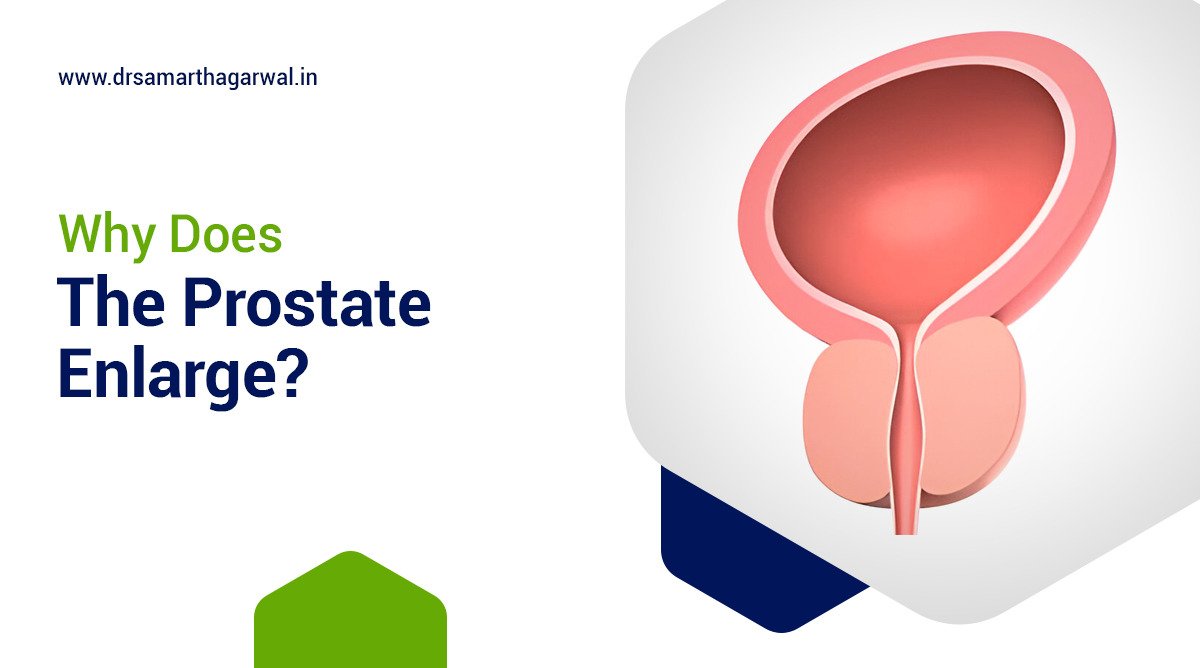
Meet the top Urologist in Siliguri

Can Kidney Stone Make You Constipated?

Kidney stones are hard mineral and salt deposits that form in the kidneys, affecting a significant portion of the population, with estimates suggesting that about 10% of people will experience them at some point in their lives. These stones can lead to severe pain and various complications, including urinary obstruction.

On the other hand, constipation is a common digestive issue characterized by infrequent bowel movements or difficulty passing stools, often caused by factors such as dehydration, a low-fiber diet, and certain medications. 

The connection between kidney stones and constipation arises from the fact that both conditions can be influenced by hydration levels and dietary choices. When kidney stones are present, they can cause pain and discomfort, which may lead individuals to alter their eating and drinking habits, potentially resulting in dehydration and subsequent constipation. 

Additionally, the pain associated with kidney stones can lead to reduced physical activity, further exacerbating constipation. Therefore, understanding the interplay between these two conditions is crucial for effective management and prevention strategies.

Mechanisms Linking Kidney Stones and Constipation

The relationship between kidney stones and constipation can be understood through various mechanisms, including anatomical and physiological connections, the role of the ureter and kidney in constipation, and how kidney stones can affect the bladder. Anatomically, the proximity of the kidneys and ureters to the intestines means that issues in one system can influence the other, as kidney stones can cause pain and inflammation that may disrupt normal bowel function.

Also when kidney stones obstruct the urinary tract, they can lead to increased pressure and discomfort, which may contribute to constipation. In cases of inflammatory bowel disease, the presence of kidney stones can further complicate digestive health, leading to a cycle of pain and altered bowel habits.

Anatomical and Physiological Connections

Understanding the mechanisms linking kidney stones and constipation involves exploring anatomical and physiological connections, particularly the role of the ureter and kidney in constipation, how kidney stones can affect the bladder, and the relationship between inflammatory bowel disease and kidney stones. These connections highlight how disturbances in one system can lead to complications in another, emphasizing the importance of a holistic view of urinary and digestive health.

Role of the Ureter and Kidney in Constipation

The ureters and kidneys play a crucial role in maintaining fluid balance and waste elimination in the body. When kidney stones obstruct the ureters, they can cause significant pain and discomfort, which may lead to changes in dietary habits and reduced fluid intake. This can result in dehydration, a common contributor to constipation. Additionally, the stress and pain associated with kidney stones can lead to decreased physical activity, further exacerbating the risk of constipation.

Kidney Stones Affecting the Bladder

Kidney stones can also impact the bladder by causing irritation and inflammation. When stones move from the kidneys to the bladder, they can lead to urinary urgency, frequency, and pain during urination. This discomfort can alter normal bladder function and may lead to a reluctance to drink adequate fluids, compounding the risk of constipation. The interplay between urinary symptoms and bowel habits underscores the interconnectedness of these systems.

Inflammatory Bowel Disease and Kidney Stones 

Inflammatory bowel disease (IBD) is characterized by chronic inflammation of the gastrointestinal tract, which can affect nutrient absorption and fluid balance. Individuals with IBD may be at an increased risk of developing kidney stones due to dehydration and dietary restrictions. The presence of kidney stones can further complicate IBD symptoms, leading to increased abdominal pain and altered bowel habits, creating a cycle that can worsen both conditions. Understanding these relationships is essential for effective management and treatment strategies.

Pain and Medication Influence

The mechanisms linking kidney stones and constipation are significantly influenced by pain and medication. The presence of kidney stones often leads to severe pain, which can adversely affect digestive health, while certain medications used to manage kidney stones may contribute to constipation. Understanding these factors is essential for addressing the interconnected nature of urinary and digestive health.

Impact of Pain on Digestive Health

The pain associated with kidney stones can be debilitating, leading to changes in dietary habits and physical activity. When individuals experience intense discomfort, they may avoid eating or drinking, which can result in dehydration and a lack of dietary fiber—two key contributors to constipation.

Additionally, the stress and anxiety related to managing pain can further disrupt normal digestive processes, leading to a cycle of discomfort that exacerbates both kidney stone symptoms and constipation.

Medications for Kidney Stones Causing Constipation

Various medications prescribed for managing kidney stones, such as pain relievers and antispasmodics, can have side effects that include constipation. Opioids, commonly used for pain management, are particularly notorious for slowing down gastrointestinal motility, making it more difficult for individuals to have regular bowel movements.

As patients focus on alleviating their kidney stone pain, they may inadvertently neglect their digestive health, leading to a compounded issue of constipation that requires careful management alongside their kidney stone treatment.

infected kidney stone treatment
Kidney stone treatment laser: Know All About

Diagnosis and Identification

Recognizing symptoms is crucial in understanding the relationship between kidney stones and constipation. Common symptoms of kidney stones include severe pain, blood in urine, and nausea, while signs of constipation may manifest as infrequent bowel movements and abdominal discomfort.

To diagnose kidney stones, medical testing and imaging techniques such as ultrasounds or CT scans are employed, which can also help identify any bowel obstructions that may contribute to constipation. Diagnostic tests specifically for kidney stones, like urinalysis, can further clarify the condition and guide appropriate treatment.

Recognizing Symptoms

Recognizing symptoms is essential for diagnosing kidney stones, especially when they contribute to constipation. Common symptoms of kidney stones include intense pain in the back or side, blood in urine, and nausea, which can overlap with signs of constipation such as infrequent bowel movements, bloating, and abdominal discomfort.

The interplay between these conditions can complicate diagnosis, as the pain from kidney stones may lead to changes in bowel habits, resulting in constipation. Understanding these symptoms is crucial for effective identification and treatment, as both conditions can significantly impact a person’s quality of life.

Medical Testing and Imaging

Medical testing and imaging play a critical role in diagnosing kidney stones and identifying any potential bowel obstructions that may contribute to constipation. Diagnostic tests for kidney stones typically include imaging techniques such as ultrasounds and CT scans, which can reveal the presence, size, and location of stones.

These tests are essential not only for confirming kidney stones but also for assessing the gastrointestinal tract to identify bowel obstructions, which can exacerbate constipation. By utilizing these diagnostic tools, healthcare providers can effectively differentiate between kidney stones and other gastrointestinal issues, ensuring appropriate treatment and management of both conditions.

Treatment Options

When addressing kidney stones that may lead to constipation, a multifaceted approach is essential. Hydration and dietary adjustments are crucial first steps; increasing fluid intake helps dilute urine and can prevent stone formation, while dietary changes, such as reducing oxalate-rich foods, can also be beneficial. In cases where stones are larger or causing significant discomfort, medical and surgical interventions, such as lithotripsy or ureteroscopy, may be necessary to remove the stones and alleviate symptoms.

Managing constipation is equally important in this context, as it can be exacerbated by kidney stones. Dietary modifications, including increasing fiber intake through fruits, vegetables, and whole grains, can promote regular bowel movements. Additionally, medical treatments for constipation, such as laxatives or stool softeners, may be recommended to relieve symptoms and improve overall digestive health. By addressing both kidney stones and constipation through these treatment options, individuals can achieve better health outcomes and enhanced quality of life.

Preventative Measures

Kidney stones can cause constipation, and vice versa. To manage and prevent these conditions, it’s essential to take proactive measures.

Preventing Kidney Stones

Preventing kidney stones requires a combination of hydration, dietary changes, and lifestyle modifications.

Hydration and Diet Tips: Aim for 8-10 glasses of water daily and limit sodium, sugar, and animal protein intake. Incorporate citrate-rich foods like lemons and oranges to inhibit stone formation.

Lifestyle Changes: Maintain a healthy weight, manage stress, and engage in regular exercise to further reduce the risk of kidney stones.

Establishing Normal Bowel Movements

Establishing normal bowel movements is crucial to preventing constipation, which can contribute to kidney stone formation.

Dietary Recommendations: Consume a high-fiber diet with fruits, vegetables, and whole grains to promote regularity. Foods like prunes, berries, and leafy greens act as natural laxatives.

Regular Physical Activity: Aim for at least 30 minutes of moderate exercise daily, such as walking or yoga, to stimulate bowel movements and enhance digestive health.

When to Consult a Urologist If kidney stone make you constipated

When experiencing kidney stones, it is crucial to recognize the indicators for seeking immediate help, especially if constipation becomes severe. Consulting a urologist is essential when kidney stones cause significant discomfort, persistent constipation, or if there are accompanying symptoms such as severe abdominal pain, blood in urine, or fever. These signs may indicate that the stones are obstructing the urinary tract or causing other complications that require professional medical intervention.

If kidney stones are causing constipation, it is vital to monitor your symptoms closely. Seek immediate medical advice if you experience severe pain, prolonged constipation, or any alarming symptoms, as these may necessitate a consultation with a urologist to address potential complications and receive appropriate treatment.

Frequently Asked Questions (FAQs)

Can kidney stones cause bowel obstruction?

Kidney stones can cause bowel obstruction, though it’s extremely rare. This occurs when a kidney stone passes through the intestinal wall and becomes lodged in the bowel. Many situations can result in intestinal obstruction, particularly when kidney stones pass through the intestinal wall. 

How does dehydration affect kidney stones and constipation?

Dehydration increases the risk of kidney stones. When you are dehydrated, your urine becomes concentrated, which can cause stone-forming crystals to stick together and form kidney stones. Dehydration can also contribute to constipation, which can be caused by a lack of fluids in the digestive system.

What diet changes can help prevent kidney stones and maintain bowel health?

To prevent kidney stones, consume calcium-rich foods, reduce sodium intake, limit animal protein, and avoid stone-forming foods like beets, chocolate, spinach, rhubarb, tea, and oxalate-rich nuts. To maintain bowel health,  increase your intake of fiber-rich foods like fruits, vegetables, and whole grains.

Dr Samarth Agarwal receiving recognition

Contact Dr. Samarth Agarwal if you have any questions or concerns about your Urinary health!

Meet the top Urologist in Siliguri

What Causes Erectile Dysfunction in Your 40s 

Erectile dysfunction (ED) is a common issue that affects many men, especially in their 40s. It means having trouble getting or keeping an erection firm enough for sex.

The incidence of erectile dysfunction in men aged 40 to 69 years old was estimated to be about 26 cases per 1,000 men annually, with the risk increasing with age, lower education, diabetes, heart disease, and hypertension, according to a study by Johannes et al. in the Journal of Urology in 2000, Johannes, C. B., Araujo, A. B., Feldman, H. A., Derby, C. A., Kleinman, K. P., & McKinlay, J. B. (2000).

The causes of ED in your 40s can be categorized into physical and psychological factors. Physical causes often stem from conditions that affect blood flow, such as high blood pressure, high cholesterol, diabetes, and heart disease. These conditions can damage the blood vessels in the penis, making it difficult to achieve an erection.

Stress, anxiety, and depression can also cause ED. These emotional issues can disrupt the brain’s signals to the penis, making it hard to get or keep an erection.

Treatment options for ED include medications, such as sildenafil (Viagra), tadalafil (Cialis), and vardenafil (Levitra), which help increase blood flow to the penis. Lifestyle changes, such as quitting smoking, reducing alcohol consumption, and exercising regularly, can also help improve ED symptoms. In some cases, counseling or therapy may be recommended to address any underlying psychological issues.

Erectile Dysfunction in Your 40s

Erectile Dysfunction (ED) is the inability to achieve or maintain an erection sufficient for satisfactory sexual performance, causing distress and impacting relationships.

Addressing ED in your 40s is crucial as it can be a sign of underlying health issues, such as cardiovascular disease or diabetes, and treating it can improve overall well-being and quality of life.

ED is often misunderstood as a natural part of aging, but it’s not an inevitable consequence of growing older. Many cases of ED are treatable, and seeking help can lead to effective solutions and improved sexual health.

Understanding Erectile Dysfunction

Erectile Dysfunction (ED) is a condition characterized by the inability to achieve or maintain an erection sufficient for satisfactory sexual performance. Common symptoms include difficulty getting or keeping an erection, reduced sexual desire, and decreased libido.

ED can be a distressing experience, affecting not only physical intimacy but also emotional well-being and relationships. Recognizing the signs and symptoms is crucial for seeking timely medical attention and effective treatment.

Difference Between ED and Other Sexual Dysfunctions

Erectile Dysfunction is distinct from other sexual dysfunctions, such as premature ejaculation, low libido, and orgasmic disorders. While these conditions can coexist, understanding the specific characteristics of ED is essential for accurate diagnosis and treatment.

Role of Blood Flow in Erections

Blood flow plays a critical role in achieving and maintaining erections. During sexual arousal, blood vessels dilate, allowing increased blood flow into the penis, causing it to become erect. Any disruption in blood flow can lead to ED, highlighting the importance of cardiovascular health.

Nervous System and Erections

The nervous system is vital for erectile function, as it transmits signals between the brain and penis. Damage to nerves or neurological conditions can impair these signals, leading to ED. Understanding the complex interplay between the nervous system and erectile function can help identify potential causes and treatment options.

Hormonal Influence on Erectile Function

Hormones, particularly testosterone, significantly impact erectile function. Hormonal imbalances or deficiencies can contribute to ED, and understanding the role of hormones can help identify underlying causes and guide treatment decisions.

Physical Causes of Erectile Dysfunction in Your 40s

Erectile Dysfunction (ED) in your 40s can be attributed to various physical factors, which are crucial to understand for effective management and treatment.

Cardiovascular Issues

Cardiovascular problems, such as blood flow issues, heart disease, and high blood pressure, can significantly contribute to ED in your 40s. These conditions can damage blood vessels, reducing blood flow to the penis and making it difficult to achieve or maintain an erection.

Erectile dysfunction (ED) is a common condition in men who are 40 years and older, with prevalence increasing with age and other co-morbidities, such as cardiovascular disease, diabetes mellitus, hyperlipidemia, and hypertension. According to Leslie, S. W., & Sooriyamoorthy, T. (2024). Erectile Dysfunction. In StatPearls [Internet]. Treasure Island (FL): StatPearls Publishing; 2024 Jan. 2024

Metabolic Disorders

Metabolic disorders, including diabetes, high cholesterol, and obesity, can also lead to ED. These conditions can damage blood vessels, nerves, and hormones, disrupting the complex process of erection.

Neurological Conditions

Neurological conditions, such as spinal cord injuries and nervous system disorders, can affect the transmission of signals between the brain and penis, leading to ED.

Hormonal Imbalances

Hormonal imbalances, including low testosterone levels and thyroid disorders, can impact erectile function, making it essential to address these underlying issues.

Medication Side Effects

Certain medications, such as those used to treat cardiovascular issues, can contribute to ED as a side effect. Understanding the potential impact of long-term medication use is vital for managing ED.

Psychological Causes of Erectile Dysfunction in Your 40s

Erectile Dysfunction (ED) in your 40s can be attributed to various psychological factors, which are essential to understand for effective management and treatment. Psychological causes can be just as significant as physical ones, and addressing them can lead to improved erectile function and overall well-being.

Stress and Anxiety

Chronic stress and anxiety can negatively impact erectile function, leading to ED. Performance anxiety, in particular, can create a cycle of fear and doubt, making it difficult to achieve or maintain an erection. Managing stress and anxiety through relaxation techniques and counseling can help alleviate ED symptoms.

Depression and Mental Health Issues

Depression and mental health issues can significantly impact sexual health, leading to ED. Treatments for mental health conditions, such as antidepressants, can also contribute to ED. Understanding the complex relationship between mental health and erectile function can help identify effective treatment strategies.

Relationship Issues

Interpersonal relationships and communication with partners can influence sexual performance and contribute to ED. Addressing relationship issues and improving communication can help resolve ED symptoms and strengthen relationships.

Lifestyle and Behavioral Factors

Lifestyle and behavioral factors play a significant role in erectile dysfunction (ED) in your 40s. Adopting healthy habits and avoiding harmful behaviors can help prevent or manage ED, improving overall well-being and sexual health.

Dietary Habits

A balanced diet rich in foods that promote blood flow, such as leafy greens, berries, and omega-3 fatty acids, can support erectile function. Conversely, diets high in processed foods, sugar, and saturated fats can negatively impact blood flow and contribute to ED.

Physical Activity

Regular exercise, especially aerobic exercises like cycling, swimming, and brisk walking, can improve cardiovascular health, boost testosterone levels, and enhance erectile function. Exercises that strengthen the pelvic floor muscles, such as Kegel exercises, can also benefit ED.

Pelvic floor exercises have been shown to improve erectile function in men with erectile dysfunction, according to a clinical trial by Dorey, G., Speakman, M. J., Feneley, R. C. L., Swinkels, A., Dunn, C. D. R. (2005). Pelvic floor exercises for erectile dysfunction. British Journal of Urology International.

Substance Use

Substance use, including excessive alcohol consumption, smoking, and illicit drug use, can significantly contribute to ED. Alcohol can impair blood flow, while smoking damages blood vessels and reduces circulation. Illicit drugs can disrupt hormonal balances and nerve function, leading to ED. Quitting or reducing substance use can help alleviate ED symptoms.

Diagnosing Erectile Dysfunction

Diagnosing erectile dysfunction (ED) in your 40s involves a comprehensive approach to identify underlying causes and develop an effective treatment plan. Accurate diagnosis is crucial for addressing physical, psychological, and lifestyle factors contributing to ED.

Initial Self-Assessment

Recognizing symptoms of ED is the first step towards diagnosis. Men experiencing difficulties getting or maintaining an erection, reduced libido, or decreased sexual satisfaction should take note of their symptoms and medical history to discuss with their doctor.

Medical Consultation

Consulting a doctor is essential for diagnosing ED. Your doctor will discuss your medical history, perform a physical examination, and provide a safe space to share sensitive information. Open communication with your doctor helps identify underlying causes and develop a personalized treatment plan.

Diagnostic Tests

Diagnostic tests, such as blood tests to check hormone levels, penile ultrasound to assess blood flow, and other diagnostic tools, may be used to identify underlying causes of ED. These tests help determine the root cause of ED, guiding treatment decisions and ensuring effective management.

Treatment Options for Erectile Dysfunction

Treatment options for erectile dysfunction (ED) in your 40s are diverse and cater to individual needs. A comprehensive approach may involve medical treatments, non-medical interventions, psychological counseling, and lifestyle changes to address physical, psychological, and behavioral factors contributing to ED.

Medical Treatments

Medical treatments for ED include phosphodiesterase inhibitors like sildenafil, which enhance blood flow, and hormone replacement therapy to address hormonal imbalances. These treatments can help restore erectile function and improve sexual satisfaction.

Non-Medical Interventions

Non-medical interventions, such as penile implants and vacuum erection devices, offer alternative solutions for ED. Penile implants provide a permanent solution, while vacuum erection devices offer a non-invasive, temporary solution.

Psychological Counseling

Psychological counseling addresses psychological causes of ED, such as stress, anxiety, and relationship issues. Therapy methods like cognitive-behavioral therapy and couple’s therapy can help individuals and couples overcome psychological barriers to erectile function.

Lifestyle Changes

Lifestyle changes, including dietary adjustments and regular physical activity, can significantly impact ED. A balanced diet, rich in fruits, vegetables, and whole grains, and regular exercise can improve cardiovascular health, boost testosterone levels, and enhance erectile function.

Preventing Erectile Dysfunction in Your 40s

Preventing erectile dysfunction (ED) in your 40s involves adopting a proactive approach to maintain overall health and well-being. By making informed lifestyle choices and staying on top of regular medical check-ups, you can reduce the risk of developing ED and promote healthy erectile function.

Maintaining a Healthy Lifestyle

Maintaining a healthy lifestyle is crucial for preventing ED. This includes following a balanced diet rich in fruits, vegetables, and whole grains, engaging in regular exercise, and avoiding harmful substances like tobacco and excessive alcohol. A healthy lifestyle can help manage stress, boost testosterone levels, and improve cardiovascular health.

Regular Medical Check-Ups

Regular medical check-ups are essential for preventing ED. Routine health screenings can help identify underlying health issues, such as diabetes, high blood pressure, and high cholesterol, which can contribute to ED. Monitoring blood pressure and cholesterol levels can help prevent cardiovascular disease, a common cause of ED. By staying on top of your health, you can address potential issues before they impact erectile function.

Coping Strategies and Support

Coping with erectile dysfunction (ED) in your 40s requires a comprehensive approach that addresses emotional, psychological, and practical aspects. Developing effective coping strategies and seeking support can help you navigate the challenges of ED and improve your overall well-being.

Emotional Support

Emotional support from loved ones, such as partners, family, and friends, can play a vital role in coping with ED. Sharing your experiences and feelings with others can help you feel less isolated and more supported. Joining support groups for men with ED can also provide a sense of community and connection with others who understand your experiences.

Long-Term Management

Long-term management of ED requires sustainable practices and realistic expectations. This involves adopting healthy lifestyle habits, such as regular exercise and balanced diet, and engaging in stress-reducing activities. Setting realistic expectations about treatment outcomes and sexual performance can also help you cope with ED and maintain a positive outlook.

Frequently Asked Questions (FAQs)

How common is erectile dysfunction in men over 40?

Erectile dysfunction (ED) is a prevalent condition in men over 40, affecting over 50% of men between the ages of 40 and 70, according to various medical research. For instance, a study showed that 52% of men aged 40–70 years had some degree of ED, with moderate-to-complete ED being encountered in 34.8% of cases. This condition is not uncommon, and it’s essential to seek medical advice if experiencing persistent difficulties.
Erectile dysfunction (ED) is a common condition in young men, with a prevalence as high as 30%, according to a review by Nguyen et al. in the journal Sexual Medicine Reviews in 2017 by H. M. T., Gabrielson, A. T., & Hellstrom, W. J. G. (2017). Erectile Dysfunction in Young Men-A Review of the Prevalence and Risk Factors. Sexual Medicine Reviews

Can lifestyle changes alone improve erectile function?

Lifestyle changes, such as increased physical activity, a healthy diet, and reduced caloric intake, have been associated with the improvement of erectile function in the general male population. Research suggests that lifestyle changes alone can significantly improve erectile dysfunction, but it’s essential to consult with a healthcare professional for personalized advice. Specifically, eating a diet rich in fruits, vegetables, fish, and whole grains, as well as regular exercise, can help reverse ED. Additionally, avoiding certain foods like processed meats, high-fat dairy, and sugary drinks may also contribute to improved erectile function.

What should I discuss with my doctor if I think I have ED?

Directly express your concerns: Start by saying, “I think I may have ED.”
Prepare for the discussion: Write down details of your experiences before your appointment.
Discuss your medical history: Your doctor will ask about your medical history, medications, and lifestyle habits.
Ask about potential causes: Ask your doctor what they think is causing your erection problems.
Inquire about tests: Ask about the tests you need to determine the cause of your ED.
Explore treatment options: Depending on the cause and severity of your ED, you might have various treatment options.
Ask about side effects and risks: Discuss potential side effects and risks of each treatment option.
Follow-up appointments: Schedule follow-up appointments to monitor your progress and adjust treatments if necessary.
Consider a referral: If you sense your doctor is uncomfortable talking about your ED, ask for a referral to a urologist.
Research and ask questions: Learn about ED, its causes, and treatments before your appointment to be well-informed.

Dr Samarth Agarwal receiving recognition

Contact Dr. Samarth Agarwal if you have any questions or concerns about your Urinary health!
Meet the top Urologist in Siliguri

Can Kidney Cancer Cause Low Hemoglobin?

Kidney cancer, also known as renal cell carcinoma, is a type of cancer that originates in the kidneys. This cancer can cause low hemoglobin levels in the body, which is a condition characterized by a decrease in the number of red blood cells or the amount of hemoglobin in the blood.

Low hemoglobin levels can lead to symptoms such as fatigue, weakness, shortness of breath, and pale skin. In some cases, kidney cancer can cause anemia, which is a condition characterized by a decrease in the number of red blood cells in the body.

According to a study published in the Journal of Clinical Oncology, anemia is a common complication of kidney cancer and can affect up to 80% of patients with advanced kidney cancer. The study found that anemia can lead to decreased physical performance, poor quality of life, and increased risk of mortality in patients with kidney cancer.

Treatment options for anemia in patients with kidney cancer include blood transfusions, erythropoietin stimulating agents, and iron supplements. In some cases, surgery to remove the tumor may also help to improve hemoglobin levels.

What is Hemoglobin?

Hemoglobin is a protein in red blood cells that carries oxygen. It is a red substance made of iron and protein, found in the blood, which carries oxygen to cells in the body from the lungs. Hemoglobin is the most common protein in red blood cells and is essential for oxygen transport in the body.

Kidney cancer can cause low hemoglobin levels, and a study by Yang et al. (2023) found that baseline and dynamic changes in hemoglobin levels can predict treatment response and prognosis in metastatic renal cell carcinoma.

In this multicenter retrospective study, the authors evaluated 276 patients with metastatic renal cell carcinoma treated with frontline immune checkpoint inhibitor (ICI) therapy, ICI and vascular endothelial growth factor (VEGF) inhibitor (VEGFI) combinations (ICI/VEGFI), or VEGFI monotherapy between 2014 and 2021. The authors found that an increase in hemoglobin was a significant independent predictor of progression-free survival amongst patients receiving ICIs.

Understanding Kidney Cancer and Anemia

How Kidney Cancer Can Lead to Anemia

Kidney cancer can lead to anemia through several mechanisms, primarily impacting red blood cell production and erythropoiesis. The disease can disrupt normal renal function, which is crucial for producing erythropoietin, a hormone that stimulates red blood cell production in the bone marrow. Additionally, chronic kidney disease (CKD), often associated with kidney cancer, can further impair erythropoiesis and lead to anemia due to reduced kidney function and the accumulation of waste products in the blood.

Kidney cancer can cause low hemoglobin levels due to microcytic hypochromic anemia, a condition characterized by small, pale red blood cells. According to Kroll, M.H., Jiji, V., & Jiji, R. (1984). Microcytic hypochromic anemia associated with renal cell carcinoma, anemia occurs in approximately one third of patients with renal cell carcinoma. The authors described a patient with classic iron deficiency anemia and renal cell carcinoma whose blood picture reverted to normal after removal of the tumor. Abundant hemosiderin in the tumor cells is evidence that tumor cells removed iron from the circulation, causing the iron deficiency anemia.

The mechanism by which kidney cancer impacts red blood cell production involves the tumor’s effect on renal function and the production of erythropoietin. As kidney cancer progresses, it can damage healthy kidney tissue, leading to decreased erythropoietin levels and, consequently, reduced stimulation of red blood cell production. In kidney cancer patients, the role of erythropoiesis becomes critical, as the body struggles to maintain adequate red blood cell levels in the face of cancer-related challenges. 

Furthermore, chronic kidney disease, which may develop as a complication of kidney cancer, exacerbates anemia by impairing the kidneys’ ability to filter waste and produce erythropoietin, leading to a cycle of worsening anemia and declining renal function. Understanding these interconnected factors is essential for managing anemia in patients with kidney cancer effectively.

Examples of kidney cancer types:

  • Renal cell carcinoma (RCC)
  • Transitional cell cancer
  • Renal sarcoma
  • Wilms tumor (in children)

Symptoms of Kidney Cancer

The following are common symptoms of kidney cancer that may indicate the presence of the disease: blood in urine, persistent flank pain, unexplained weight loss, loss of appetite, tiredness or fatigue, fever or night sweats, high blood pressure, anemia, high calcium levels, and bone pain. Additionally, there are several types of kidney cancer, including renal cell carcinoma, transitional cell cancer, renal sarcoma, and Wilms tumor.

  1. Blood in Urine (Hematuria): Hematuria is often one of the first noticeable symptoms of kidney cancer. It occurs when cancerous cells invade blood vessels in the kidney, leading to bleeding. This symptom can appear intermittently and may be visible to the naked eye or detected through a urinalysis. It is important to note that hematuria can also be caused by other conditions, such as urinary tract infections or kidney stones.
  2. Persistent Flank Pain: Flank pain refers to discomfort in the side of the body, typically between the ribs and the hip. In kidney cancer, this pain can result from the tumor pressing against surrounding tissues or nerves. It may be dull or sharp and can persist over time, often worsening as the cancer progresses.
  3. Unexplained Weight Loss: Many cancer patients experience unexplained weight loss due to a combination of factors, including increased metabolic demands from the tumor, loss of appetite, and changes in how the body processes nutrients. In kidney cancer, this symptom may occur as the disease advances and the body struggles to maintain energy levels.
  4. Loss of Appetite: A decrease in appetite can occur due to the body’s response to cancer, including the release of certain hormones and inflammatory substances. This symptom may develop gradually and can be exacerbated by other symptoms, such as nausea or pain, making it difficult for patients to maintain a healthy diet.
  5. Tiredness or Fatigue: Fatigue is a common symptom in cancer patients and can result from several factors, including anemia, the body’s increased energy expenditure to fight the cancer, and the psychological impact of dealing with a serious illness. In kidney cancer, fatigue may be persistent and not relieved by rest.
  6. Fever or Night Sweats: Fever and night sweats can occur as a result of the body’s immune response to cancer. The presence of a tumor can trigger systemic inflammation, leading to elevated body temperature. Night sweats may also be related to hormonal changes or the body’s attempt to regulate temperature during periods of stress.
  7. High Blood Pressure: Kidney cancer can lead to high blood pressure (hypertension) due to the tumor’s effect on the kidneys’ ability to regulate blood pressure. Additionally, the production of certain hormones by the tumor can constrict blood vessels, further contributing to elevated blood pressure levels.
  8. Anemia: Anemia in kidney cancer patients can result from several factors, including the cancer’s impact on erythropoietin production (a hormone that stimulates red blood cell production) and the potential for chronic blood loss. Anemia can lead to symptoms such as fatigue, weakness, and pallor.
  9. High Calcium Levels: Kidney cancer can cause hypercalcemia (high calcium levels) due to the release of parathyroid hormone-related peptide (PTHrP) from the tumor, which mimics the action of parathyroid hormone. This can lead to symptoms such as nausea, vomiting, confusion, and increased thirst.
  10. Bone Pain: Bone pain may occur if kidney cancer metastasizes (spreads) to the bones. The presence of cancerous cells in the bone can cause localized pain, which may worsen with movement. Additionally, high calcium levels associated with kidney cancer can also contribute to bone discomfort.

Diagnostic Methods for Kidney Cancer and Anemia

Blood Tests for Kidney Cancer and Hemoglobin Levels

Diagnostic methods for kidney cancer often include various blood tests, which can also provide insights into hemoglobin levels, a critical factor in diagnosing anemia. Among the types of blood tests, the Complete Blood Count (CBC) is essential as it measures red blood cells, hemoglobin, and other components, helping to identify anemia that may arise due to kidney dysfunction or cancer. Interpreting blood test results is crucial; low hemoglobin levels can indicate anemia, while abnormal kidney function markers may suggest the presence of kidney cancer. Thus, understanding these diagnostic methods is vital for effective management and treatment of both conditions.

Blood tests play a pivotal role in diagnosing kidney cancer and assessing hemoglobin levels, with the Complete Blood Count being a primary tool for identifying anemia. The interpretation of these results is essential, as low hemoglobin can signal anemia, while abnormal kidney function indicators may point to kidney cancer. Together, these diagnostic methods provide a comprehensive view of a patient’s health, highlighting the intricate relationship between kidney cancer and anemia.

Imaging Studies

Understanding kidney cancer and anemia involves recognizing the diagnostic methods that are essential for identifying these conditions. Imaging studies, particularly CT scans and MRIs, play a crucial role in diagnosing kidney cancer by providing detailed images of the kidneys and surrounding tissues. These imaging techniques help in identifying renal masses, which can indicate the presence of tumors. The impact of these masses is significant, as they can lead to complications such as anemia, often due to the cancer’s effect on kidney function or the body’s response to the tumor. Therefore, the interplay between imaging studies and the identification of renal masses is vital for diagnosing kidney cancer and understanding its potential to cause anemia.

Imaging studies like CT scans and MRIs are fundamental diagnostic methods for kidney cancer, allowing for the identification of renal masses that may lead to anemia. These imaging techniques provide critical insights into the structure and function of the kidneys, helping to assess the extent of cancer and its implications on overall health. The relationship between kidney cancer and anemia underscores the importance of accurate imaging in guiding treatment decisions and managing patient outcomes effectively.

Biopsy and Histopathological Examination

Understanding kidney cancer and anemia requires a comprehensive look at the diagnostic methods used to identify these conditions, particularly through biopsy and histopathological examination. A renal biopsy is a critical procedure that involves extracting a small sample of kidney tissue to examine for cancerous cells. This examination is vital for confirming a diagnosis of kidney cancer, as it provides definitive information about the type and aggressiveness of the tumor. Additionally, the relationship between kidney cancer and hemoglobin levels is significant; tumors can affect kidney function, leading to anemia due to decreased erythropoietin production, a hormone essential for red blood cell formation. Thus, the insights gained from a renal biopsy not only aid in diagnosing cancer but also help in understanding its impact on hemoglobin levels and overall patient health.

A renal biopsy and histopathological examination are essential diagnostic methods for kidney cancer, providing crucial information about the presence and characteristics of tumors. This procedure is important not only for confirming cancer but also for understanding its implications on hemoglobin levels, as kidney dysfunction related to cancer can lead to anemia. The interplay between these diagnostic techniques and their findings highlights the importance of thorough evaluation in managing kidney cancer and its associated effects on anemia.

Role of a Urologist in Diagnosis

Understanding kidney cancer and anemia involves recognizing the critical role of a urologist in the diagnostic process. A urologist conducts a comprehensive history and physical examination to assess symptoms and risk factors associated with kidney cancer. This initial evaluation is essential for determining the need for further diagnostic methods, such as imaging studies or biopsies.

Patients should be prepared to ask their healthcare provider specific questions regarding their symptoms, family history, and any relevant lifestyle factors that could influence their risk for kidney cancer and anemia. This proactive approach not only aids in the accurate diagnosis of kidney cancer but also helps in understanding how the disease may contribute to anemia through impaired kidney function.

The role of a urologist in diagnosing kidney cancer is pivotal, as they perform thorough history-taking and physical examinations to identify potential symptoms and risk factors. This foundational assessment guides the need for additional diagnostic methods and encourages patients to engage actively with their healthcare providers by asking pertinent questions.

By fostering open communication and comprehensive evaluations, patients can better understand the relationship between kidney cancer and anemia, leading to more effective management and treatment strategies.

Treatment Options for Kidney Cancer and Managing Low Hemoglobin

Kidney cancer presents unique challenges, particularly when compounded by low hemoglobin levels, which can significantly affect a patient’s overall health and treatment tolerance. Understanding the treatment options and effective management strategies is essential for improving patient outcomes and quality of life.

Treatment Approaches for Kidney Cancer

Treatment for kidney cancer encompasses a range of modalities designed to eliminate or control cancerous cells. Nephrectomy involves the complete removal of the kidney, while Partial Nephrectomy conserves healthy tissue by excising only the tumor. Targeted Therapy utilizes drugs that specifically inhibit cancer cell growth by blocking molecular pathways, while Immunotherapy harnesses the body’s immune system to fight cancer.

Although Chemotherapy is less effective for kidney cancer, it may be used in certain cases, and Radiation Therapy can alleviate symptoms or target metastases. These approaches are vital not only for cancer control but also for managing anemia, as effective cancer treatment can improve hemoglobin levels and overall patient health.

Managing Anemia in Kidney Cancer Patients

Anemia management in kidney cancer patients is critical, as low hemoglobin can exacerbate fatigue and reduce treatment efficacy. Erythropoiesis-Stimulating Agents (ESAs), such as epoetin alfa, are commonly prescribed to boost red blood cell production. Blood Transfusions provide immediate relief for severe anemia, while Iron Supplements help replenish iron stores essential for hemoglobin synthesis. Additionally, Diet and Lifestyle Adjustments—including a diet rich in iron (e.g., red meat, leafy greens) and vitamin B12—can enhance red blood cell production. These interventions not only alleviate symptoms but also empower patients to better tolerate ongoing cancer therapies.

Monitoring and Follow-Up

Ongoing monitoring and follow-up are essential for kidney cancer patients, particularly those with low hemoglobin levels. Regular Blood Tests assess hemoglobin levels, while Imaging Studies (like CT scans) evaluate tumor response and detect any progression. Proactive management of Potential Side Effects from treatments—such as fatigue, nausea, or infection—ensures timely interventions, minimizing complications. By maintaining a close watch on patient health, healthcare providers can tailor treatment plans and supportive care, ultimately optimizing outcomes for those battling kidney cancer and its associated anemia.

Impact of Kidney Cancer on Overall Health

Kidney cancer not only affects the renal system but also has extensive implications for a patient’s overall health. The disease can lead to systemic complications, particularly when compounded by factors like low hemoglobin levels, which can significantly alter a patient’s quality of life and long-term health outcomes.

Complications Related to Low Hemoglobin

Low hemoglobin levels, frequently seen in kidney cancer patients, can lead to debilitating Fatigue and Weakness, which hinder daily activities and diminish productivity. This fatigue can also strain Social Interactions, leading to isolation and mental health issues such as depression and anxiety. Furthermore, low hemoglobin can worsen existing conditions like Cardiovascular Disease, increasing the risk of heart complications due to inadequate oxygen delivery to tissues. Effective management of anemia through interventions like ESAs and dietary changes is essential to enhance energy levels and improve overall well-being, allowing patients to engage more fully in life.

Prognosis and Long-Term Management

The Prognosis for kidney cancer patients is significantly better with early detection, which allows for more effective treatment options, including surgery and targeted therapies. Long-Term Health Monitoring is crucial, involving regular assessments of kidney function, imaging studies, and blood tests to track hemoglobin levels and detect any recurrence early. This proactive management approach not only addresses potential side effects of treatments but also facilitates timely adjustments to therapy, thereby minimizing complications. By prioritizing ongoing care, healthcare providers can help patients maintain a better quality of life and preserve their overall health in the face of kidney cancer.

FAQ

Can kidney cancer be detected through routine blood tests?

Routine blood tests, such as a complete blood count (CBC) and blood comprehensive metabolic panel (CMP), can show signs in the blood that are linked with kidney cancer. However, these tests cannot diagnose kidney cancer directly. Instead, they provide important information about kidney function and overall organ health. For a definitive diagnosis, doctors often rely on imaging tests like ultrasounds, CT scans, or MRI scans.

How does kidney cancer affect the body’s ability to produce red blood cells?

Kidney cancer may lead to a drop in red blood cell count (anemia) due to slow bleeding into the urine. This anemia may cause easy fatigability or dizziness. Additionally, kidney cancer can cause high levels of calcium in the blood (hypercalcemia), which may cause weakness, fatigue, slowed reaction times, and constipation. These effects are primarily due to the kidneys’ role in producing a hormone that regulates red blood cell production.

Can anemia be a symptom of other types of cancer besides kidney cancer?

Yes, anemia can be a symptom of various types of cancer, not just kidney cancer. Gastrointestinal cancers, such as stomach or colon cancer, can cause anemia due to internal bleeding. Additionally, anemia can be associated with many other types of cancer, including cancers of the pancreas, lung, and uterus, among others. Anemia may also be caused by inflammation or chronic disease related to cancer.

How does chemotherapy affect hemoglobin levels in kidney cancer patients?

Chemotherapy for kidney cancer, can cause a drop in hemoglobin levels due to its effect on all rapidly growing cells, including red blood cells. This often leads to anemia, a condition characterized by a lower than normal number of red blood cells or lower than normal hemoglobin in the blood. The severity of anemia can vary among patients, and it may require treatment to manage symptoms and maintain quality of life during cancer treatment.

Can dietary changes help manage low hemoglobin levels in kidney cancer patients?

Yes, dietary changes can help manage low hemoglobin levels in kidney cancer patients. Foods rich in iron, such as red meat, poultry, fish, beans, and dark green leafy vegetables, can boost hemoglobin levels. Additionally, consuming vitamin C-rich foods, like citrus fruits and strawberries, can aid in iron absorption. A dietician’s guidance can play a significant role in managing anemia related to kidney cancer.

Are there any alternative treatments for kidney cancer-related anemia?

Kidney cancer-related anemia is primarily treated with iron substitution, erythropoiesis-stimulating agents (ESA), and in refractory cases with red blood cell transfusions. However, alternative treatments may include targeted drug therapy for kidney cancer, as some of these drugs can help manage anemia symptoms. For instance, the American Cancer Society mentions that targeted drugs work differently on renal cell carcinoma (kidney cancer) than standard chemotherapy drugs. Additionally, the National Kidney Foundation suggests that drugs called erythropoiesis stimulating agents (ESAs) can help your body make red blood cells, which may alleviate anemia symptoms. It’s essential to consult with a healthcare provider for personalized treatment recommendations.

How does kidney cancer affect the body’s overall iron levels?

Kidney cancer, particularly renal cell carcinoma, is associated with increased iron accumulation in the cancer cells. This iron accumulation intensifies with worsening kidney cancer pathology, including size, stage, and grade. Conversely, iron levels in the renal epithelial cells decrease, leading to reduced erythropoiesis and anemia. This is due to cancer-induced hormone secretion that impairs iron absorption and recycling, restricting circulating iron levels.

In healthy individuals, iron is essential for producing red blood cells and maintaining overall good health. However, kidney cancer can disrupt this process, leading to anemia and related symptoms such as looking pale, having less energy, cold feet and hands, and irritability. It is crucial for healthcare providers to monitor iron levels in kidney cancer patients, as anemia can have significant impacts on their overall health and treatment outcomes.

Can kidney cancer patients donate blood?

Kidney cancer patients are generally not eligible to donate blood due to changes in their hormone levels that decrease blood production. This is a safety measure to protect both the donor and the recipient. However, eligibility may vary on a case-by-case basis, depending on the type of cancer, treatment, and the time since completion of treatment. It’s essential to consult with healthcare providers for specific guidelines.

How does kidney cancer affect the body’s ability to fight infections?

Advanced stage kidney cancer can impact the body’s ability to fight infections. In such cases, CD8+ T cells, a type of immune cell, become “exhausted” and unable to perform their usual function. Additionally, an increase in anti-inflammatory or “M2-like” macrophages, which suppress the immune system, is observed in advanced stage disease. Furthermore, the immune system’s focus on the cancer can divert it from other important tasks, like fighting infections. The presence of a virus can also enhance inflammation within a tumor, potentially further weakening the immune system’s ability to fight infections.

Dr Samarth Agarwal image 8

For kidney cancer-related concerns, please contact Dr. Samarth Agarwal.

Meet the top Urologist in Siliguri

Why would a urologist do a cystoscopy?

Cystoscopy is a medical procedure performed by a urologist to examine the inside of the bladder and the urethra. This procedure involves the insertion of a thin, flexible tube called a cystoscope into the bladder through the urethra.

The purpose of a cystoscopy is to diagnose, monitor, and treat conditions affecting the bladder and urethra. Some indicators that a urologist might recommend a cystoscopy include blood in the urine, incontinence, overactive bladder, and painful urination.

Cystoscopy can help identify the cause of these symptoms, as well as detect abnormalities such as bladder stones, tumors, and polyps. It can also be used to monitor the progression of bladder cancer and to assess the effectiveness of treatment.

Why did my urologist order a cystoscopy?

The urologist ordered cystoscopy for bladder and urethra examination. Commonly used for diagnosing, monitoring, and treating bladder and urethra conditions.

Cystoscopy may be done in a testing room, using a local anesthetic jelly to numb your urethra, or as an outpatient procedure with sedation. Cystoscopy in a hospital under general anesthesia is an option. The procedure type depends on the reason.

Your doctor might recommend cystoscopy to investigate causes of signs and symptoms such as blood in the urine, incontinence, overactive bladder, and painful urination. Cystoscopy can also help determine the cause of frequent urinary tract infections. However, cystoscopy generally isn’t done while you have an active urinary tract infection.

Cystoscopy can diagnose bladder diseases and conditions, such as bladder cancer, bladder stones, and bladder inflammation (cystitis). It can also treat bladder diseases and conditions, such as removing very small bladder tumors during the procedure. Cystoscopy can also diagnose an enlarged prostate by revealing a narrowing of the urethra where it passes through the prostate gland, indicating an enlarged prostate (benign prostatic hyperplasia).

A hollow tube with a lens, is inserted into the urethra and advanced into the bladder during a cystoscopy. The cystoscope has a lens on the end that works like a telescope to magnify the inner surfaces of your urethra and bladder. Your doctor might place a special video camera over the lens to project the images onto a video screen.

Complications of cystoscopy can include infection, bleeding, and pain during urination. Your doctor might prescribe antibiotics to take before and after cystoscopy, especially if you have trouble fighting off infections. You may also be asked to wait to empty your bladder before the procedure in case a urine sample is needed.

After the cystoscopy, you might be allowed to resume your daily routine. Side effects after cystoscopy might include bleeding from your urethra, a burning sensation during urination, and more frequent urination for the next day or two. You can relieve some of the discomfort by drinking water, taking an over-the-counter pain reliever, placing a warm, damp washcloth on the opening to your urethra, and taking a warm bath (unless your doctor asks you to avoid baths).

After cystoscopy, your doctor may discuss results immediately. If biopsy collected for bladder cancer test, sample sent to lab. Upon test completion, doctor informs results.

Can you see cancer with cystoscopy?

Cystoscopy helps to diagnose bladder cancer by allowing a urologist to examine the inside of the bladder for any suspicious growths or tumors. If abnormalities are found, a biopsy may be taken for further examination. However, it’s important to note that cystoscopy is not 100% accurate and other diagnostic tests may also be required.

What conditions does a cystoscopy diagnose?

A cystoscopy is a medical procedure used to diagnose various conditions affecting the urinary tract. This list includes bladder cancer, polyps, bladder stones, scarring from frequent UTIs, congenital problems, urinary tract injury, structural issues, and infections or blockages.

A cystoscopy can diagnose the following conditions:

  1. Bladder cancer or tumor
  2. Polyps or overgrowth of normal tissue
  3. Bladder stones
  4. Scarring and damage caused by frequent urinary tract infections (UTIs)
  5. Problems of the urinary tract that may be present at birth (congenital)
  6. Urinary tract injury
  7. Structural problems that may block the flow of urine
  8. Infection, narrowing, blockage, or bleeding in the urinary tract.

What are the indications for cystoscopy?

The indications for cystoscopy include investigating various urinary symptoms, diagnosing and treating bladder diseases, and monitoring conditions affecting the bladder and urethra. Cystoscopy is generally not performed while a patient has an active urinary tract infection.

  1. Investigating causes of signs and symptoms such as blood in the urine, incontinence, overactive bladder, painful urination, and frequent urinary tract infections.
  2. Diagnosing bladder diseases and conditions like bladder cancer, bladder stones, and bladder inflammation (cystitis).
  3. Treating bladder diseases and conditions, such as removing small bladder tumors during the procedure.
  4. Diagnosing an enlarged prostate.
  5. Determining the cause of frequent urinary tract infections.
  6. Monitoring and treating conditions affecting the bladder and urethra.

Cystoscopy is generally not done while a patient has an active urinary tract infection.

What type of anesthesia will be used during the cystoscopy?

During a cystoscopy, anesthesia options may include local anesthetic jelly for the urethra, sedation, general anesthesia, or a combination, depending on the reason for the procedure and the preference of the patient and doctor.

How long will the recovery process of cystoscopy take?

Most people recover within a day or two from a cystoscopy. However, recovery time may vary depending on the type of cystoscopy performed and individual health conditions. It’s essential to follow your doctor’s advice for a smooth recovery.

What are the potential risks and complications of cystoscopy?

Cystoscopy may lead to complications such as infection (UTI), bleeding, abdominal pain, a burning sensation during urination, formation of scar tissue in the urethra, and in rare cases, damage to the bladder or urinary retention. Serious complications are rare, but there is also the risk of infection, bleeding, and complications from anesthesia.

Meet Dr Samarth Agarwal

For assistance with any symptoms related to urological conditions, please contact Dr. Samarth Agarwal

Meet the top Urologist in Siliguri

What is the difference between urologist and sexologist

Urology and sexual health are two distinct yet interconnected fields of medicine. A urologist is a medical professional who specializes in the diagnosis and treatment of conditions related to the urinary tract and male reproductive system. On the other hand, a sexologist is a medical professional who specializes in human sexuality, addressing a spectrum of concerns related to sexual health, function, and relationships.

Urologists are primarily concerned with the diagnosis and treatment of conditions affecting the urinary tract and male reproductive system. This includes conditions such as urinary tract infections, kidney stones, prostate problems, and erectile dysfunction. Urologists may also perform surgeries to treat these conditions.

Sexologists, on the other hand, are medical professionals trained in human sexuality. They address a spectrum of concerns related to sexual health, function, and relationships. This includes issues such as sexual dysfunction, sexual health education, and sexual therapy. Sexologists use a variety of techniques, including counseling, non-medicinal methods, and medications, to treat sexual health issues.

See answers to many more questions here: FAQ

What’s the difference between urologist and sexologist?

Urologists primarily focus on urological and male reproductive health issues, while sexologists specialize in broader aspects of human sexuality and relationship dynamics.

This table provides a comparison of the key aspects of the fields of urology and sexology. Highlighting the differences and similarities between the two disciplines in terms of their areas of expertise, qualifications, focus, treatment approaches, patient population, diagnostic techniques, treatment settings, educational background, and career paths.

AspectsUrologistSexologist
Area of ExpertiseUrology (study of the urinary tract system and male reproductive system)Human sexuality, sexual dysfunction, sexual health, and relationships
QualificationsMedical degree (MD) or doctor of osteopathic medicine (DO), residency training in urologyMaster’s or doctoral degree in human sexuality, sex therapy, or a related field
FocusUrinary tract issues, kidney stones, bladder problems, prostate issues, erectile dysfunction, and other urological concernsSexuality, sexual function, desire, pleasure, intimacy, and relationships, as well as sexual dysfunction and trauma
Treatment ApproachesMedication, surgery, minimally invasive proceduresTalk therapy, sex therapy, couple’s therapy, and sex coaching
Patient PopulationBoth male and female patients, with a focus on treating urological issuesMostly adults, with a focus on sexuality and relationships
Diagnostic TechniquesUrinalysis, cystoscopy, blood tests, imaging studies (e.g. CT, MRI)Questionnaire, interview, observation, and behavioral assessment
Treatment SettingsHospital, clinic, officePrivate practice, hospital, university, or research setting
Educational BackgroundMedical school, urology residencyGraduate school (MA, MS, or Ph.D.) in human sexuality, sex therapy, or a related field

Who is better, an urologist or a sexologist?

An urologist and a sexologist are different medical professionals, each specializing in distinct areas. An urologist focuses on the male and female urinary system and male reproductive system, while a sexologist specializes in sexual health, behavior, and relationships.

The choice between a urologist and a sexologist depends on the nature of your health concerns. Urologists specialize in the urinary tract and male reproductive system, addressing issues.

The choice between a urologist and sexologist depends on your specific health concerns:

• Consult a urologist for issues related to the urinary tract or male reproductive system. They focus on physical conditions and can perform surgeries if needed.

• See a sexologist for broader sexual health, relationship issues, or emotional/psychological aspects of sexuality. They take a more holistic approach considering psychological and social factors.

Consider your symptoms and whether they seem more physical or psychosocial in nature to determine which specialist is most appropriate for your situation.

Do you see a sexologist or urologist for ED? 

For erectile dysfunction (ED), you should primarily consult a urologist. They are specialists trained in diagnosing and treating ED, and can recommend appropriate treatment options. However, in some cases, a sexologist may also be involved for counseling and guidance. It’s essential to discuss your concerns with your primary care physician, who can help you find the right specialist for your needs.

When to consult a Sexologist?

Here are some common reasons people seek the help of a sexologist, including difficulty achieving orgasm, erectile dysfunction, pain during or after intercourse, loss of interest in sex, and seeking guidance on sexual health and wellness. Other reasons include struggling with relationships and intimacy, desire to improve sexual satisfaction, and need for counseling to overcome sexual dysfunctions.

  1. Difficulty in achieving orgasm
  2. Erectile dysfunction
  3. Pain during or after intercourse
  4. Loss of interest in sex
  5. Fears or inhibitions due to past abuse or trauma
  6. Inability to hold an erection
  7. Small penis size causing anxiety and affecting performance
  8. Problems with relationships and intimacy
  9. Seeking guidance on sexual health and wellness
  10. Desire to improve sexual satisfaction and communication with partner
  11. Struggling to find answers to sexual problems
  12. Failed relationships due to sexual issues
  13. Need for counseling to overcome sexual dysfunctions
  14. Desire to understand and address physiological, mental, or emotional causes of sexual problems
  15. Need for a sexologist’s expertise to overcome confidence issues related to sexual performance.

When should you see a Urologist?

24

Here are ten signs that may indicate it’s time to see a urologist, including recurring urinary tract infections, incontinence issues, pelvic pain, kidney stones, prostate problems, sexual dysfunction in men, blood in urine, infertility, pelvic organ prolapse, and urinating when you don’t want to.

If you’re experiencing any of these symptoms, it’s important to see a urologist for a proper diagnosis and treatment.

  1. Frequent urinary tract infections (UTIs): If you have recurring UTIs, especially two or more in six months or three or more in a year, you should consider seeing a urologist.
  2. Incontinence issues: If you’re experiencing urine leakage or the need to go frequently or urgently, a urologist can help.
  3. Pelvic pain: Unexplained pelvic pain can be a sign of a serious condition and should be evaluated by a doctor.
  4. Kidney stones: Symptoms of kidney stones include sharp pain in the lower abdomen, burning sensation or pain while urinating, frequent urination, feeling like you’re not emptying your bladder completely, blood in urine, and nausea or vomiting.
  5. Prostate problems: Men should see a urologist for issues such as difficulty emptying the bladder, a weak stream, painful urination, blood in urine, or any other prostate-related symptoms.
  6. Sexual dysfunction in men: Urologists are experts in sexual and reproductive health for men, including treatment of low testosterone, erectile dysfunction, male infertility, and vasectomy.
  7. Blood in urine: Any blood in the urine, in the absence of an infection, should be evaluated as soon as possible.
  8. Infertility: If you’re having trouble conceiving, a urologist can help investigate and treat any underlying issues.
  9. A feeling that something is falling down into your vagina or heaviness in that area: These symptoms can indicate pelvic organ prolapse, which is a condition where the pelvic organs drop from their normal position.
  10. Urinating when you don’t want to, like at night or when you sneeze, laugh or exercise: This can be a sign of a urinary problem and should be evaluated by a urologist.

If you’re experiencing any of these symptoms, it’s important to see a urologist for a proper diagnosis and treatment.

Dr Samarth Agarwal receiving recognition

If you reside in Siliguri and are experiencing any urology symptoms, we recommend scheduling a consultation with Dr. Samarth Agarwal today!

Types of Stress incontinence

Stress incontinence is a type of urinary incontinence where urine leaks involuntarily when pressure is placed on the bladder, such as when coughing, sneezing, or exercising. It is a common condition that can affect people of all ages, but it is more prevalent in women, particularly after childbirth or menopause.

Stress incontinence occurs when the pelvic floor muscles, which support the bladder and urethra, become weak or damaged. This can lead to leakage when the bladder is under pressure. Common causes of stress incontinence include childbirth, aging, obesity, and certain medical conditions like diabetes or multiple sclerosis.

Treatment options for stress incontinence vary depending on the severity of the condition and the individual’s needs. These can include lifestyle modifications such as weight loss, avoiding caffeine and alcohol, and pelvic floor muscle exercises. In more severe cases, surgical procedures, such as a sling procedure, may be necessary to support the urethra and prevent leakage.

What are the different types of stress urinary incontinence?

  1. Urethral Hypermobility: Increased abdominal pressure causes the bladder and urethra to descend. Without the support of a hammock-like structure, the urethra cannot be compressed against itself, leading to leakage.

    Treatment options for urethral hypermobility focus on strengthening the pelvic floor muscles and supporting the urethra. This may include:
    • Pelvic floor muscle exercises (Kegel exercises): These exercises strengthen the muscles that support the bladder and urethra, helping to control urine flow.
    • Surgery: Surgical procedures such as a urethral sling can be used to support the urethra and prevent leakage.
    • Minimally invasive procedures: Bulking injections, which involve injecting a material into the urethra to add bulk and support, can also be effective.
    • Lifestyle interventions: Avoiding caffeine, alcohol, and smoking, managing weight, and limiting fluid intake before bedtime can also be helpful.
  2. Treatment for urethral hypermobility often involves a combination of these approaches. It’s important to consult a healthcare professional for a proper diagnosis and personalized treatment plan.
  3. Intrinsic Sphincter Deficiency: Dysfunction in the urinary sphincter can either prevent complete closure or cause it to open unexpectedly under pressure.

    ISD can be diagnosed through a thorough medical history, physical examination, and urodynamic testing. Treatment options for ISD vary depending on the severity of the condition and the individual patient’s needs.

    The mainstay treatment involves a mid-urethral sling, a surgical procedure that provides support to the urethra and helps improve continence.

    Another treatment option is the injection of a periurethral bulking agent, which thickens the urethral tissue and helps tighten the sphincter. These injections are typically done in an outpatient setting under anesthesia and take 15-20 minutes.

    In some cases, a combination of treatments may be necessary to achieve optimal results. While these treatments can be effective, it’s important to discuss the risks and benefits with a qualified healthcare professional to determine the best course of action.

The key distinction between these two types of stress urinary incontinence is the underlying cause – urethral hypermobility is due to weakened pelvic floor muscles and support structures, while intrinsic sphincter deficiency is caused by issues with the urinary sphincter itself.

What are the most common cause of stress incontinence?

Stress incontinence, the involuntary leakage of urine during physical exertion, can be caused by a variety of factors. This list explores the most common causes, from weakened pelvic floor muscles to urethral issues and lifestyle choices.

Understanding these classification is crucial for individuals experiencing stress incontinence, as it helps them identify potential contributing factors and seek appropriate treatment options.

Here are the most common causes of stress incontinence, based on the provided information:

  1. Pelvic floor weakness: This is a primary cause, often linked to:
    • Pregnancy and childbirth: The strain of pregnancy and delivery can weaken pelvic floor muscles.
    • Menopause: Estrogen decline during menopause weakens the urethra, contributing to incontinence.
    • Age: As we age, our pelvic floor muscles naturally weaken.
  2. Urethral problems: These can be caused by:
    • Prostate surgery in men: This can lead to loss of urethral support.
  3. Other factors:
    • Obesity: Extra weight puts additional pressure on the bladder.
    • Chronic cough: Smoking, lung conditions, etc., can put stress on the bladder.
    • Constipation: Straining during bowel movements can weaken pelvic muscles.
    • High-impact activities: Repetitive stress from certain exercises can contribute to weakness.

It is important to note that stress incontinence can affect both men and women, although it is significantly more common in women.

What is the best treatment for stress urinary incontinence?

Stress urinary incontinence can be a frustrating and embarrassing condition, but there are effective treatments available. This list explores the best options, from simple behavioral changes to more invasive procedures, helping you find the right approach to manage your symptoms and regain control of your bladder.

  1. Behavioral Therapies: These non-invasive approaches can effectively address symptoms and often serve as a first line of treatment.
    Pelvic floor muscle exercises (Kegels): Strengthening the muscles can significantly improve bladder control.
    Bladder training: Following a specific urination schedule helps train the bladder and reduce episodes of leakage.
    Fluid and dietary modification: Consuming the right amount of fluid and choosing bladder-friendly foods can alleviate symptoms of stress urinary incontinence.
  2. Medications: These options are typically employed when conservative methods fail, and often require a physician’s prescription.
    Duloxetine (Cymbalta): An antidepressant that’s sometimes prescribed to relieve the symptoms of stress urinary incontinence.
    Other medications: In some cases, doctors may prescribe alternative medications to alleviate symptoms related to bladder control and irritation.
  3. Devices: For patients who may not respond well to conservative treatment or medication, the following medical devices could offer relief:
    Pessaries: Vaginal insertable devices provide temporary support for the urethra and bladder neck, relieving episodes of leakage.
    Urethral inserts: Similar to pessaries, urethral inserts temporarily strengthen the urethra to improve bladder control.
  4. Surgery: For severe and persistent stress urinary incontinence, surgery might be necessary.
    Mid-urethral slings: The placement of a tape-like support around the urethra is a commonly performed surgery. 

Colposuspension: In this surgery, the urethra and bladder neck are lifted back into position and secured for better bladder control.

How to stop stress incontinence?

Making a few lifestyle changes like losing weight, stopping smoking, and limiting drinks before bedtime may help reduce symptoms of stress incontinence according to the National Institute on Aging. Pelvic floor exercises (Kegels) are also recommended by Verywell Health. For severe cases, surgery might be considered as indicated by the National Institutes of Health. Discussing with a healthcare provider is advised to determine the best approach.

Is there medicine for stress incontinence?

There is no medicine specifically approved to treat stress incontinence in the United States. While some providers may prescribe duloxetine, it is not specifically marketed for this purpose. However, there are several experimental medications currently in development.

Should I be worried about stress incontinence?

Stress incontinence is common, especially in women over 65. While it’s not a normal part of aging, it’s a treatable condition. If you experience urine leakage during activities like coughing or sneezing, consult a doctor.

What causes stress incontinence in males?

Stress incontinence in males typically occurs due to weakened pelvic floor muscles, which are responsible for controlling urine flow. This weakness can be caused by factors like aging, prostate surgery, or certain medical conditions.

Is stress incontinence chronic?

Stress incontinence can be chronic, meaning it lasts for a long time. While it’s often associated with aging and childbirth, other factors like obesity and certain medical conditions can also contribute to its persistence.

Dr Samarth Agarwal receiving recognition

Don’t let urological issues impact your quality of life. Take the first step towards optimal health and well-being by consulting with one of Siliguri’s top urologists.

Contact Dr. Samarth Agarwal today to schedule your consultation and reclaim your urological health.

Erectile Dysfunction vs Testicular Hypofunction

Erectile dysfunction (ED) is a common sexual disorder characterized by the inability to achieve or maintain an erection sufficient for satisfactory sexual activity. Testicular hypofunction, on the other hand, refers to reduced function of the testicles, which can lead to low testosterone levels and, in turn, contribute to ED.

The symptoms of ED include difficulty achieving or maintaining an erection, reduced sexual desire, and inability to have sexual intercourse. These are forms of sexual dysfunction often linked to hypogonadism. ED can be caused by various factors, such as age, lifestyle, and underlying medical conditions. Late-onset hypogonadism can also be a contributing factor.

Testicular hypofunction, also known as hypogonadism, can cause symptoms such as reduced sex drive, fatigue, depression, and osteoporosis. Hypogonadism can be caused by a variety of factors, including genetic disorders, infections, and tumors.

A 2011 study by J Barkin found that the prevalence of hypogonadism in men with ED was 28%. This suggests that testicular hypofunction may play a significant role in the development of ED.

Erectile Dysfunction vs Testicular Hypofunction

In the following table, we compare and contrast the aspects of Erectile Dysfunction (ED) and Testicular Hypofunction (Low Testosterone). We discuss their defining conditions, terminology, gonad activation, testosterone level ranges, types, common symptoms, their association with each other, impact on erections, healthy erections, common causes, psychological causes. We also discuss the link between low testosterone and anxiety, and the connection to Coronary Artery Disease, Diabetes, and Metabolic Syndrome. These conditions affect the overall quality of life.

Defining ConditionDifficulty in achieving or maintaining an erectionLow Testosterone production in male body
Condition TerminologyRefers to the impairment of sexual functioningHypogonadism or Low Testosterone
Gonad ActivationTesticular function is below normal levelsInvolves underactive gonads (testes in males)
Testosterone Level RangeNormal range of testosterone: 300-1,000 ng/dL or 10-35 nmol/LSerum testosterone concentration falls below range
Type of HypogonadismAffects the testicles (primary) or elsewhere in body (secondary)Primary – physical issues affecting testes. Secondary – issues in pituitary gland or brain responsible for testosterone production.
Common SymptomsLow sexual desire, reduced sperm count, reduced muscle mass, increased body fat, reduced bone strength, difficulty falling asleep, poor concentration, mood changes or depressionSimilar symptoms in addition to erectile dysfunction
Erectile Dysfunction AssociationLinked with low testosterone, ED symptoms include low sex drive, ED caused by reduced blood flow and nerve issues.Direct link not clearly established; overlaps with conditions causing ED.
Impact on ErectionDepends on sexual desire, sexual stimulation, and blood flowTestosterone plays a role in maintaining sex drive and erections, critically affecting sexual function and overall quality of life.
Healthy ErectionBlood flow into erectile tissue during sexual stimulationNormal blood flow for hard, firm erection suitable for penetrative sex.
Common ED CausesHeart disease, hypertension, diabetes, metabolic syndrome affecting blood flow and nerve function.Heart disease, hypertension, diabetes, and metabolic syndrome may affect male hypofunction.
Psychological ED CausesSexual performance anxiety, depression, guilt related to sex, low self-esteemRelated to depression, which can impact sexual functioning.
Link to Coronary Artery Disease, Diabetes & Metabolic SyndromeAssociated with these conditions that may affect blood flow to the penis and cause EDA link exists between low testosterone levels and these conditions.
AspectsErectile DysfunctionTesticular Hypofunction

Is There a Link Between Hypogonadism and Erectile Dysfunction?

Yes, there is a direct link between hypogonadism and erectile dysfunction (ED).

Hypogonadism, a condition characterized by low testosterone levels, is associated with ED. Testosterone plays a crucial role in male sexual function, and its deficiency can lead to impaired erectile function and other sexual dysfunctions.

Hypogonadism and ED are interconnected, and addressing testosterone deficiency can improve erectile function. Men experiencing ED should be evaluated for hypogonadism, and if diagnosed, testosterone replacement therapy can help manage both conditions. It is essential to consult a healthcare professional for accurate diagnosis and treatment.

Mechanism of Action

Testosterone stimulates the production of nitric oxide, a compound that relaxes smooth muscles in the corpus cavernosum of the penis, allowing blood to flow in and causing an erection. In hypogonadism, the reduced testosterone levels lead to a decrease in nitric oxide production, thereby impairing erectile function.

Numerous studies have demonstrated the association between hypogonadism and ED. For instance, a review published in the Canadian Journal of Urology (Barkin, 2011) reported that testosterone replacement therapy can improve erectile function in men with ED and hypogonadism.

Another study found an increasing prevalence of hypogonadism with age, with 49% of men in their eighties having low total testosterone levels. This suggests a potential link between low testosterone and age-related conditions like ED. Also Chiles, K. A. (2016) Hypogonadism and erectile dysfunction as harbingers of systemic disease. Translational Andrology and Urology, 5(2), 195-200.) emphasizes that advancing age, obesity, smoking, and a sedentary lifestyle are common risk factors for both HG and ED, further strengthening the association between these conditions. 

Can Testicular Problems Cause Erectile Dysfunction?

Testicular problems, such as atrophy (shrinkage) due to hormonal changes, injury, or trauma, can indirectly lead to erectile dysfunction (ED). This is because low testosterone levels, a hormone crucial for maintaining erectile function, may result from these testicular issues. Additionally, conditions like epididymitis, which can occur post-trauma, may cause ED due to long-term inflammation affecting the epididymis. However, it’s essential to note that not all cases of testicular pain are related to ED.

What are signs of low testosterone in males?

Males with low testosterone may experience reduced sex drive, erectile dysfunction, loss of body hair, less beard growth, loss of lean muscle mass, extreme fatigue, obesity, symptoms of depression, and possible low sperm count leading to infertility.

8 Common Symptoms of Low Testosterone Associated with Testosterone Deficiency:

  1. Reduced sex drive.
  2. Reduced erectile function.
  3. Loss of body hair.
  4. Less beard growth.
  5. Loss of lean muscle mass.
  6. Feelings of extreme tiredness/fatigue.
  7. Obesity (being overweight).
  8. Symptoms of depression.

Other symptoms may include decreased energy, irritability, and fuzzy thinking. Some men may also notice a drop in libido, loss of interest in sex, and changes in mood. Additionally, low testosterone can lead to muscle changes, increased body fat, thinning body hair, and loss of bone mass.

How Do You Test for Testicular Hypofunction?

Measure the early morning serum total testosterone level, which should be repeated if the value is low. Additionally, follicle-stimulating hormone (FSH), luteinizing hormone (LH), and prolactin might also need to be measured. Initial testing for hypogonadism in individuals with testes involves the immunoassay method for total testosterone. Secondary hypogonadism may also be evaluated during this process. Testosterone exhibits diurnal variation, peaking in the morning (between 8-10 am), thus it should be measured at 8 am, fasting. Further tests may include examination of seminal fluid, karyotyping, and testicular biopsy, as necessary.

Does Testosterone Improve Erectile Dysfunction?

Yes, testosterone can improve erectile dysfunction (ED) in some men. This improvement is primarily due to testosterone’s role in the male sexual response, including triggering an erection and enhancing sexual desire.

However, it’s important to note that not all men with ED have low testosterone levels. Some men with normal testosterone levels may still experience ED. Therefore, testosterone therapy may not be beneficial for everyone with ED.

In clinical studies, testosterone therapy has shown improvement in erectile function for men with ED and low testosterone levels. For instance, a study published in the Journal of Sexual Medicine in 2017 found that testosterone therapy improved erectile function in men with ED who were unresponsive to PDE5 inhibitors and had mild ED.

However, testosterone therapy may not be effective for all men with ED. A study published in the American Journal of Medicine in 2006 found that testosterone therapy did not improve erectile function in men with normal testosterone levels.

37

For any Erectile dysfunction problems in Siliguri, you can consult with Dr. Samarth Agarwal.

Does Kidney Stone Cause High Blood Pressure

Kidney stones are hard deposits that form in the kidneys when minerals in the urine crystallize. They can cause significant pain and discomfort, and in some cases, can lead to complications like infections or kidney damage. While the direct link between kidney stones and high blood pressure (hypertension) is not definitively proven, research suggests that there may be an association.

High blood pressure, also known as hypertension, is a condition in which the force of blood pushing against the artery walls is consistently too high. This can strain the heart and damage blood vessels over time. It can lead to serious health problems like heart attacks, strokes, and kidney failure.

The exact causes of kidney stones are not fully understood, but certain factors can increase the risk of developing them, including dehydration, high intake of certain foods, and certain medical conditions like obesity, diabetes, and gout. While research suggests that kidney stones may increase the risk of developing hypertension, it’s important to note that many other factors contribute to high blood pressure.

Kidney stones, particularly those composed of uric acid, can contribute to high blood pressure.

According to KC, Manish and Leslie, Stephen W.’s 2023 work in “Uric Acid Nephrolithiasis” on StatPearls, uric acid nephrolithiasis, a form of kidney stone disease, often correlates with elevated uric acid levels in the bloodstream, known as hyperuricemia.

This condition can be influenced by dietary factors, genetics, and certain medical conditions, such as metabolic syndrome, obesity, diabetes mellitus, and hypertension.

However, in general kidney stones themselves do not directly cause high blood pressure, but the intense pain they cause can trigger a temporary rise in blood pressure.

According to Favus, Murray J. and Feingold, Kenneth R.’s 2018 work in “Kidney Stone Emergencies”, kidney stone colic, or pain caused by kidney stones, often induces hypertension and tachycardia due to the intense pain.

Can kidney problems cause high blood pressure?

Yes, kidney problems can cause high blood pressure, often referred to as renal hypertension. This occurs when the blood vessels leading to the kidneys become narrowed, reducing blood flow. The kidneys, in response to this diminished blood supply, release hormones that increase blood pressure.

A strong association between kidney stones and high blood pressure (hypertension) exists, indicating that kidney stones might contribute to the development of hypertension.

According to Rahman, I. A. et al.’s (2021) systematic review and Bayesian meta-analysis titled “Association between metabolic syndrome components and the risk of developing nephrolithiasis,” they discovered a significant association between hypertension and the development of nephrolithiasis (kidney stones). Their meta-analysis showed a pooled odds ratio (OR) of 1.613 (95% CI: 1.213–2.169) for hypertension and nephrolithiasis, implying that individuals with hypertension have an elevated risk of developing kidney stones.

Renal hypertension is a treatable form of high blood pressure, but it’s crucial to diagnose and address the underlying kidney issue to effectively manage blood pressure. When kidney function is compromised, the body can’t efficiently eliminate waste and excess fluid, further contributing to elevated blood pressure. This creates a dangerous cycle where kidney damage leads to increased blood pressure, which in turn worsens kidney health, potentially leading to kidney failure.

It’s important to note that high blood pressure can also damage the kidneys. This occurs because high blood pressure puts extra stress on the blood vessels in the kidneys, ultimately leading to kidney dysfunction and potentially chronic kidney disease. Regular monitoring of blood pressure and early intervention are vital to prevent complications related to both kidney problems and high blood pressure.

What are the first warning signs of kidney stones? 

The first warning sign of kidney stones is usually severe pain in the lower back or side, often radiating to the groin or abdomen.

Kidney stones form when minerals and salts in the urine crystallize and harden, causing pain and potentially blocking the flow of urine. The pain associated with kidney stones can be excruciating and may come and go in waves. Other early warning signs include:

  • Blood in the urine: This can be a sign that the stone has damaged the lining of the urinary tract.
  • Nausea and vomiting: These symptoms can occur as a result of the pain caused by the stone.
  • Fever and chills: These symptoms suggest that an infection may be developing.
  • Frequent urination: This may be a sign that the stone is blocking the flow of urine.
  • Cloudy or foul-smelling urine: These symptoms also suggest an infection may be present.

If you experience any of these symptoms, it’s important to see a doctor right away. Kidney stones are a common and painful condition, but they are treatable. Treatment options depend on the size and location of the stone and may include medications, shock wave therapy, or surgery. It’s important to consult with your doctor for the best treatment options.

Can kidney function affect blood pressure?

Yes, kidney function can significantly affect blood pressure. When kidneys are not functioning properly, they struggle to remove waste and excess fluid from the body, leading to an increase in blood volume and pressure.

This complex relationship between kidney function and blood pressure is a two-way street. High blood pressure can damage the kidneys, while impaired kidney function can worsen blood pressure.

This is because healthy kidneys play a crucial role in regulating blood pressure by controlling sodium and water balance in the body. When the kidneys are compromised, they release hormones that further elevate blood pressure. 

This can lead to a dangerous cycle of escalating blood pressure and kidney damage. Consequently, maintaining healthy blood pressure is essential to protecting kidney function, and vice versa. Early detection and management of either condition can prevent further damage and complications.

How to control high BP of kidney patients?

Controlling high blood pressure in kidney patients requires a multifaceted approach that focuses on managing the underlying kidney disease and reducing blood pressure. This involves medication, lifestyle changes, and regular monitoring.

Controlling high blood pressure in kidney patients is crucial because it can significantly worsen kidney function and lead to further complications. When blood pressure is high, it puts extra strain on the kidneys, which can damage the delicate blood vessels and reduce their ability to filter waste products. This can eventually lead to kidney failure, requiring dialysis or a kidney transplant.

A healthcare professional will work with the patient to determine the best course of treatment, which may involve a combination of:

  • Medication: Specific blood pressure medications are prescribed to lower blood pressure and protect the kidneys. These medications are carefully chosen, taking into account the patient’s overall health and kidney function.
  • Lifestyle Changes:
    • Dietary Changes: A healthy diet low in sodium and saturated fat is essential. This may involve reducing processed foods, limiting salt intake, and increasing fruits, vegetables, and whole grains.
    • Regular Exercise: Regular physical activity, such as walking, swimming, or biking, can help lower blood pressure.
    • Weight Management: Maintaining a healthy weight can reduce stress on the kidneys and heart.
    • Stress Management: Managing stress through techniques like yoga, meditation, or deep breathing can benefit blood pressure and overall health.
  • Regular Monitoring: Regular monitoring of blood pressure, kidney function, and other relevant markers is essential. This allows for timely adjustments to treatment and early detection of any complications.

It is important to remember that controlling high blood pressure in kidney patients is a collaborative effort between the patient and their healthcare team. By adhering to the recommended treatment plan, patients can improve their quality of life and reduce the risk of further kidney damage.

Are kidney stones a risk factor for developing high blood pressure in the future?

While kidney stones themselves don’t directly cause high blood pressure, they can be a risk factor for developing hypertension in the future.

Kidney stones can lead to complications that indirectly contribute to high blood pressure. For instance, if a kidney stone obstructs the flow of urine, it can cause kidney damage and inflammation. This damage can impair the kidneys’ ability to regulate blood pressure, leading to an increase in blood pressure. Additionally, persistent kidney stones can increase the risk of chronic kidney disease (CKD), which is a major risk factor for hypertension.

The link between kidney stones and hypertension has been observed in several studies. A research paper published in the Clinical Journal of the American Society of Nephrology found a higher risk of developing hypertension among people with first-time symptomatic kidney stones. The exact mechanism by which kidney stones lead to hypertension is not entirely understood, but it likely involves a combination of factors, including kidney damage, inflammation, and hormonal imbalances.

Kidney stone treatment laser: Know All About

Is there anything I can do to prevent both kidney stones and high blood pressure?

While it’s impossible to completely prevent both kidney stones and high blood pressure, adopting a healthy lifestyle can significantly reduce your risk of developing both conditions.

Maintaining a healthy weight, eating a balanced diet, and engaging in regular physical activity are crucial for preventing both kidney stones and high blood pressure. Limiting your sodium intake, as per the National Kidney Foundation, can specifically help lower your risk of high blood pressure. Additionally, staying hydrated by drinking plenty of water, as advised by the Mayo Clinic in their article on kidney stones, is essential to prevent stone formation. Maintaining a healthy weight can also lower your risk of both conditions, as excess weight puts strain on your kidneys and heart.

Moreover, avoiding smoking and excessive alcohol consumption can also reduce your risk. It’s also important to remember that genetics play a role, so family history should be considered. Consulting with a healthcare professional for personalized guidance and regular check-ups is recommended to monitor your risk factors and ensure early detection of any potential issues.

23

Kidney Stone is a serious issue that warrants professional consultation. If you are in Siliguri then Contact Dr. Samarth Agarwal for any Urologgy related symptoms and diseases.

Percutaneous Nephrolithotomy vs Lithotripsy

 Percutaneous nephrolithotomy and lithotripsy are surgical interventions used to treat kidney stones, a prevalent issue among populations worldwide. This article elucidates the differences and individual attributes of these techniques, focusing particularly on their effectiveness as stone-removal therapies.

Percutaneous nephrolithotomy (PCNL) refers to the technique of removing renal stones through small incisions or catheter insertion with the aid of various tools and methods such as sound waves (ultrasonic) or a pneumatic hammer.

Its application is chiefly considered when the stone burden is severe and stones are positioned in the middle calyces or more basally, making stone localization more challenging.

Lithotripsy, on the other hand, focuses on breaking up renal stones. Lithotripsy relies on Extracorporeal shock wave lithotripsy (ESWL), ureteroscopy (RIRS), and laser-lithotripsy through a small scope passing directly into the kidney under camera guidance, depending on factors like the location and nature of the stones.

What is the difference between percutaneous nephrostomy and lithotripsy?

This table compares the key aspects of Percutaneous Nephrolithotomy (PCNL) and Extracorporeal Shock Wave Lithotripsy (ESWL), two methods used for treating kidney stones. The table highlights their differences in purpose, success rate, stones types, equipment used, invasiveness, advantages, and limitations.

AspectPercutaneous Nephrolithotomy (PCNL)Extracorporeal Shock Wave Lithotripsy (ESWL)
PurposeProcedure for removal of larger renal stonesNon-invasive method to fragment renal or ureteral stones
Success rateInitial success and lower re-treatment ratesLess initial success compared to PCNL
Stones typesSuitable for larger dense or moderate-sized stonesMore suitable for smaller or simpler stones
Equipment usedRigid telescopes and special instrumentsSpecialized machines to produce high energy shockwaves
InvasivenessRelatively more invasive due to surgical procedureMinimally invasive procedure, similar to a scan
AdvantageOffers complete removal of stonesEasier and less recovery time
LimitationsCan lead to complications, requires expertiseFrequently stones need repeated procedures; results vary, some cases failure

What size kidney stone requires percutaneous nephrolithotomy?

Percutaneous nephrolithotomy is recommended for kidney stones larger than 2 centimeters (2 cm) or kidney stones that block more than one branch of the collecting system of the kidney, known as staghorn kidney stones.

Success rate of Percutaneous Nephrolithotomy?

On average success rate of Percutaneous Nephrolithotomy is more than 80%

According to Robin Joshi’s 2019 study ‘Success rate of percutaneous nephrolithotomy in renal stone’, the success rates of stone clearance were 97.8 %, 95.3%, 80% and 50% for Guy’s stone score 1, 2, 3 and 4 respectively.

According to Ujwal Kumar, Vinay Tomar, Sher Singh Yadav, Shivam Priyadarshi, Nachiket Vyas, Neeraj Agarwal, and Ram Dayal in a 2018 study published in Urol Ann, they evaluated the success rate of percutaneous nephrolithotomy (PCNL) using Guy’s Stone Score (GSS) and STONE score. The study included 445 patients who underwent PCNL between July 2015 and December 2016. The success rate in their study was 86.29%. Both the GSS and STONE score were significantly associated with the success rate of the procedure. Both the scoring systems correlated with operative time and postoperative hospital stay. Of the total cases, 102 patients (22.92%) experienced complications.

Success rate of Percutaneous Lithotripsy?

The average success rate of Percutaneous Lithotripsy is around 80%.

According to Ufuk Ozturk, et al’s 2013 study titled “Comparison of Percutaneous Nephrolithotomy (PCNL), Shock Wave Lithotripsy (SWL), and Retrograde Intrarenal Surgery (RIRS) for 1- to 2-cm Lower Pole Kidney Stones”, the success rates were 76, 94, and 73%, respectively, in Shock Wave Lithotripsy (SWL), Percutaneous Nephrolithotomy (PCNL), and Retrograde Intrarenal Surgery (RIRS).

Which is best PCNL or RIRS?

The selection of Percutaneous Nephrolithotomy (PCNL) or Retrograde Intrarenal Surgery (RIRS) depends on various factors, including stone size, location, the patient’s medical history, and anatomical factors. RIRS has increasingly become the first choice for doctors when treating kidney stones in patients with health complications due to its numerous benefits.

According to recent research, RIRS can provide successful results in stones greater than 2 cm, thanks to advancements in technology. It is especially suitable for patients who have earlier attempts to remove renal stones have failed, stones that are too large in size, the presence of tumors in the kidney, kidney strictures, patients who are children, patients with bleeding disorders, obese patients, and those with complex anatomical kidneys.

The advantages of RIRS over PCNL are apparent when considering the recovery time and chances of post-operative complications. Patients undergoing RIRS typically experience quicker recovery times compared to PCNL patients, who may encounter longer recovery times due to increased complications like blood transfusion and multi-follow-ups.

However, PCNL can still be recommended when the stones are larger than 2cm, as RIRS instruments and gadgets are extremely expensive, and multiple RIRS sessions may be required to remove large, hard, or brittle stones.

Both PCNL and RIRS procedures need to be evaluated on a case basis to determine which method is best suited for individual patients. Ultimately, it’s crucial to consult with an expert urologist to discuss your options, as both procedures can help manage and treat kidney stones effectively.

What size kidney stone for lithotripsy?

Kidney stones smaller than 2 cm (20 mm) are ideal for extracorporeal shockwave lithotripsy (SWL). However, stones that are approximately 1-2 cm in size may be considered controversial and the treatment option could be SWL or ureteroscopic lithotripsy, as per medical evaluation. Larger stones might require more extensive procedures like percutaneous nephrolithotomy (PCNL), minimally invasive percutaneous endoscopic lithotripsy, or surgical techniques such as miniperc-PNL or tubeless.

32

Contact Dr. Samarth Agarwal if you have any questions or concerns about your Urinary health!
What Causes Erectile Dysfunction?

Erectile dysfunction (ED) is a prevalent sexual disorder marked by the failure to achieve or sustain an erection suitable for sexual intercourse. This condition significantly impacts a man’s self-esteem, relationships, and quality of life.

The symptoms of erectile dysfunction include the persistent inability to get an erection, difficulty maintaining an erection during sexual activity, and a decrease in sexual desire.

Erectile dysfunction can be caused by various factors, both physical and psychological. Physical causes may include heart disease, high cholesterol, high blood pressure, diabetes, and damage to the nerves or blood vessels in the penis. Psychological factors such as stress, anxiety, depression, and relationship issues can also contribute to ED.

Research has shown that certain medications, alcohol, and drug use can also cause ED. For instance, a study published in the Journal of Sexual Medicine in 2017 found that antidepressants were associated with an increased risk of ED.

What are the causes of erectile dysfunction in men? 

Erectile dysfunction (ED) in men is often a result of physical or psychological factors.

Physical Causes

  1. Heart disease, high cholesterol, high blood pressure, diabetes, obesity, metabolic syndrome, and certain prescription medications can restrict blood flow to the penis, making it difficult to achieve or maintain an erection.
  2. Conditions affecting the nervous system, such as Parkinson’s disease, multiple sclerosis, and injuries to the pelvic area or spinal cord, can also cause ED.
  3. Peyronie’s disease, a condition characterized by the development of scar tissue inside the penis, can lead to ED.
  4. Alcoholism, tobacco use, and substance abuse can damage blood vessels and nerves, contributing to ED.
  5. Sleep disorders and treatments for prostate cancer or an enlarged prostate can also cause ED.
  6. Low testosterone levels can affect sexual function and contribute to ED.

Psychological Causes

  1. Anxiety, stress, and depression can interfere with sexual feelings and cause or worsen ED.
  2. Relationship problems, feelings of guilt, or performance anxiety can also contribute to ED.

It is essential to consult a healthcare provider if experiencing ED, as it may be a symptom of an underlying condition that requires treatment.

How to avoid erectile dysfunction?

To avoid erectile dysfunction (ED), adopt a healthy lifestyle and address underlying conditions that may contribute to this issue.

Adopt a Healthy Lifestyle

  • Maintain a Healthy Weight: Obesity increases the risk of ED. Aim for a healthy weight to reduce the likelihood of developing ED and associated conditions like diabetes and high blood pressure.
  • Quit Smoking: Smoking damages blood vessels and can lead to ED. Seek help to quit smoking for overall health benefits.
  • Exercise Regularly: Regular physical activity improves blood flow and overall cardiovascular health, which can help prevent ED.
  • Eat a Balanced Diet: A Mediterranean diet rich in fruits, vegetables, lean proteins, and healthy fats may help reduce the risk of ED.

Manage Underlying Conditions

  • Control Blood Pressure and Cholesterol Levels: High blood pressure and cholesterol levels can lead to ED. Manage these conditions through diet, exercise, and medication if necessary.
  • Diabetes Management: Diabetes increases the risk of ED. Maintain blood sugar levels within the target range to reduce the risk of ED.
  • Treat Sleep Apnea: Sleep apnea has been linked to ED. If you have sleep apnea, getting treatment can improve erectile function.
  • Address Mental Health Issues: Depression, anxiety, and stress can contribute to ED. Seek help for mental health issues to reduce the risk of ED.

Monitor and Manage Alcohol Consumption: Excessive alcohol consumption can lead to ED. Limit alcohol intake for overall health benefits.

Treat Erectile Dysfunction if Diagnosed: If ED occurs despite adopting a healthy lifestyle, consult a healthcare provider for evaluation and treatment options.

How can I test for erectile dysfunction at home? 

To test for erectile dysfunction (ED) at home, one can perform the Nocturnal Penile Tumescence (NPT) self-test, which involves monitoring your penis for erections during sleep. If erections occur, this can indicate that the issue might be psychological rather than physical.

Firstly, obtain self-testing strips or markers meant for this purpose. Place these marks or strips on your penis prior to going to bed. The testing strip has an indicator which turns blue when it absorbs moisture from an erection, while the mark left by a pen or pencil remains when the erection happens.

After waking up in the morning, check the marks or testing strips. A continuous line of blue (in the case of testing strips) or a pen mark that hasn’t smudged (from a mark made with a pencil) might indicate that you have experienced normal nocturnal erections and thus may not be dealing with erectile dysfunction. Conversely, a lack of signs in the morning may suggest issues with your erectile function and encourage you to consult a healthcare provider.

However, it is crucial to understand that this self-test does not substitute for a professional medical consultation, diagnosis, or treatment. Self-test kits can be inaccurate and inconsistent, leading to either unnecessary anxiety or a false sense of assurance. It is best to speak with a healthcare provider if experiencing concerns related to erectile function or performance.

What are the erectile dysfunction symptoms?

Symptoms of erectile dysfunction (ED) commonly include:

  1. Inability to achieve an erection.
  2. Inability to maintain an erection for sex.
  3. Occasional successful erections but not all the time.

These symptoms should not be ignored as they may indicate an underlying health issue. For comprehensive diagnosis and treatment options, consult a healthcare professional.

How is erectile dysfunction diagnosed?

Erectile dysfunction (ED) diagnosis involves a comprehensive evaluation that combines a thorough medical and sexual history review, a physical examination, and—in some cases—additional tests to determine the underlying cause.

Typically, a healthcare provider will perform an initial consultation to gather information regarding your symptoms, medical history, medication use, and lifestyle factors. Discussing your sexual health and concerns, along with your partner’s perspectives if applicable, is important in identifying potential causes and effective treatment options.

A physical exam allows the healthcare provider to detect any obvious physical abnormalities related to ED, such as disorders in the genital area. Other tests may be ordered based on preliminary findings, including:

  • Blood tests: To assess hormonal imbalances, diabetes, heart disease, and cholesterol levels that could contribute to ED
  • Urine tests: For determining kidney function and potential kidney-related issues
  • Psychological evaluations: Mental health assessments to identify depressive disorders, anxiety, or performance stress
  • Ultrasounds or injections: To observe the penile blood flow and measure penile function

If diabetes is suspected, additional tests (HbA1c, OGTT) may also be performed to confirm the diagnosis, since ED is a common comorbidity in diabetic males. The diagnostic process may involve involving specialized healthcare professionals, such as cardiologists, neurologists, and endocrinologists, depending on preliminary findings.

Urologists, primary care physicians, or internists are often qualified to diagnose and manage ED as the first point of contact. In extreme cases or when other specialists are unable to diagnose the cause, a multi-disciplinary approach may be sought for a more accurate diagnosis. Consult with Dr. Samarth Agarwal at Siliguri for personalized recommendations and guidance.

How to cure ED permanently?

To permanently cure erectile dysfunction (ED), the following steps are recommended:

  1. Consult a family doctor: Your family doctor can evaluate your overall health and address any concerns about ED or associated symptoms, such as premature or delayed ejaculation. Your family doctor may refer you to an urologist for medical diagnosis as well.
  2. Evaluate underlying health conditions: Identify and manage any underlying health conditions like diabetes, heart disease, or high blood pressure, as these can contribute to ED.
  3. Lifestyle modifications: Make changes to your lifestyle, such as:
    • Adopting a heart-healthy diet low in saturated fats, processed foods, and cholesterol
    • Regular exercise
    • Limiting alcohol consumption
    • Quitting smoking
    • Reducing stress levels and improving mental health through counseling or stress management techniques
  4. Address psychological factors: ED can result from stress, anxiety, depression, or relationship issues. Seek counseling or therapy to address these psychological factors and improve sexual function.
  5. Oral medications: Use medications such as sildenafil (Viagra®), vardenafil (Levitra®), tadalafil (Cialis®) or avanafil (Stendra®) to improve blood flow and increase the ability to achieve and maintain an erection.
  6. Penile low-intensity focused shockwave therapy (LiSWT): This non-invasive treatment uses sound waves to improve blood flow and can take up to two months to show improvement.
  7. Injectable medications: Directly inject medications like alprostadil (Caverject®), papaverine (Papacon®), phentolamine (Regitine®) or a combination of multiple medications into the penis to create an erection.
  8. Penis pumps: Use a penis pump to manually create an erection, which usually starts working almost immediately.
  9. Testosterone replacement therapy: If low testosterone levels are contributing to ED, consider testosterone replacement therapy, which can help in around four weeks.
  10. Penile implant procedure: If other treatments are unsuccessful, consider a penile implant procedure to help maintain an erection. The device doesn’t affect sensation, peeing, or orgasm.

By addressing these factors, men with ED can increase the chance of curing the condition permanently. Always consult a medical professional for proper evaluation, diagnosis, and treatment. Please do not experiemnt with prescription drugs on your own.

Can erectile dysfunction be prevented?

Erectile dysfunction can be prevented to some extent by adopting healthy lifestyle changes. Regular exercise is highly beneficial for vascular health, contributing to the improvement of ED symptoms. Weight loss is essential since carrying excess weight is a leading driver of ED. Reducing stress, staying sexually active, eating a balanced diet, and quitting smoking or reducing alcohol consumption are also recommended to minimize the chances of ED.

In some instances, treating underlying medical conditions could help reverse ED. For individuals dealing with chronic conditions like diabetes, cardiovascular disease, or nerve disorders, proper management of these health issues might prevent or alleviate ED. In other cases, medications or other direct treatments may be necessary to overcome ED.

What are Some Erectile Dysfunction Treatment?

Treatment options for ED include:

  1. Oral Medications: These are the first line of treatment for ED. PDE5 inhibitors, such as Sildenafil (Viagra), Tadalafil (Cialis), Vardenafil, and Avanafil (Stendra), work by relaxing the muscles in the penis and increasing blood flow.
  2. Alprostadil: This medication is administered through self-injection or urethral suppository. It helps to increase blood flow to the penis.
  3. Penis Pumps: These devices are used to draw blood into the penis to create an erection. They are battery-powered and relatively safe when used as directed.
  4. Surgery: In some cases, surgery may be necessary. This could include implanting a penile prosthesis or vascular reconstructive surgery.
  5. Hormone Therapy: In cases where ED is caused by hormonal imbalances, hormone replacement therapy may be recommended.
  6. Counseling: Psychological counseling can help treat ED caused by stress, relationship issues, or depression.
  7. Lifestyle Changes: Regular exercise, quitting smoking, and limiting alcohol consumption can help improve ED symptoms.
  8. Dietary Changes: Certain foods, such as those rich in nitrates, can interfere with ED medications. Therefore, dietary modifications may be necessary.
  9. Vacuum Constriction Devices: These devices are used to draw blood into the penis and maintain an erection.
  10. Behavioral Therapies: Techniques such as mindfulness, biofeedback, and sexual counseling can help improve ED symptoms.

When to see a doctor for erectile dysfunction?

If you experience persistent signs and symptoms with getting or maintaining an erection firm enough for sexual intercourse, it is advisable to consult a healthcare provider. Seek medical attention if you have concerns about your erections, experience other sexual problems like premature or delayed ejaculation, have diabetes, heart disease, or any other known health condition that might be linked to erectile dysfunction, or have other symptoms along with erectile dysfunction.

Erectile dysfunction (ED) can be a symptom of various underlying health conditions, such as cardiovascular disease, diabetes, hypertension, neurological conditions, mental health conditions, and injuries. Early diagnosis and treatment can help manage ED and improve overall health.

Urologists are healthcare professionals who specialize in treating conditions related to the urinary tract and reproductive system. They can diagnose and treat ED, providing prescription medication and invasive treatment plans like implants and surgery when necessary. In some cases, treating an underlying condition may be enough to reverse ED.

37

Contact Dr. Samarth Agarwal if you have any questions or concerns about your Urinary health!

What Is the Most Common Cause of Prostatitis

The most common cause of Prostatitis is a bacterial infection in the prostate. Bacteria can get into the prostate when infected urine flows backwards from the urethra, which is not contagious through sexual contact. This is the leading cause of the chronic pelvic pain syndrome (CPPS), which occurs in approximately 1 out of 3 people assigned male at birth (AMAB).

Bladder infections, bladder stones, sexually transmitted infections (STIs), using a urinary catheter, prostate biopsy, prostate stones, urinary retention or urinary blockage, UTIs, and injury to the pelvic area are also the causes of prostatitis.

What is prostatitis?

Prostatitis is a disorder associated with inflammation of the prostate gland, found in men. This condition usually causes painful or difficult urination and pain in the groin, pelvic area, or genitals. While bacterial infections cause some, but not all, cases of prostatitis, there are four types, namely:

  1. Acute Bacterial Prostatitis (ABP): ABP causes the prostate to swell due to a bacterial infection and occurs in a bimodal distribution, mostly affecting young men (under 35 years) and elderly men (over 50 years).
  2. Chronic Bacterial Prostatitis (CBP): CBP develops gradually, causing irritative and obstructive symptoms but generally without the acute bacterial infection-related signs found in ABP.
  3. Chronic Prostatitis or Chronic Pelvic Pain Syndrome (CPPS): CPPS usually occurs in men younger than 50 and has no definite underlying bacterial cause. Symptoms such as pain in the lower abdomen, groin, penis, scrotum, or upper thighs, along with burning and frequent urination, persist or worsen.
  4. Asymptomatic Inflammatory Prostatitis: Men affected with asymptomatic inflammatory prostatitis do not show any symptoms or prostate-related issues; its presence is discovered only when medical tests are conducted for other urological or reproductive concerns.

Bacterial infections in the prostate may spread from other parts of the urinary or reproductive systems. Risk factors for prostatitis include previous prostatitis, infection of the urinary or reproductive system, HIV infection or AIDS, and the use of a urinary catheter or having a prostate biopsy.

Complications of acute or chronic prostatitis can include bacterial infection of the blood (bacteremia), inflammation of the coiled tube attached to the back of the testicle (epididymitis), pus-filled cavity in the prostate (prostatic abscess), infection that spreads to the upper pelvic bone or lower spine, and complications of chronic prostatitis/chronic pelvic pain syndrome, such as sexual dysfunction.

Diagnosis: Prostatitis may be diagnosed through a combination of methods, including digital rectal exam, urinalysis, prostate-specific antigen (PSA) testing, and transrectal ultrasound. Treatment for bacterial prostatitis involves antibiotics, while nonbacterial prostatitis may require other therapies, such as alpha-blockers, nonsteroidal anti-inflammatory drugs (NSAIDs), and physical therapy for pelvic floor muscle dysfunction. Acute bacterial prostatitis is a medical emergency, and prompt treatment is crucial. Prevention measures include practicing safe sex to prevent prostatitis caused by sexually transmitted infections and maintaining good urinary hygiene.

Treatments: Treatment approaches include behavioral modifications (such as maintaining hydration, avoiding specific foods, taking OTC pain relievers, warm baths, and Pelvic floor physical therapy), Antibiotics, Muscle Relaxants, Anti-inflammatory pain medication, and Prostate rehabilitation exercises (such as Kegels and stretches for pelvic floor muscles). Sometimes, a system called “UPOINT” may be used which classifies symptoms into six categories and treats those that are specifically present (Urinary, Psychological/Social, Organ specific, Infection, Neurological and Tenderness).

Risk Factors: Some potential factors include age, having previously suffered from prostatitis, conditions that predispose individuals to UTIs (urinary tract infections), having HIV or AIDS, use of catheters or invasive medical procedures, and prostate biopsies.

Frequency: Chronic pain syndrome affects 1 in 3 men who have been assigned a male at birth (AMAB) and is one of the most frequent urinary tract issues in men. It can be experienced at any point in adult life, with older adults (over 50) more prone due to enlarged prostate.

Complications: Prostatitis can lead to more serious problems if left untreated. Complications of bacterial prostatitis include spread of the infection to other body parts, such as blood (sepsis), reproductive organs, or the bladder, and the development of a pus-filled prostate cavity (prostatic abscess). Chronic forms of prostatitis may cause depression, sexual dysfunction, and infertility. However, there is no proven connection between prostatitis and prostate cancer.

Management and Prevention: Seek prompt medical attention if you experience urinary or genital discomfort, as this can signify prostatitis. Treatment involves addressing any underlying bacterial infection with appropriate antibiotics, managing symptoms through lifestyle changes, and providing education to reduce stress. It’s essential to practice good genital hygiene and engage in safe sexual practices to lower the risk of prostatitis and UTIs.

What are two causes of prostatitis?

Two primary causes of prostatitis include bacterial infections and nonbacterial inflammation.

Acute bacterial prostatitis, a severe form, is often caused by common strains of bacteria such as Escherichia coli. These bacteria can spread from other parts of the urinary or reproductive systems to the prostate.

Nonbacterial prostatitis may result from stress, nerve inflammation or irritation, injuries, or previous urinary tract infections. The condition lacks bacterial presence in urine or seminal fluid.

Acute bacterial prostatitis requires immediate medical attention as it may cause complications like sepsis. In contrast, chronic nonbacterial prostatitis can be difficult to treat, and the main goal is to control symptoms rather than cure the condition.

What are the symptoms of prostate infection?

The most common signs and symptoms of prostatitis can include:

  • Chronic bacterial prostatitis symptoms:
    • Frequent urination (up to 8 times a day).
    • Pain during urination.
    • Pain in the genital area, groin, lower abdomen, or lower back.
    • Painful ejaculation.
    • Urinary retention (inability to empty the bladder completely).
    • Trouble starting a urine stream.
    • Urinary blockage (complete inability to urinate).
  • Chronic prostatitis/chronic pelvic pain syndrome symptoms:
    • Pain in the pelvic area.
    • Pain between the scrotum and anus.
    • Central lower abdomen pain.
    • Pain in the penis.
    • Pain in the scrotum.
    • Pain in the lower back.
    • Pain during or after ejaculation.
    • Pain in the urethra during or after urination.
    • Urinary frequency (8 or more times a day).
    • Urinary urgency (inability to delay urination).
    • A weak or interrupted urine stream.
  • Acute bacterial prostatitis symptoms:
    • Severe symptoms that require immediate medical care.
    • Frequent urination.
    • Urinary urgency.
    • Fever.
    • Chills.
    • A burning feeling or pain during urination.
    • Pain in the genital area, groin, lower abdomen, or lower back.
    • Nocturia (frequent urination during sleep).
    • Nausea and vomiting.
    • Body aches.
    • Urinary retention.
    • Trouble starting a urine stream.
    • A UTI (urinary tract infection) as shown by bacteria and infection-fighting cells in the urine.

How can I avoid getting prostatitis again?

To avoid getting prostatitis again, it is essential to take a proactive approach in maintaining prostate health. Here are some measures to lower the risk of recurrence:

  1. Maintain good hygiene: Keep the genital area clean by washing daily with warm water and a mild, unscented soap. Always wipe from front to back to avoid transferring bacteria from the anus to the urethra.
  2. Regularly empty the bladder: It is crucial to urinate as soon as the urge arises to prevent bacteria from building up in the urinary tract.
  3. Hydrate adequately: Staying well-hydrated helps ensure that urine flows freely and bacteria are flushed out regularly.
  4. Practice safe sex: Using condoms during sexual activity can help prevent sexually transmitted infections (STIs) that may lead to prostatitis.
  5. Manage stress and engage in regular exercise: Stress has been linked to an increased risk of prostatitis. Regular exercise can help reduce stress levels and improve overall well-being.
  6. Take antibiotics as prescribed: If you have been diagnosed with bacterial prostatitis, it is vital to complete the full course of antibiotics, even if symptoms improve before the prescribed treatment period ends. This will help ensure that the infection is fully eradicated and reduce the risk of recurrence.
  7. Consider lifestyle modifications: Some studies suggest that a diet rich in fruits, vegetables, and low-fat dairy products may help reduce the risk of prostatitis. Limiting spicy foods, alcohol, and caffeine may also be beneficial.

It is essential to consult a healthcare provider for guidance on managing prostatitis and preventing recurrence. Regular check-ups can help identify any potential issues early and ensure proper treatment.

How do you flush out a prostate infection?

To effectively address a prostate infection (Prostatitis), follow these recommendations:

  1. Seek medical care: Consult a healthcare provider to diagnose and recommend appropriate treatment options for the prostate infection. The diagnosis will typically involve physical exams, urine tests, blood tests, and possibly a prostatic specimen test.
  2. Antibiotics: If the prostate infection is bacterial in nature, antibiotics will likely be prescribed, sometimes for 6-8 weeks, depending on the severity and the type of bacteria responsible for the infection.
  3. Prostate massage: In certain instances, a healthcare provider may gently massage the prostate during a rectal examination to release prostate fluid into the urethra. The fluid is then expelled through urine for further bacterial testing.
  4. Stay hydrated: Drinking plenty of water is recommended, as it increases urine output, aiding in the flushing of bacteria from the urinary system.
  5. Lifestyle modifications: Some changes in your daily routine can help alleviate prostatitis symptoms, such as limiting alcohol, caffeine, spicy or acidic foods, and maintaining a balanced diet.
  6. Heat therapy: Soaking in a warm bath or using a heating pad might provide relief from the discomfort and inflammation caused by prostatitis.
  7. Ongoing support: If treatment is prolonged, it may be necessary to have regular follow-up visits with your healthcare provider to ensure the infection has been eliminated and that there are no complications.

Can you live a normal life with prostatitis?

Prostatitis, particularly the chronic abacterial type, is a troublesome condition that can negatively impact a man’s quality of life. Despite being able to live a relatively normal life, it can be difficult, and the people around them are often unaware of the challenges they face.

The symptoms can cause severe pain and frequent mood fluctuations, leading to social isolation, restrictions at work, and difficulties during leisure activities. Accepting the situation and developing coping strategies are essential challenges for individuals suffering from chronic abacterial prostatitis.

Prompt healthcare attention is essential if you’re experiencing pain or changes while urinating, as delaying treatments may make the condition more severe. Consulting medical professionals like Dr. Samarth Agarwal can prevent complications in prostatitis, and make sure it does not impact an individual’s quality of life.

What Does The Kidney Stone Look Like when Passed

Kidney stone treatment usually involves a combination of medication and self-care techniques to help manage the symptoms and facilitate the passage of the stone. The goal of treatment is to alleviate the discomfort and pain associated with kidney stones, as well as reduce the risk of complications.

The symptoms of kidney stones include severe pain in the side or back, nausea and vomiting, frequent urination, and painful urination.

The causes of kidney stones are numerous, including bowel disease, family history, and diet. The formation of kidney stones can also be influenced by certain medical conditions, such as inflammatory bowel disease, and may be more likely to occur in people with a family history of kidney stones.

Treatment options for kidney stones vary depending on the size and location of the stone. Small stones may pass on their own with the help of medication and fluids, while larger stones may require surgical intervention.

What Color Is a Kidney Stone when You Pass It?

The color of a kidney stone when you pass it is usually brown or yellow.

Passing a kidney stone can be a painful and uncomfortable experience. Kidney stones are little crystals that form into pebble-like stones in the kidneys, and they can vary in size, shape, and color. The color of a kidney stone when you pass it is predominantly brown or yellow, and they can be smooth or jagged. When a kidney stone forms, it can move around within the kidney or pass into the tube that connects the kidney to the bladder.

If you have kidney stones, you may not have any symptoms until the stone moves around in your kidney or passes into your ureter. The symptoms of kidney stones can include severe pain in the side or back, pain that spreads to the lower abdomen or groin, frequent or painful urination, and nausea and vomiting. If you suspect you have a kidney stone, it is essential to seek medical attention to receive proper diagnosis and treatment.

Treatment for kidney stones often depends on the size and location of the stone. Small stones can pass on their own, but larger stones may require medical treatment, such as medication to help break down the stone or surgery to remove the stone. To prevent kidney stones from forming, it is crucial to drink plenty of water, limit your intake of oxalate-rich foods, and reduce your sodium intake. Additionally, eating a diet low in animal protein and high in citrus fruits and vegetables can help prevent kidney stones.

Do You Bleed when You Pass a Kidney Stone?

Yes, you may experience blood in your urine when passing a kidney stone.

Passing a kidney stone can be a painful and uncomfortable experience. Kidney stones are clusters of crystals that form from various substances in your urinary tract. Stones pass out of your body in your urine on their own, but they can cause blood in the urine, which is a common symptom of kidney stones. In fact, most people with kidney will have blood in their urine, also known hematuria.

When a kidney stone is passing through your urinary tract, it can cause and inflammation, leading to bleeding. The bleeding may be visible, or it may only be detected a urine test. In addition to bleeding, you may experience severe pain in your lower back, belly, or, as well as nausea and vomiting.

It is essential to seek medical attention if you experience blood in your urine, as it can be sign of a kidney stone or other underlying condition. A healthcare provider may prescribe medication for your and any nausea or vomiting, and may recommend testing to determine the cause of the bleeding.

In some cases, a kidney stone may not cause any symptoms at all, and the first indicator of a problem may be blood in the urine. However, if you experience severe pain, nausea, vomiting, or fever in addition to blood in your urine, seek medical attention promptly.

Do You Pee when Passing a Kidney Stone?

Yes, you may experience blood in your urine when passing a kidney stone.

Passing a kidney stone can be a painful and uncomfortable experience. Kidney stones are clusters of crystals that form from any other substances in your urinary tract. Stones pass out of your body in your urine on their own, but they can cause blood in the urine, which is a common symptom of kidney stones. In fact, most people with kidney will have blood in their urine, also known hematuria.

When a kidney stone is passing through your urinary tract, it can cause and inflammation, leading to bleeding. The bleeding may be visible, or it may only be detected a urine test. In addition to bleeding, you may experience severe pain in your lower back, belly, or, as well as nausea and vomiting.

How do I know if I passed my kidney stone?

You have passed a kidney stone if you experience a decrease in pain intensity and changes in urine color.

Stages of passing a kidney stone:

The process of passing a kidney stone can be divided into four stages: 1) the stone moves into the ureter, 2) it travels down the ureter, 3) it reaches the bladder, and 4) it expelled from the body. Each stage is accompanied by distinct symptoms and characteristics.

What Are the Different Types of Kidney Stones?

There are four main types of kidney stones: calciumalate stones, which are the most common type, accounting for about 80% of all stones; uric acid stones, often with gout and diets high in meat and; struvite stones, often caused urinary tract infections; and cystine stones, a rare type caused by a genetic disorder.

How Long Do Kidney Stone Symptoms Last?

Kidney stone symptoms can last anywhere from 20 minutes to several weeks, with waves of severe pain lasting 20-60 minutes, and the entire process taking one to four weeks for the stone to pass.

How Do I Know if My Kidney Is Bleeding?

If your kidney is bleeding, you may blood in your urine, which can appear red, pink, or brown. You might not see it with the naked eye, but a urine test can detect it. 

Are Kidney Stones Visible when Passed?

Kidney stones are not usually visible when passed, as they can be small and fragments may be tiny, in some cases, they can be seen in the toilet bowl or in a strainer.

Dr Samarth Agarwal receiving recognition

Contact Dr. Samarth Agarwal if you suspect that you have kidney stones.

Meet the top Urologist in Siliguri

What causes blood in urine?

Blood in urine, also known as hematuria, can be a concerning sign, particularly when it appears without an apparent cause. The presence of blood in urine can occur due to a variety of reasons, each with unique symptoms and potential complications.

In the urinary system, the kidneys filter blood and eliminate waste products through urine. This fluid then flows through the ureters to the bladder, where it is stored before being expelled through the urethra. When blood appears in the urine, it often indicates some issue within this system.

Several factors can lead to hematuria, including urinary tract infections (UTIs), kidney stones, kidney disease, or bladder or kidney cancer. For instance, UTIs, which are typically caused by bacteria, can irritate the lining of the urinary tract, resulting in bleeding. Similarly, kidney stones may cause bleeding when they block the urinary tract or are being passed through it.

What Is Blood in Urine (Hematuria)?

Blood in urine, known as hematuria, is the medical term for the presence of blood cells in urine (peeither grossly visible or microscopically present). It is a condition characterized by either visible redness or pinkness of the urine or detection under a microscope. Various underlying causes are possible, including kidney disorders, urinary tract infections, stones, or cancer, among others.

What causes hematuria or blood in urine?

Hematuria, or blood in urine, can be caused by various factors such as infections in or inflammation of the bladder, kidney, urethra, or prostate; trauma; and urinary tract stones or a recent urinary tract procedure. It may also occur due to vigorous exercise, kidney diseases, or certain medications.

The following are some possible causes of blood in urine (hematuria), including various kidney and urinary tract problems, bleeding disorders, and medication side effects. Seek immediate medical attention if accompanied by other symptoms and consult a healthcare provider for proper diagnosis and treatment.

Blood in urine (hematuria) may be caused by problems in the kidneys or urinary tract, including:

  1. Cancer of the bladder or kidney
  2. Infection of the bladder, kidney, prostate, or urethra
  3. Inflammation of the bladder, urethra, prostate, or kidney (glomerulonephritis)
  4. Injury to the bladder or kidney
  5. Kidney or bladder stones
  6. Kidney disease after strep throat (post-streptococcal glomerulonephritis)

In other cases, the blood may be coming from sources such as:

  1. Bleeding disorders (such as hemophilia)
  2. Blood clots in the kidneys
  3. Blood-thinning medicines (such as aspirin or warfarin)
  4. Sickle cell disease
  5. Thrombocytopenia (low numbers of platelets)

Blood in urine should never be ignored. Seek immediate medical attention if accompanied by other symptoms like discomfort with urination, frequent urination, unexplained weight loss, urgent urination, fever, nausea, vomiting, shaking chills, or pain in the abdomen, side, or back.

Dr. Samarth Agarwal may conduct a physical exam, ask questions, and perform tests like urinalysis, urinary cytology, urine culture, 24-hour urine collection, kidney biopsy, and CT scan of the abdomen to determine the cause and appropriate treatment.

Blood in Urine Diagnosis and Tests

A urinalysis, specifically the blood in urine test, examines various aspects of urinary health, assessing the presence of blood cells and other substances.

Common symptoms, such as painful urination and abdominal pain, may trigger this test, while a clean catch method ensures an accurate sample. 

Diagnosis of Blood in Urine (Hematuria)

  1. Urinalysis: A basic urine test to detect the presence of blood.
  2. Dipstick test: A urine test strip that changes color when blood is present, though it may give false positives.
  3. Microscopic examination: Examination of urine under a microscope to identify red blood cells.
  4. Imaging tests: If a cause is not found, further tests like CT MRI scan, or ultrasound exam may be necessary.
  5. Cystoscopy: An examination of the bladder and urethra using a thin, flexible tube with a camera.
  6. Urine culture: To identify an infection causing the hematuria.
  7. Blood tests: PT, PTT, or INR tests may be conducted to check for abnormal clotting.
  8. Intravenous pyelogram (IVP): An X-ray of the kidneys, ureters, and bladder after injection of a contrast material.
  9. Renal function tests: To evaluate kidney function, if necessary.
  10. Genetic testing: To identify hereditary causes, such as Alport syndrome or IgA nephropathy.

Symptoms and Causes of Blood in Urine

  1. Gross hematuria: Visible blood in urine.
  2. Microscopic hematuria: The presence of blood in urine is only detected under a microscope.
  3. Common causes include urinary tract infections, kidney stones, tumors, or inflammation.
  4. Less common causes include glomerulonephritis, lupus, or bleeding disorders.
  5. Certain medications, such as aspirin or cyclophosphamide, can cause hematuria.
  6. Strenuous exercise or straining during bowel movements can sometimes cause temporary hematuria.
  7. Dehydration may lead to concentrating urine and causing it to appear dark, potentially giving the appearance of blood in urine.

What is the most common reason blood in urine?

Urinary tract infections (UTIs), found in the bladder, ureters, or kidneys, are the most common cause of blood in urine. Common types include bladder infections (cystitis) and kidney infections (pyelonephritis).

What is the treatment for blood in your urine?

Treatment for blood in urine may involve taking antibiotic medicines to clear a urinary tract infection. For an enlarged prostate, urologists typically use two classes of medications. Other causes may require different treatments, as advised by healthcare providers. In case of suspected blockage due to blood clots, doctors often insert a flexible tube in the urinary tract.

Can lack of water cause blood in urine?

Lack of water does not typically cause blood in urine (hematuria) due to dehydration alone. However, severe or frequent dehydration may potentially contribute to hematuria in combination with other underlying conditions like urinary tract infections, bladder injuries, or kidney stones. Staying well-hydrated is beneficial for your urinary tract, and dark-colored urine indicates dehydration.

Is blood in urine serious?

Blood in urine (hematuria) is often harmless but can be a sign of a serious illness. When visible, it’s called gross hematuria, and when not, it’s called microscopic hematuria. In most cases, it’s caused by an infection, which can be easily treated. However, untreated hematuria could lead to bigger problems, especially if the cause is more serious. Seek medical advice to ensure proper diagnosis and treatment.

How much blood in urine is normal?

A normal range for red blood cells (RBC) in urine is up to four RBCs per high-power field. However, different laboratories may have different ranges for a “normal” result. Hematuria, or blood in urine, affects an estimated 2% to 30% of the U.S. adult population, with any blood in the urine considered abnormal.

Where does blood in urine come from?

Blood in urine (hematuria) can originate from any part of the urinary tract, such as the kidneys, ureters, bladder, or urethra. Causes may include infections, kidney disease, urinary tract stones, or certain cancers like bladder or kidney cancer. Exercise, unexplained individual variation, and urinary tract infections are also possible reasons for blood in urine. Consult your local medical authority for advic

What causes blood in urine after dialysis?

Blood in urine after dialysis is often caused by renal cell carcinoma, angiomyolipoma, vascular diseases such as polyarteritis nodosa (PAN), or spontaneous hematomas, especially in those with acquired renal cystic disease (ARCD). Less commonly, kidney or bladder cancer, irritation or swelling in the urinary system, or medication side effects like rifampin, warfarin, and chloroquine could be the culprit. Always consult a healthcare professional for a definitive diagnosis.

Which STI causes blood in urine?

Chlamydia and gonorrhea are the STIs that most commonly cause blood in urine. These infections can result in a swelling of the urethra in men, leading to blood in the urine, and may also affect women, potentially leading to endometriosis. Babies born to infected mothers can also get lung and eye infections.

Dr Samarth Agarwal receiving recognition

Consult with the best urology specialist in Siliguri for any urology symptoms.

Glandular Hypospadias Surgery

What Is the Treatment for Glanular Hypospadias?

Glanular hypospadias is a type of congenital condition where the urethral opening is in the head of the penis. The treatment for glandular hypospadias typically involves surgery, which is usually performed between 6-12 months of age. The surgery involves straightening the penis, reconstructing the urethra, and performing a circumcision. Some types of glandular hypospadias may require more than one surgery. After surgery, the child may be prescribed antibiotics to prevent infection and an antispasmodic medication to suppress muscle spasms. 

What Are the Complications of Glandular Hypospadias?

Complications of glandular hypospadias include urethrocutaneous fistula, urethral stricture, urethral diverticulum, and recurrent curvature. A urethrocutaneous fistula is a hole between the urethra and the outer skin of the penis, causing urine to exit the body at the wrong location.

A urethral stricture is a narrowing of the urethra caused by scarring, making urination difficult and leading to abnormal urine stream direction. A urethral diverticulum is a widening of the urethra near the repair area, causing urine to gather and potentially leading to urinary tract infections. Recurrent curvature is a re-curving of the penis after hypospadias repair.

  1. Opening location: Glandular hypospadias typically involves an opening near the head of the penis.
  2. Pyuria: This is a condition where there is pus in the urine causing a burning sensation while urinating. It is a common complication of hypospadias.
  3. Obstructive urethral strictures: Urethral strictures, or narrowing of the urethra, are more common in patients with hypospadias and can lead to urinary symptoms such as pain or inability to empty the bladder completely.
  4. Fistulas: Fistulas are common, painful channels that form between the skin and the urinary tract. They can develop after surgery and are open to the skin, which can cause urinary contamination and increase the risk of infection.
  5. Urethral irritation and continence issues: These are some of the most common complications of hypospadias repair. Patients may experience discomfort and pain in the urinary tract, and urinary tract infections are more common. Incontinence is not as common as it is in distal hypospadias repairs, but it is still possible.
  6. Phimosis: This is a condition where the foreskin, the skin that covers the head of the penis, is too tight. It can occur in some patients with hypospadias and may require circumcision.
  7. Anesthesia complications: Anesthesia is used during hypospadias surgery to help manage pain and reduce discomfort. Complications such as aspiration, respiratory depression, and cardiac arrest can occur during and after anesthesia.
  8. Postoperative bleeding: Bleeding can occur after hypospadias surgery, and it is usually noticed in the first few days. It may require additional intervention such as blood transfusions in severe cases.
  9. Trauma and scrotal wounds: These can occur if the surgery is not performed properly, and they can be painful and can require wound healing and infection prevention measures.
  10. Penile relocation: In some rare cases, the hypospadias is so severe that the penis needs to be realigned and positioned differently. This may lead to additional complications such as penile retraction, intolerance to sexual activity, and decreased sexual function.

What Are the Three Types of Hypospadias?

  1. There are three main types of hypospadias, each depending on the location of the urethral opening:
    • Glanular Hypospadias: The opening is on the head of the penis.
    • Subcoronal Hypospadias: The opening is just below the head of the penis.
    • Distal Hypospadias: The opening is located midway between the head and the middle of the shaft.
  2. More severe cases of hypospadias can be classified as Midshaft, Penoscrotal, or Perineal Hypospadias.
    • Midshaft Hypospadias: The opening is halfway down the side of the penis.
    • Penoscrotal Hypospadias: The opening is where the shaft meets the scrotum.
    • Perineal Hypospadias: The opening is along the center of the divided scrotum.

Which Type of Hypospadias Needs Surgery?

The types of hypospadias that need surgery are glandular, coronal, penile, and perineal.

Hypospadias is a congenital condition where the urethral opening is not located at the tip of the penis. The type and severity of hypospadias determine whether surgery is necessary. In most cases, hypospadias requires surgery to correct the defect and restore normal urinary function.

There are several types of hypospadias, categorized based on the location of the urethral opening. The types that typically require surgery are:

  • Glanular (balanic) hypospadias: Thethral opening is located in the head of the penis, but not at the tip.
  • Coronal hypospadias: The urethral opening is located at the base of the glans penis.
  • Penile hypospadias: The urethral opening is located along the shaft of the penis.
  • Perineal hypospadias: The urethral opening is located on the scrotum or perineum.

Surgery is usually recommended for hypospadias to correct the defect and prevent potential complications, such as urinary tract infections, sexual dysfunction, and psychological distress. The type and complexity of the surgery vary depending on the severity and location of the defect.

In some cases, mild hypospadias may not require surgery, but this decision is made on a case-by-case basis by a healthcare provider or urologist. The goal of surgery is to create a normal urinary stream, improve sexual function, and enhance the overall quality of life for individuals with hypospadias.

What Is the Difference Between Glandular and Coronal Hypospadias?

The table below provides a detailed comparison of Glandular Hypospadias and Coronal Hypadias, two types of hypospadias, highlighting their differences in location, type, effect on urination, surgical correction, incidence, severity, and symptoms.

Glandular HypospadiasCoronal Hypospadias
Opening of the urethra is located on the head the penis, but not at the tip.Opening of the urethra is located just below the head of the penis.
Milder form of hypospadiasMore severe form of hypospadias
Does not cause misdirected urinary stream or curvature of the penisCan cause misdirected urinary stream and curvature the penis
Most common form of hypospadiasLess common form of hypospadias
Includes distal hypospadiasIncludes subcoronal and midshaft hypadias
Classified as anterior or minor hypospadiasClassified as midshaft hypospadias
Less severe curvature of the penisMore severe curvature of the penis

What Is the Success Rate of Hypospadias Surgery? 

The success rate of Hypospadias Surgery is above 90% as per many recent studies.

According to AlTaweel, Waleed M., et al’s 2017 study ‘Hypospadias repair during adulthood: series’, the overall success rate was 95% with no difference between primary and secondary repair.

According to Uygur, M. Cemil, et al’s 2002 ‘Factors affecting outcome of one-stage anterior hypospadias repair: analysis of cases’, one-stage anterior hypospadias repair was successful in 95% of, with a complication rate of 18%. According to Diao, B., et’s 2006 study ‘Anterior hypadias: one-stage repair. Report of 40 cases, 40 cases of anterior hypospadias were successfully repaired using a one-stage procedure.

How Long Does Hypospadias Surgery Take to Heal?

Hypospadias surgery typically takes around 6 weeks to fully heal. Mild cases may take 60-120 minutes to repair, while more severe cases can take 3 hours or more. After surgery, a urinary catheter is usually needed for 5-14 days, and full healing takes around 6 weeks.

How Long Does Hypospadias Surgery Take? 

Hypospadias surgery can take anywhere from 60 minutes to three hours, depending on the severity of the condition. Mild cases may take around 60-120 minutes, while more severe forms can take longer.

43

If you are concerned about Hypospadias surgery then please consult with the top urologist surgeon in Siliguri, Dr. Samarth Agarwal today!

Penile Urethral Stricture – Causes, Coping Strategies 

A penile urethral stricture is a narrowing of the urethra, the tube that carries urine from the bladder out of the body. This narrowing can cause difficulty urinating, pain, and other problems.

Several factors can cause a penile urethral stricture, including scar tissue from injury or infection, inflammation, and certain medical procedures. Treatment options depend on the severity of the stricture and may involve minimally invasive procedures, surgery, or medications.

A study published in the Journal of Urology by C. Chen et al. in 2018 found that the most common causes of urethral stricture are related to inflammation and infections.

You might also like: Urethral Stricture Disease Symptoms

What Are the Causes of Urethral Stricture?

The most common causes of urethral stricture are scar tissue formation, which can be the result of a number of different factors. The scar tissue can form due to infections, injury, prostate surgeries, or even genetic conditions.

Other known causes of urethral stricture include…

  • Polymyalgia rheumatica (PMR)
  • Radiation therapy for cancer
  • Lichen sclerosus
  • Pelvic floor dysfunction

Urethral stricture can also be a result of unknown causes.

Symptoms of urethral stricture include weak urine stream, bladder that doesn’t empty completely, spraying or dribbling urine, pain during urination, and difficulty urinating.

If you suspect you have urethral stricture, it’s essential to see a healthcare professional. They can perform tests, such as a urine test, ultrasound, or urethral catheterization, to diagnose the condition and determine the best course of treatment.

You might also like: Urethral stricture Treatments

Management of complex urethral strictures 

The management of complex urethral strictures, specifically within the male urethral stricture disease domain, primarily involves urethral reconstruction. Treatment options vary based on the length of the stricture, its location—be it bulbar urethra, posterior urethra, or complex anterior urethral strictures—and the history of any urethral trauma. Urethral reconstruction aims to restore the urethral lumen’s patency, often employing the urethral plate when viable.

In instances where the urethral plate is not usable, grafts or flaps may be required to repair the strictured segment. For recurrent urethral strictures, a thorough assessment identifies the cause, guiding the choice between repeated urethral dilation, urethroplasty, or the insertion of a urethral catheter as interim management.

Stricture recurrence demands meticulous evaluation to decide on either endoscopic treatment or open surgical repair, factoring in the previous interventions’ outcomes. The selection of treatment for urethral stricture hinges on the stricture’s characteristics and the presence of any complicating factors, such as stricture length and associated urethral meatus involvement. Therefore, the management of urethral stricture, particularly complex strictures, requires a tailored approach designed to minimize stricture recurrence and preserve urinary function. 

What is the success rate of penile urethral strictures? 

The study “Urethroplasties for male urethral strictures: Success rates and predictive factors,” published in Prog Urol in 2022, analyzed the success rates and predictive factors of urethroplasty treatment for male urethral strictures at a single center. The overall success rate was 69% (CI95% [60-79]) for a mean follow-up of 62.85 months. The study also identified suprapubic urinary drainage as a predictive factor that improved surgery outcomes.

Can urethral stricture be treated without surgery?

Yes, urethral strictures can be treated non-surgically in certain cases. For mild strictures with minimal scar tissue, doctors may recommend active surveillance, urethral dilation, or an endoscopic incision in the urethra instead of surgical procedures. However, without treatment, urinary and testicular infections can occur and may worsen over time. It’s important to consult a healthcare professional for an accurate evaluation and treatment plan.

How long does it take for an urethral stricture to heal? 

The healing time depends on the severity of the stricture and the type of treatment used.

Here’s a breakdown of factors affecting healing time:

  • Severity of Stricture: Generally, smaller strictures treated with dilation (widening the urethra) heal faster, within days or weeks. More severe strictures requiring urethroplasty (surgical repair) take longer to heal, with recovery often involving catheter use for several weeks and restrictions on activity for a few months.
  • Treatment Method: Dilation is a minimally invasive procedure with a shorter recovery time. Urethroplasty is a surgery requiring more healing time.

Following doctor’s instructions regarding post-treatment care plays a vital role in healing and reducing the risk of recurrence. This may involve avoiding strenuous activity, using medications, and attending follow-up appointments to monitor progress.

Can I live with urethral stricture?

Yes, you can live a full and normal life with a urethral stricture.  While there is no cure, effective treatments exist to manage the condition and prevent complications.  Early diagnosis and treatment are key for optimal outcomes. 

How do I know if I have urethral stricture?

You may have a urethral stricture if you experience changes in your urination habits, particularly difficulty passing urine.

Some specific symptoms to watch for include:

  • A weak or slow urine stream
  • Feeling like you can’t empty your bladder completely
  • Frequent urination or a sudden urge to urinate
  • Pain or burning during urination
  • Blood in the urine

If you experience any of these symptoms, it’s important to see a doctor to get a diagnosis and discuss treatment options.

Meet the best Urologist in Siliguri

Strictures can significantly hinder your ability to live a normal life. If you are having any symptoms then Dr. Samarth Agarwal can help you.

What Does a Urologist Do for Prostate Cancer?

Prostate cancer treatment by urologists involves various therapies aimed at managing the disease and improving patient outcomes. One of the primary purposes of urologists in prostate cancer care is to provide effective treatment options that offer significant benefits to patients.

Urologists play a crucial role in identifying the most suitable treatment approach tailored to each patient’s specific needs. This personalized approach ensures that patients receive optimal care, maximizing the potential benefits of therapy while minimizing risks and side effects. Additionally, urologists guide patients through the decision-making process, offering valuable insights and expertise to help them make informed choices regarding their treatment.

What does a urologist do to detect prostate cancer?

A urologist employs various diagnostic tools to detect prostate cancer, primarily relying on procedures such as a digital rectal examination (DRE), prostate-specific antigen (PSA) test, and prostate biopsy. After conducting a physical examination and possibly ordering a PSA test, the urologist may perform a biopsy by removing a small tissue sample from the prostate for microscopic analysis. This biopsy helps determine if cancer cells are present and allows for the assignment of a Gleason score, indicating the likelihood of cancer spreading. Additional imaging tests like transrectal ultrasound or magnetic resonance imaging (MRI) may be utilized to guide the biopsy or assess the extent of cancer if diagnosed.

Prostate cancer diagnosis often begins with a thorough medical history review and physical examination to identify potential symptoms and risk factors. If warranted, the urologist may recommend further testing, including imaging studies like MRI or computed tomography (CT) scans to assess the cancer’s spread. Genomic testing of prostate cancer cells may also be performed to determine specific gene mutations, aiding in treatment planning.

Collaboration with specialists such as oncologists and radiologists is common to develop comprehensive treatment strategies tailored to each patient’s unique condition. Overall, timely detection facilitated by a urologist’s expertise and diagnostic tools is crucial for initiating appropriate treatment and improving prostate cancer outcomes.

Obstructing Kidney Stone Treatment

You might also like: Obstructing Kidney Stone Treatment

How is prostate cancer treated?

Prostate cancer is treated through various methods, including hormone therapy, surgery, and radiation therapy.

Treatment for prostate cancer depends on factors such as cancer stage, patient health, and treatment goals. In early stages, active surveillance or surgery like prostatectomy may be recommended. Hormone therapy, either alone or combined with chemotherapy, is common for advanced stages. Radiation therapy, including external and internal approaches, is also utilized. Other emerging treatments like cryotherapy and immunotherapy are being investigated. Multidisciplinary teams, involving urologists, oncologists, and other specialists, collaborate to tailor treatment plans. Regular monitoring and adjustments are crucial for managing side effects and ensuring treatment efficacy. Consultation with healthcare providers and consideration of clinical trials may further inform treatment decisions, aiming for optimal outcomes in prostate cancer care.

Urethral Stricture Disease Symptoms

You might also like: Urethral Stricture Disease Symptoms

Determining whether prostate cancer is aggressive

Determining whether prostate cancer is aggressive involves assessing the cancer cells’ characteristics, such as their grade, which indicates the likelihood of rapid spread.

When diagnosed with prostate cancer, determining its aggressiveness is crucial for treatment decisions. Doctors evaluate factors like Gleason score and PSA levels. A higher grade suggests more aggressive cancer prone to spreading. Genomic tests like Decipher analyze gene activity, aiding in prognosis. Sharp increases in PSA levels often indicate aggressive cancer. Monitoring techniques, including PSA doubling time, help track disease progression. Incorporating genomic information enhances risk assessment, guiding personalized treatment plans for better outcomes. Early detection and comprehensive evaluation by a urologist are essential for optimal prostate cancer care.

How often should you get checked for prostate cancer?

Prostate cancer screenings are crucial for early detection and treatment. The American Cancer Society advises men aged 50 to start screenings, but for African American men or those with a family history, screenings should begin at age 45. Digital Rectal Exams (DRE) and Prostate-Specific Antigen Tests (PSA) are common screening methods. If abnormalities are detected, further tests like biopsies may be necessary. Treatment options vary and may include surgery, radiation therapy, chemotherapy, or active surveillance. Consultation with a doctor is essential to determine the best course of action based on individual factors such as age, health status, and cancer stage. Regular discussions with healthcare providers ensure appropriate screening intervals and personalized care plans.

What increases the risk of prostate cancer?

Increased age, especially after 50, is a significant risk factor for prostate cancer, with around 60% of cases diagnosed in individuals over 65. Additionally, being African-American or having a family history of prostate cancer elevates the risk, while high-fat diets, obesity, and certain genetic factors may also contribute to increased susceptibility.

Prostate cancer risk escalates with age, notably after 50, constituting about 60% of diagnoses in individuals aged 65 or older. Furthermore, African-American ethnicity or a family history of prostate cancer amplifies vulnerability. Lifestyle factors such as high-fat diets and obesity, along with genetic predispositions, can further influence susceptibility.

Home Treatment for BPH

You might also like: Home Treatment for BPH

FAQ

How quickly does prostate cancer spread?

Prostate cancer typically grows slowly over many years, with signs often appearing later as the cancer progresses.
Prostate cancer generally exhibits slow growth over an extended period, often remaining asymptomatic in its early stages. However, once symptoms manifest, such as difficulty urinating or blood in urine, the cancer may have advanced. Detection methods include the PSA test and prostate biopsy. Treatment decisions depend on factors like tumor aggressiveness and metastasis.
Options range from active surveillance for low-grade cancers to surgery, radiation therapy, hormone therapy, chemotherapy, and targeted drug therapy for more advanced cases. Regular monitoring post-treatment is crucial, considering the possibility of recurrence or metastasis. Patients may also require pain management and emotional support, especially in cases of advanced cancer. Additionally, understanding risk factors, such as age and family history, aids in early detection and prevention strategies. Overall, individualized approaches are essential for optimal care of prostate cancer patients, ensuring the best possible outcomes and quality of life.

What is the most accurate test for prostate cancer?

The most accurate test for diagnosing prostate cancer is a biopsy. This procedure involves collecting a tissue sample from the prostate gland and examining it for cancerous cells, making it a reliable method for diagnosis. Biopsies are often recommended when abnormalities are detected during other screening tests like digital rectal exams (DRE) or when PSA levels are elevated.
Doctors typically use a combination of tests to diagnose prostate cancer, including PSA testing and imaging scans like MRI. PSA testing measures the level of prostate-specific antigen in the blood, while imaging scans help identify abnormalities in the prostate gland. Although these tests can indicate the presence of cancer, a biopsy is necessary for confirmation due to its higher accuracy.
Recent advancements in testing, such as the Prostate Health Index (PHI) and genetic testing, offer additional options for assessing prostate cancer risk and detecting the disease. The PHI test, approved by the FDA, provides a more accurate assessment of cancer risk based on blood markers. Similarly, genetic testing aims to identify individuals at high risk of developing prostate cancer, enhancing early detection and personalized treatment strategies.

What is the number 1 symptom of prostate cancer?

The number 1 symptom of prostate cancer is a weak or interrupted flow of urine, often accompanied by other signs such as frequent urination, especially at night, trouble emptying the bladder completely, and pain or burning during urination.

What is the most common cure for prostate cancer?

The most common cure for prostate cancer is radiation therapy, which is often recommended for men with early-stage prostate cancer. It’s especially suitable for older men or those with underlying health conditions. External beam radiation therapy is one of the main types used, delivering radiation from outside the body to target the cancerous cells.

Dr Samarth Agarwal receiving recognition

When it comes to prostate cancer treatment, Dr. Samarth Agarwal is the go-to urologist in Siliguri. If you have any questions or concerns about your prostate health, don’t hesitate to reach out to him. With his expertise and dedication to excellence, Dr. Agarwal ensures that you receive the best possible care for your condition.

Common Symptoms of Urinary Problems

Urinary problems can significantly impact an individual’s well-being, and understanding the symptoms is crucial for timely intervention. Addressing these concerns often involves therapeutic approaches tailored to the specific symptoms experienced. One noticeable symptom of urinary problems is an increased frequency of urination, accompanied by a sense of urgency, which can disrupt daily activities. Additionally, individuals may experience discomfort or pain during urination, indicating potential issues with the urinary tract.

When it comes to treating urinary problems, various options exist based on the specific diagnosis. Management may involve antibiotics for infections, lifestyle modifications to address contributing factors, or more advanced interventions like surgical procedures, depending on the severity of the condition. Seeking professional medical advice and a thorough diagnosis is crucial for determining the most suitable course of action.

What are the Symptoms of Urinary Problems?

Urinary Incontinence

Urinary incontinence, the loss of bladder control, ranges from occasional leaks during activities to sudden, strong urges resulting in involuntary urine loss.

Urinary incontinence is a common issue, impacting people’s lives emotionally and socially. It can manifest as stress incontinence, occurring during activities like coughing, or urge incontinence, involving sudden intense urges. Causes include age-related changes, pregnancy, childbirth, and chronic conditions. Lifestyle factors like diet and exercise influence incontinence risk. Temporary incontinence can result from diuretic substances or urinary tract infections. Treatment options encompass lifestyle adjustments, pelvic floor exercises, and medical interventions like bulking agents or surgery. Seeking professional advice is crucial for effective management and an active, confident life.

Urinary Retention

Urinary retention is a condition where the bladder cannot empty urine fully, leading to acute or chronic issues.

Urinary retention, whether acute or chronic, manifests through symptoms like abdominal pain, difficulty urinating, and incomplete bladder emptying. Diagnosis involves medical history, physical exams, and various tests. Causes include blockages, medications, or nerve problems.

Treatment varies, from catheterization for acute cases to medications, surgery, or self-care. Preventive measures include prompt bathroom use and pelvic floor exercises. Complications may arise, such as UTIs or bladder damage. Prompt medical attention is crucial, and treatment depends on the type and cause of urinary retention.

Pain or burning sensation when urinating

Burning during urination may indicate a Urinary Tract Infection (UTI), a common condition affecting both men and women.

A UTI occurs when bacteria grow in the kidneys, bladder, or urethra, leading to symptoms like pelvic pain, frequent and painful urination, cloudy or reddish urine. Prompt treatment with antibiotics is crucial to relieve symptoms and prevent complications, such as kidney infections. Risk factors include sexual activity, weakened immune system, and certain contraceptives. Lifestyle changes can help prevent UTIs. If experiencing changes in urination or unexplained pelvic pain, seeking medical attention is essential for proper diagnosis and treatment, ensuring a swift return to normalcy.

Needing to urinate more often than usual

Frequent urination, defined as the need to urinate more often than average (seven to eight times) throughout the day, can result from various causes, such as urinary tract infections, overactive bladder, or pregnancy. Treatment depends on the underlying cause.

Frequent urination, clinically termed as voiding dysfunction, can stem from multiple factors. Common causes include urinary tract infections (UTIs), pregnancy-related pressure on the bladder, or overactive bladder syndrome. In some cases, pelvic floor issues, neurogenic bladder conditions, or even prostate problems contribute to this symptom. Specific conditions like interstitial cystitis or bladder prolapse may also lead to increased urination frequency. It’s essential to consider age-related norms, lifestyle factors, and possible triggers like caffeine intake. 

Cloudy or strong-smelling urine

Cloudy urine may result from various factors, including dehydration, urinary tract infections (UTIs), kidney stones, diabetes, prostate issues, and vaginitis.

Cloudy urine, characterized by a murky or milky appearance, often signifies an imbalance in the composition of urine components. Dehydration can concentrate urine, causing cloudiness, while UTIs, common in women, manifest with symptoms like burning during urination. Kidney stones may lead to cloudy urine, accompanied by severe pain. Diabetes, specifically when uncontrolled, can result in sweet-smelling urine. Prostate problems and vaginitis also contribute to cloudiness. If persistent, especially with accompanying symptoms, consulting a healthcare professional for proper diagnosis and treatment is crucial.

Blood in urine

Blood in urine, known as hematuria, may signal various issues. While often harmless, it could indicate serious conditions like kidney, bladder, or prostate cancer. It’s crucial to identify the cause for appropriate treatment.

Hematuria, visible or microscopic, may result from factors like urinary tract infections, injuries, medications (e.g., cyclophosphamide), or even inherited conditions like sickle cell anemia. Additionally, conditions such as BPH or urinary stones may contribute. Diagnosis involves various tests like urinalysis and imaging. Treatment depends on the underlying cause, ranging from addressing infections with antibiotics to managing serious conditions like cancer through multidisciplinary approaches. Early detection is vital for better outcomes. If you observe blood in your urine, consult a healthcare provider promptly to determine the cause and appropriate intervention.

Lower abdominal pain or discomfort

Experiencing lower abdominal pain or discomfort accompanied by painful urination may indicate various underlying issues, such as urinary tract infections (UTIs), sexually transmitted infections (STIs), or conditions like interstitial cystitis.

Lower abdominal pain and painful urination can be attributed to different causes. A common culprit is urinary tract infection (UTI), often caused by bacteria. Symptoms include a burning sensation during urination, an intense urge to urinate, and changes in urine appearance. STIs like gonorrhea and chlamydia can also lead to these symptoms. In more severe cases, conditions like interstitial cystitis may be responsible, characterized by recurring discomfort or pain in the bladder and pelvic area.

Prompt medical attention is crucial, especially for pregnant individuals or those with immune system disorders. Treatment typically involves antibiotics for infections, pain management, and, in specific cases, lifestyle changes. Regular hydration and proper hygiene practices can contribute to prevention. Seeking timely medical advice is essential for accurate diagnosis and effective management, preventing potential complications associated with persistent symptoms.

Dr Samarth Agarwal with team

You might be interested in this: BPH Benign prostatic hyperplasia symptoms

How to Choose the Right Urologist for You ?

How to find the best urologist in Siliguri?

Gather Referrals from Trusted Sources

When embarking on the quest to find the best urologist in Siliguri, start by tapping into your personal health network. Reach out to your primary care physician—they understand your medical history and needs and can recommend a urologist who aligns with your specific conditions. Friends, relatives, and colleagues might also chime in with their experiences.

Researching Urologists’ Credentials and Experience

Diligence in researching a urologist’s credentials and experience sets the stage for quality healthcare. Visit online medical directories like the American Board of Medical Specialties to confirm board certification in urology—a clear indicator of a professional’s expertise. Then, delve into their work history, focusing on how many years they’ve been in practice and the number of procedures they’ve performed.

The percentage of patients considering a urologist’s experience as a key factor in choosing their healthcare provider varies, with studies indicating a significant emphasis on attributes such as surgical ability (84.6%), experience (72.4%), and knowledge (61%). Additionally, a majority (68.4%) prioritize the efficiency and training of the urologist in their decision-making process.

Assessing Hospital Affiliations and Performance

When choosing your urologist, their hospital affiliations matter significantly. The hospital is where they will likely perform surgeries and handle your treatments, so you’ll want to ensure it’s a place with a sterling reputation for urological care. Check the hospital’s infection rates, patient survival statistics, and other quality metrics. Places like the Maharaja Agrasen Hospital in Siliguri may be on your radar, known for its comprehensive urology department and patient care.

Gender-Specific Preferences

Choosing a urologist with whom you’re comfortable discussing intimate health issues is essential, and for many, this includes gender considerations. If you have gender-specific preferences, it’s more than okay to choose based on what makes you feel at ease. Urologists are trained to treat both males and females and understand the nuances related to each gender’s urologic health.

Preferences were influenced by factors like perceived embarrassment, limited daily activities, and worrisomeness, with patients preferring a male urologist in some cases and a female urologist in others.

Communication and Comfort Levels With the Urologist

Feeling comfortable with your urologist’s communication style is key to a positive healthcare experience. They should be attentive, empathetic, and responsive to your concerns, explaining medical conditions and treatments in plain language. Choose someone who values your input and respects your decision-making process. Remember, a comfortable dialogue can greatly impact the quality of care and your overall satisfaction.

Accessibility and Location Convenience

The convenience of your urologist’s location is more than just a perk; it can be crucial for ongoing health management. A clinic close to home or work in Siliguri minimizes travel time and disruption to your daily schedule, especially if multiple or urgent appointments are needed. Evaluate the clinic’s proximity, ease of scheduling, and available transportation. A practical choice would be a clinic like Anandaloke Multispeciality Hospital, known for its accessibility and patient convenience.

Making an Informed Decision

Reading Patient Reviews and Testimonials

Seeing through the lens of other patients can greatly inform your decision. Reviews and testimonials often reflect real-life experiences with the urologist’s practice style, bedside manner, and the clinic’s environment. Be thorough yet discerning when reading through these, as they may range from highly subjective to objectively helpful. Balance your findings with your personal needs and preferences for a well-rounded view.

Scheduling Consultations to Meet the Urologists

Once your research has narrowed down the list, the best way to finalize your choice is through one-on-one consultations. This is your opportunity to evaluate the urologist’s bedside manner, confirm their willingness to explore multiple treatment options and assess the clinic’s overall atmosphere and staff courtesy. If you feel uncomfortable or unacknowledged in any way, trust your instincts and consider other options on your list.

Understanding the Role of a Urologist

Which problem is treated by a urologist?

Urologists specialize in treating a myriad of conditions affecting the urinary tract and male reproductive system. They handle urinary tract infections (UTIs), kidney stones, incontinence, and various forms of cancer within the urologic organs. For men, they also address concerns like prostate enlargement, erectile dysfunction, and infertility. Trust them to provide expert care whether your symptoms are acute or you’re managing a chronic condition.

Here are the most common urological condition and their symptoms.

Urological ConditionSymptoms
Urinary Tract Infections (UTIs)– Burning sensation during urination
– Frequent urge to urinate
– Cloudy or strong-smelling urine
– Pelvic pain or discomfort
Kidney Stones– Severe pain in the back or side
– Blood in urine
– Frequent urination
Benign Prostatic Hyperplasia– Difficulty starting urination
(BPH)– Weak urine stream
– Frequent urination, especially at night
Erectile Dysfunction (ED)– Difficulty achieving or maintaining
an erection
– Reduced sexual desire
Overactive Bladder– Frequent, urgent need to urinate
– Involuntary loss of urine (urge
incontinence)

FAQs: Your Questions Answered

How do I verify a urologist’s qualifications?

To verify a urologist’s qualifications in India, you can follow these steps:

  1. Check the Indian Medical Register (NMC): Enter the urologist’s name, qualification, or registration details to verify their registration with the Medical Council of India.

Verify Urological Society Membership (Urological Society of India): Visit the official website of the Urological Society of India to cross-reference the urologist’s membership, as reputable urologists often associate with professional societies.

Does the gender of the urologist matter?

The gender of the urologist may matter to you for personal comfort, especially when discussing sensitive health issues. It’s important to choose a urologist with whom you feel at ease, and for some, this could mean selecting one that aligns with their own gender. However, all urologists are trained to treat conditions of both males and females professionally.

How can I understand more about a hospital’s performance in urology?

To understand more about a hospital’s performance in urology, consider looking at objective data like surgery success rates, patient satisfaction scores, and complication statistics. Hospitals often publish this information online or provide it upon request. Furthermore, you can consult healthcare rating organizations and read patient reviews for insights on urological care quality.

Should I consider the location of the urologist when making my decision?

Considering the location is practical; you’ll want a urologist who is easily accessible, especially during emergencies. Choose a urologist within a reasonable distance for convenience and to ensure that you can maintain consistent, quality care without the hassle of long commutes.

How important are patient reviews in choosing a urologist?

Patient reviews are quite important in choosing a urologist as they offer insights into others’ experiences. They can provide information on aspects such as the urologist’s communication style, trustworthiness, and the effectiveness of treatments. However, reviews should be one of many factors considered, alongside credentials, experience, and personal comfort with the urologist.

Recognizing and Addressing Urinary Tract Infections in Siliguri.

Urinary Tract Infections (UTIs) can disrupt daily life, but with early recognition and proper treatment, individuals in Siliguri can find relief and restore urinary health. This blog sheds light on the common symptoms of UTIs and emphasizes the importance of seeking specialized care from Dr. Samarth Agarwal, renowned as the best urologist in the region, for effective management.

Spotting the Telltale Signs of UTIs:

  1. Frequent Urination
  2. Pain or Burning Sensation
  3. Cloudy or Bloody Urine
  4. Strong-Smelling Urine
  5. Pelvic Discomfort
  6. Fatigue or Malaise
  7. Fever or Chills

Seeking Expert UTI Treatment in Siliguri with Dr. Samarth Agarwal: When confronted with symptoms of UTIs, seek timely evaluation and treatment from Dr. Samarth Agarwal. As the premier urologist in Siliguri, Dr. Agarwal offers expert guidance and tailored treatment plans to address UTIs effectively.

Don’t overlook the symptoms of UTIs – recognize the signs early and seek specialized care from Dr. Samarth Agarwal, the leading urologist in Siliguri. Schedule a consultation today to reclaim urinary health and well-being.