Treatment For Underactive Bladder

Treatment For Underactive Bladder

Underactive bladder, also known as UAB, can be a challenging condition that affects daily life, leading to discomfort and frustration. It is often linked to detrusor underactivity and neurogenic bladder conditions.

Understanding what it is, its causes, symptoms, and proper diagnosis, including the use of AI in diagnosis and systematic review of bladder functions, is essential for managing this issue effectively.

A range of treatment options is available, from lifestyle changes and bladder training techniques to medications, clean intermittent catheterization, and surgical interventions.

This article explores these treatments, including conservative treatment options and pharmacotherapy for DU, in detail, offering insights into how to better manage underactive bladder symptoms and improve quality of life.

What Is an Underactive Bladder?

An Underactive Bladder (UAB) refers to a condition characterized by the inability of the bladder to contract adequately, leading to reduced urinary flow rate and incomplete bladder emptying, often evidenced by high post-void residual volumes. Proper diagnosis of DU often involves tests such as pressure-flow study, electromyography, and cystometry. This dysfunction may stem from various factors, including detrusor underactivity, which is frequently associated with neurogenic factors and neurological conditions such as Multiple Sclerosis, Guillain-Barre Syndrome, Diabetes, and Parkinson’s Disease. Proper understanding of UAB, including its epidemiology and diagnostic criteria, is essential for effective treatment and management.

What Are the Treatment Options for an Underactive Bladder?

Regarding managing an Underactive Bladder (UAB), various treatment options are available, each tailored to the individual’s specific needs and underlying causes, such as detrusor underactivity, neurogenic bladder, or neurological conditions. Treatment pathways generally include pharmacotherapy for DU, conservative treatment options, and interventions like clean intermittent catheterization (CIC), which can significantly improve bladder function, urinary flow rate, and quality of life.

Lifestyle Changes

Lifestyle changes play a crucial role in managing underactive bladder symptoms, positively impacting urinary flow rate and overall bladder health. Simple modifications, such as increasing fluid intake, establishing regular bathroom schedules, and incorporating bladder training techniques, can enhance bladder contractility and reduce post-void residual volumes.

Making mindful adjustments can significantly improve daily life. For instance, UAB treatment guidelines often suggest,

  • adopting pelvic floor exercises can strengthen the bladder muscles,
  • while dietary changes, such as reducing caffeine and alcohol, can also provide relief.
  • Maintaining a healthy weight is another vital factor, as excess weight can add pressure on the bladder, contributing to complications of underactive bladder.

Keeping a bladder diary helps track patterns and identify triggers that affect bladder function, offering a practical approach to systematic review of bladder activities and improving sensitivity and specificity in symptom tracking. By focusing on lifestyle modifications, individuals can experience better control and comfort, leading to a notable difference in their quality of life.

Bladder Training

Bladder training techniques are essential for those experiencing underactive bladder symptoms, as they effectively help improve bladder control and reduce incidents of urinary retention. These methods focus on gradually increasing the intervals between urination, which can assist in overcoming bladder outlet obstruction, enhancing overall bladder contractility, and reducing post-void residual volumes.

Incorporating a variety of strategies can play a significant role in improving outcomes for individuals facing these challenges. Initially, setting a regular schedule for restroom visits allows users to understand their body’s signals better and adapt accordingly. Additionally, positive reinforcement such as acknowledging progress can motivate persistent practice. Techniques may include:

  • Timed voiding: Planning restroom breaks at consistent intervals.
  • Bladder diary: Keeping track of fluid intake and urination patterns.
  • Pelvic floor exercises: Strengthening muscles to support better bladder function.

These approaches not only foster gradual improvements but also give the power to individuals in managing their symptoms more effectively, fostering a sense of control and well-being.

Medications

Pharmacotherapy for an underactive bladder often includes medications like Acotiamide, Tadalafil, Alpha-Blockers, and Prostaglandin E2, which aim to enhance bladder contractility and improve urinary flow rate. Alpha-blockers are also utilized to alleviate symptoms caused by bladder outlet obstruction, thus providing a multifaceted approach to managing UAB effectively.

The primary goal of these treatments is to optimize urinary function and diminish discomfort associated with the condition, following the UAB treatment guidelines.

Nerve Stimulation

Nerve stimulation, particularly sacral neuromodulation, is an innovative treatment option for individuals with underactive bladder, especially those with underlying neurological conditions. This method works by delivering electrical impulses to the sacral nerves, promoting improved bladder contractility and urinary flow rates.

This approach harnesses the body’s natural pathways to alleviate symptoms effectively. By stimulating the nerves, patients often experience a significant reduction in urgency, frequency, and incontinence, thereby enhancing their quality of life. Notably, nerve stimulation techniques also act as alternative solutions when traditional therapies such as medications yield insufficient results.

  • These techniques are less invasive compared to surgical options, providing a viable choice for many.
  • The adaptability of nerve stimulation, including sacral neuromodulation and stem cell therapy, allows for adjustments based on individual patient needs.
  • Clinical trials continue to affirm the long-term benefits and safety of these methods.

As awareness of these options increases, more individuals may find relief and regain control over their daily lives.

Surgery

In severe cases of underactive bladder, where both conservative and pharmacological treatments have failed, surgical intervention may be a viable option, especially when other treatments have failed to yield satisfactory results. Surgical options aim to address underlying complications of underactive bladder, including detrusor underactivity, and can significantly improve bladder function and patient quality of life.

For patients facing persistent symptoms, various surgical interventions can be considered based on individual circumstances, complications of underactive bladder, and overall health. These may include:

  • Implantable Devices: Devices like sacral neuromodulation can help stimulate bladder activity and enhance voiding.
  • Augmentation Cystoplasty: This procedure involves enlarging the bladder using a patch of intestine, addressing issues caused by a smaller bladder.
  • Urethral Stricturoplasty: Repairing narrowed areas can help restore normal urine flow.

As with all surgical procedures, there are potential risks that patients must consider, including infection, bleeding, and complications related to anesthesia.

It’s crucial for individuals to discuss these options thoroughly with their healthcare provider to determine the most appropriate course of action tailored to their unique case.

Catheterization

Clean intermittent catheterization (CIC) is often employed as a primary strategy for managing underactive bladder, particularly in cases of significant bladder contractility issues and chronic post-void residual volumes. This method allows for regular bladder emptying, reduces urinary retention, and minimizes complications associated with underactive bladder.

According to Miyazato M, Yoshimura N, Chancellor MB. The other bladder syndrome: underactive bladder. Rev Urol. 2013, the use of catheterization is part of an overall management strategy for underactive bladder (UAB). This strategy is focused on three main goals: prevention of upper tract damage, avoidance of overdistension, and reduction of residual urine. By addressing these issues, catheterization plays a crucial role in managing the symptoms and potential complications of UAB.

The implementation of CIC offers numerous advantages that extend beyond basic bladder management. It enhances patient autonomy by enabling individuals to conduct self-catheterization discreetly, leading to improved quality of life. Regular use of this technique diminishes the risk of urinary tract infections, a common concern for those experiencing underactive bladder.

  • By maintaining optimal bladder levels, CIC supports kidney health and overall urinary system functionality.
  • Patients often report experiencing increased comfort and less anxiety regarding potential leaks or accidents.

The thoughtful adoption of CIC not only streamlines the management of underactive bladder but also mitigates complications, allowing individuals to navigate daily life with greater confidence.

What Are the Causes of an Underactive Bladder?

Understanding the causes of an Underactive Bladder (UAB) is crucial for effective diagnosis and treatment, as they can range from neurogenic factors, such as neurological conditions, to myogenic factors linked to aging, and iatrogenic factors resulting from medical procedures. Various conditions, such as Multiple Sclerosis, Diabetes, and the effects of Radical Prostatectomy, can all contribute to detrusor underactivity and neurogenic bladder, significantly impacting bladder function.

Neurological Conditions

Neurological conditions are significant contributors to underactive bladder, often resulting in a neurogenic bladder characterized by diminished bladder contractility and requiring comprehensive UAB treatment guidelines for effective management.

These disorders can impede the brain’s communication with the bladder, leading to impaired control over urination. Diagnostic criteria often involve advanced tests such as sensitivity and specificity analysis to ensure accurate diagnosis. Patients with such conditions may experience a range of symptoms, including frequent urinary infections and an inability to fully empty the bladder.

  • Multiple Sclerosis disrupts nerve transmission, affecting both motor and sensory functions. It can also impact bladder function, contributing to underactive bladder symptoms.
  • Parkinson’s Disease can cause rigidity and tremors that impact bladder muscle coordination, and is a significant factor in the diagnosis of DU (Detrusor Underactivity).
  • Guillain-Barre Syndrome triggers inflammation that compromises nerve signals, contributing to urinary retention and underactive bladder.

Diagnosis often involves a combination of clinical evaluations, patient history, and specialized tests such as urodynamics to assess bladder pressure and flow, including AI in diagnosis to enhance accuracy. Understanding these connections is crucial for tailoring effective management strategies for affected individuals, guided by UAB treatment guidelines.

Medications

Certain medications may contribute to the development of an underactive bladder, falling under the category of iatrogenic factors or iatrogenic DU. Pharmacotherapy for unrelated conditions can inadvertently lead to reduced bladder contractility, resulting in urinary retention and other bladder dysfunctions. Pharmacotherapy for DU may include specific drugs aimed at improving bladder function.

Understanding this complex relationship highlights the significance of patient awareness regarding their prescriptions. For example, medications such as anticholinergics, commonly used for treating overactive bladder or allergies, are known to inhibit bladder muscle contractions. Similarly, alpha-blockers and other drugs can affect bladder function. This inhibition can exacerbate symptoms of an underactive bladder, causing unnecessary distress. Similarly, opioids, often prescribed for pain management, can lead to significant urinary retention, contributing to an overall decline in quality of life. Medications like Acotiamide and Tadalafil are also being studied for their potential effects on bladder function.

  • Antidepressants: These can also interfere with normal bladder function.
  • Blood pressure medications: Some may further complicate urinary issues.

By recognizing these potential side effects, individuals can engage in more informed discussions with their healthcare providers, paving the way for more suitable treatment strategies.

Pelvic Surgery

Pelvic surgery, particularly procedures like Radical Prostatectomy, can result in an underactive bladder due to iatrogenic factors affecting the nerves and muscles responsible for bladder function. Neurogenic factors may also play a significant role in these scenarios.

These alterations can disrupt the intricate balance necessary for effective bladder performance, leading to a range of complications. For many individuals, this can manifest as detrusor underactivity, causing significant urinary retention. Consequently, the patient may experience an array of symptoms, including frequent urination, urgency, and discomfort, all of which could diminish quality of life.

  • Identifying the precise mechanisms that trigger these issues is critical for developing effective treatment protocols, including Clean Intermittent Catheterization.
  • Health professionals often recommend various management strategies, from behavioral modifications to pharmacological therapies, including the use of Prostaglandin E2.
  • In some cases, pelvic floor therapy may be introduced to enhance bladder function and alleviate the symptoms associated with an underactive bladder. In more advanced cases, Stem Cell Therapy is being explored as a treatment option.

By closely monitoring post-operative conditions, healthcare providers can offer tailored solutions that address the unique challenges faced by these patients.

Aging

Aging is a natural process that can lead to various changes in bladder function, contributing to underactive bladder symptoms. Myogenic factors associated with aging often result in decreased bladder contractility and increased post-void residual volumes, complicating urinary health.

As individuals grow older, the gradual decline in bladder muscle efficiency can significantly impact their daily lives. The reduced ability to initiate and sustain urination often manifests as increased frequency of urination and a sensation of inadequately emptying the bladder.

Several key factors contribute to this challenging transition, including myogenic factors and neurogenic factors:

  • Neurological Changes: Aging may lead to alterations in neural pathways that control bladder function.
  • Hormonal Fluctuations: Changes in hormone levels can also affect bladder sensitivity and function.
  • Medications: Many older individuals are prescribed medications that might inadvertently affect their bladder health.

Understanding these elements is crucial for managing symptoms effectively and improving quality of life.

41

Contact Dr. Samarth Agarwal if you have any questions or concerns about your Urinary health!

Meet the top Urologist in Siliguri

Pyocele Treatment

Pyocele, a condition involving the accumulation of pus in a cavity, often poses significant health challenges, particularly in cases linked to severe infections like epididymitis or urinary tract infection.

Understanding its causes, symptoms, and treatment options is crucial for effective management. This article explores the nuances of pyocele, differentiating it from similar conditions like abscesses and hydroceles, and provides insights into diagnostic procedures, hematological analysis, and common blood test results, including severe leukocytosis and elevated systemic inflammatory markers.

We discuss treatment guidelines, the role of antibiotics, potential complications, such as testicular damage and sepsis, and preventive measures. Join us as we unpack this important topic, including case presentations and management protocols.

What Is a Pyocele?

A pyocele is a pathological condition characterized by the accumulation of pus within the tunica vaginalis of the scrotum, often resulting from infections such as epididymitis or urinary tract infections, with bacterial pathogens like Escherichia coli playing a significant role. The presence of pyocele can lead to severe pain, scrotal swelling, and, if untreated, the risk of complications such as sepsis, testicular damage, or even septic shock increases significantly. Understanding this condition is crucial for timely medical intervention, surgical intervention, and effective treatment outcomes.

What Are the Causes of Pyocele?

Pyocele is primarily caused by infectious processes such as epididymitis, which can occur due to bacterial pathogens like Escherichia coli, or as a complication of urinary tract infections, often necessitating emergent surgical intervention. Additionally, a trauma history or underlying conditions like benign prostatic hyperplasia, as seen in older patients such as a 66-year-old male, can contribute to the development of pyocele, highlighting the multifactorial nature of this condition.

What Is the Difference Between Pyocele and Abscess?

While both pyocele and abscess represent localized collections of pus, the key difference lies in their location and underlying causes; pyocele specifically refers to pus accumulation in the scrotum, whereas abscesses can form in various body tissues. This distinction is critical in determining the appropriate management approaches and potential surgical intervention required for each condition.

To better understand these two conditions, it’s essential to explore their definitions and characteristics further. A pyocele often arises due to infections or trauma affecting the scrotum, leading to swelling, pain, and sometimes fever, which necessitates rigorous medical assessment. In contrast, abscesses can occur in multiple sites, including skin, organs, or dental areas, often resulting from bacterial infections or foreign bodies. Both require prompt attention, but their treatment strategies can differ.

  • Characteristics of abscess:
  • Swelling that can occur anywhere in the body
  • Redness and warmth over the affected area
  • In severe cases, systemic illness

Treatment typically involves drainage and antibiotics, with the need for surgical intervention more common in pyocele due to its specific location. Understanding these differences helps healthcare providers tailor effective management for each patient.

What Is the Difference Between Pyocele and Hydrocele?

Pyocele and hydrocele are both conditions affecting the scrotum but differ significantly in their nature; pyocele involves the presence of pus, often due to infection, while hydrocele entails the accumulation of serous fluid, typically benign in nature. Understanding these differences is essential for accurate diagnosis and appropriate management options, often involving scrotal ultrasound for differentiation.

Both conditions are generally painless but can present with some discomfort depending on their severity. Common symptoms associated with these ailments include swelling in the scrotal area and potential changes in skin color.

  • Causes of Pyocele: It usually arises from infections, trauma, or complications of other scrotal diseases.
  • Causes of Hydrocele: Typically results from fluid buildup due to congenital conditions, inflammation, or injury.

In terms of treatment, while hydrocele may often resolve without intervention, pyocele generally requires antibiotics or surgical drainage. An accurate diagnosis through scrotal ultrasound is crucial in determining the appropriate approach for each condition, ensuring that patients receive timely and effective care.

What Are the Pathology Outlines of Pyocele?

The pathology of pyocele encompasses various elements, including the presence of inflammatory cells, neutrophil predominance, and systemic inflammatory markers indicative of infection, which may lead to severe complications such as testicular damage. Understanding these pathology outlines is critical in guiding clinical analysis and subsequent treatment strategies.

The histological examination reveals a plethora of neutrophils infiltrating the affected tissues, often accompanied by necrosis and fibrin deposition. Such findings are pivotal as they suggest not only an acute inflammatory response but also highlight the potential for systemic implications, particularly when correlated with elevated systemic inflammatory markers.

  • Systemic inflammatory markers, including C-reactive protein, might be significantly elevated, indicating the body’s reaction to the infection.
  • Persistent inflammation can lead to fibrosis and ultimately compromise testicular function.

Therefore, meticulous histological analysis provides invaluable insights that are critical for tailoring patient management, recommending interventions that could range from antibiotic therapy to surgical drainage, depending on the severity and extent of the condition.

What Are the Symptoms of Pyocele?

Symptoms of pyocele commonly include severe pain in the scrotum and noticeable scrotal swelling, which can significantly impact a patient’s quality of life, prompting clinical observation and adherence to management protocols. These symptoms often signal the need for immediate medical assessment to prevent complications.

What Are the Common Blood Test Results for Pyocele?

Blood test results for pyocele often reveal severe leukocytosis and elevated markers such as erythrocyte sedimentation rate and C-reactive protein, indicating an ongoing inflammatory process. These laboratory investigations are essential for confirming the diagnosis and guiding treatment decisions.

When exploring blood tests associated with pyocele, it’s vital to recognize how these markers contribute to understanding the patient’s condition.

For instance,

  • Severe leukocytosis signifies an acute immune response, typically pointing to an infection or inflammation in the body.
  • The elevated erythrocyte sedimentation rate reflects ongoing inflammation, suggesting that the body is fighting an underlying issue.
  • Increases in C-reactive protein also indicate inflammation, providing further clarity on the severity of the infection.

Taken together, these findings not only assist healthcare providers in diagnosing, but also play a crucial role in determining the urgency of treatment interventions. Understanding these test results give the power tos both clinicians and patients in addressing pyocele effectively.

How Is Pyocele Diagnosed?

Diagnosing pyocele typically involves a combination of clinical examination, patient history, and diagnostic imaging techniques such as scrotal ultrasound, which can help differentiate it from other scrotal conditions. Timely medical intervention based on accurate diagnosis is crucial to prevent complications and guide treatment.

What Are the Treatment Options for Pyocele?

Treatment options for pyocele can vary significantly, ranging from conservative management strategies to more aggressive surgical intervention, depending on the severity and underlying causes of the condition. A well-structured antibiotic regimen is often part of the treatment protocol, especially in cases linked to bacterial infection.

What Are the Guidelines for Pyocele Treatment?

Pyocele treatment guidelines emphasize the importance of timely intervention, often requiring emergent surgical procedures in severe cases, alongside appropriate antibiotic treatment to address underlying infections.

Following structured management approaches, including conservative management and surgical group evaluations, can significantly influence treatment outcomes and patient recovery. When considering the overall strategy, both surgical and medical options should be carefully evaluated.

  • Emergent surgical intervention may be necessary if there are signs of rupture or severe infection.
  • In less critical situations, a thorough assessment involving imaging studies might be performed to guide treatment.
  • Initiating a regimen of antibiotic treatment is essential early in the management process to combat the infection effectively.
  • In addition, monitoring the patient’s response to therapy is crucial to adjusting the management plan as needed.

Consultation with a multidisciplinary team can enhance the efficacy of pyocele management, ensuring all aspects of patient care are addressed.

How Is Antibiotic Treatment Used for Pyocele?

Antibiotic treatment for pyocele is a critical component of management, with options like meropenem treatment employed to effectively target bacterial pathogens, particularly in cases linked to severe infections.

In the intricate landscape of pyocele management, selecting the appropriate antibiotic becomes essential for improving patient recovery rates. Various options are available, but meropenem stands out due to its broad-spectrum activity against multidrug-resistant organisms.

  • Clinical analysis indicates that meropenem’s effectiveness is particularly pronounced in organisms that often evade standard treatment, such as *Escherichia coli* and *Neisseria gonorrhea*.
  • Other antibiotics such as piperacillin-tazobactam may also be considered, depending on culture results.

By tailoring the regimen to individual patient profiles, healthcare providers can enhance the chances of resolution and minimize complications.

What Are the Possible Complications of Pyocele in Urological Emergencies?

Pyocele can lead to a range of possible complications, including septic shock, testicular damage, and scrotal pain, particularly if left untreated or inadequately managed. In severe cases, pyocele may contribute to the development of Fournier’s gangrene, a life-threatening condition requiring immediate medical intervention. Notably, the condition may arise in patients with a significant medical history of hypertension or Parkinson’s disease.

How Can Pyocele Be Prevented?

Preventing pyocele primarily involves implementing effective prevention strategies aimed at reducing the risk of underlying causes, such as urinary tract infections and maintaining proper scrotal hygiene. Education regarding early symptoms, particularly in non-sexually active individuals, and timely medical intervention can significantly mitigate the incidence of pyocele.

IMG 3078

Contact Dr. Samarth Agarwal if you have any questions or concerns about your Urinary health!

Meet the top Urologist in Siliguri

Prostatic Urethral Calculus Treatment

Prostatic urethral calculi, or prostate stones, can lead to discomfort and various complications if left untreated. These stones are often composed of calcium deposits.

Understanding what causes these stones, recognizing their symptoms, and knowing how they are diagnosed and treated is essential for effective management. Symptoms occur when urinary problems arise due to these stones.

What Is a Prostatic Urethral Calculus?

A prostatic urethral calculus, commonly known as a prostate stone, is a mineral deposit that forms within the prostatic urethra, which can lead to various urinary problems and discomfort. These stones are primarily composed of calcium phosphate or other calcium deposits and can occur when there is an imbalance in the urinary system, resulting in the formation of urinary calculi. The presence of these stones can significantly impact men’s health, leading to complications such as urinary tract infections, chronic prostatitis, and potentially increasing the risk of prostate cancer if left untreated.

What Causes Prostatic Urethral Calculi?

The causes of prostatic urethral calculi, or prostate stones, are multifaceted and often linked to various urinary problems, including benign prostatic hyperplasia (BPH) and the aging processes that affect the male urinary system. Factors such as dehydration, dietary habits, and underlying medical conditions can lead to the accumulation of calcium phosphate in the prostatic urethra, creating an environment conducive to stone formation.

What Are the Risk Factors for Developing Prostatic Urethral Calculi?

Various risk factors can contribute to the development of prostatic urethral calculi, including age, lifestyle, and existing health conditions, with men in middle-age being particularly susceptible to prostate stones and related urinary problems. Men experiencing chronic prostatitis or a history of prostate cancer may find themselves at an even greater risk as these conditions can lead to significant alterations in the prostate’s structure and function, creating an environment ripe for stone formation.

The interplay of these factors underscores the importance of awareness and preventive measures.

  • Age plays a pivotal role; as men grow older, the prostate tends to enlarge, which may further complicate urinary function.
  • Lifestyle choices, including diet and hydration levels, can also impact the risk; a diet low in hydration and high in certain minerals may predispose individuals to stone formation.
  • Personal medical history, such as previous urinary tract infections or conditions affecting urinary flow, contributes significantly to this vulnerability.

Understanding these elements can give the power to men to seek medical advice promptly and adopt lifestyle changes that may reduce their risk of prostatic urethral calculi, ultimately leading to improved urinary health.

What Are the Symptoms of Prostatic Urethral Calculi?

Symptoms of prostatic urethral calculi can vary widely but often include pelvic pain, lower back pain, urinary urgency, and difficulties urinating. These symptoms occur when stones obstruct the urinary flow. Men experiencing these symptoms may also be at risk of urinary tract infections, highlighting the importance of recognizing these signs early to seek appropriate treatment. The presence of prostate stones can lead to significant discomfort and complications if not addressed promptly.

What Other Conditions Can Have Similar Symptoms to Prostatic Urethral Calculi?

Conditions such as kidney stones, Schistosoma haematobium infection, and prostate enlargement may also present similar symptoms.

Several conditions can present symptoms similar to those of prostatic urethral calculi, including chronic prostatitis, prostate cancer, and urinary tract infections, making accurate diagnosis crucial. Many men may experience urinary urgency, difficulties urinating, or pelvic pain due to prostate enlargement as well, which can complicate the clinical picture.

The overlapping symptoms can often lead to confusion, as patients may not be aware of the underlying issues that contribute to their discomfort.

  • Chronic prostatitis can mimic the painful symptoms associated with prostatic urethral calculi.
  • Prostate cancer may initially present with similar signs, including urinary obstructive symptoms.
  • Urinary tract infections can also result in painful urination and increased urgency, further clouding the clinical diagnosis.

Given these complexities, it becomes essential for individuals experiencing such symptoms to seek a comprehensive medical evaluation. A thorough history and diagnostic tests can help distinguish these conditions, ensuring appropriate treatment and management strategies are employed.

By doing so, unnecessary complications or delays in care can be avoided, enhancing overall health outcomes.

How Is Prostatic Urethral Calculi Diagnosed?

Diagnosing prostatic urethral calculi typically involves a combination of patient history, physical examination, and advanced imaging techniques such as an ultrasound scan, computed tomography (CT), and transrectal ultrasound. These diagnostic methods help urologists visualize the presence of prostate stones and assess their size and location, facilitating an effective treatment plan tailored to the patient’s needs.

What Tests Are Used to Diagnose Prostatic Urethral Calculi?

To accurately diagnose prostatic urethral calculi, healthcare providers commonly utilize a variety of tests, including an ultrasound scan, computed tomography (CT), and transrectal ultrasound, which provide valuable information regarding the stones’ presence and characteristics. A pelvic ultrasound may also be used to assess surrounding structures. These tests are critical in discerning the nature of urinary problems and paving the way for appropriate treatment options.

Among these, the ultrasound scan is often the first step; it uses sound waves to create images of the prostate, helping to visualize stones and assess any swelling or inflammation.

The computed tomography (CT) scan, known for its detailed imaging, allows healthcare providers to locate stones with precision, evaluating their size and exact position, which is essential for determining treatment plans.

Meanwhile, the transrectal ultrasound offers a unique perspective by inserting a small probe into the rectum, enabling close examination of the prostate and any surrounding structures.

This multi-faceted approach to testing is invaluable, as it not only aids in confirming the presence of prostatic urethral calculi but also assists in identifying related urinary tract issues, thus ensuring that the right therapeutic measures can be executed effectively.

What Are the Treatment Options for Prostatic Urethral Calculi?

In terms of treating prostatic urethral calculi, several options are available, depending on the severity of the condition and the symptoms presented. Treatment needed may range from non-invasive to surgical methods. Treatment may range from medication, such as antibiotics to address related urinary tract infections, to more invasive procedures using surgical instruments, including transurethral resection of the prostate, which can effectively remove larger stones and alleviate related urinary problems.

Medications

Medications, particularly antibiotics, play a crucial role in the treatment of prostatic urethral calculi, especially when urinary tract infections are present. These medications can help mitigate symptoms and prevent further complications associated with prostate stones. The antibiotics option is often necessary to treat infections.

In the context of managing prostatic urethral calculi, it’s essential to understand that antibiotics serve not only to eliminate existing infections but also to reduce inflammation, making patients more comfortable during recovery. The choice of antibiotic often depends on the specific bacteria identified through urine cultures, emphasizing a tailored approach in treatment.

Here are some common medications used:

  • Fluoroquinolones
  • Trimethoprim-sulfamethoxazole
  • Cephalosporins

While medication can effectively address symptoms and combat infections, surgical intervention using a surgical instrument may become necessary if the calculi cause severe blockages or do not respond to medical management. In such cases, procedures like cystolitholapaxy or transurethral resection of the prostate using an endoscope may be warranted to relieve the patient’s discomfort and prevent more serious complications.

Urethral Dilation

Urethral dilation is a less invasive treatment option for prostatic urethral calculi that aims to relieve urinary problems by widening the urethra, facilitating easier passage of urine and minimizing discomfort.

This procedure can also help in cases where stones need to be managed without surgical intervention.

This technique is particularly beneficial for patients experiencing chronic urinary obstruction, as it reduces the need for more invasive surgical solutions that come with higher risks and longer recovery times. By allowing for a smoother urinary flow, urethral dilation can enhance the quality of life for individuals suffering from frequent urinary issues, such as a weak stream or pain during urination. Along with its effectiveness, the procedure can often be performed on an outpatient basis, meaning patients can return home the same day.

  • Ideal candidates for this treatment include those with:
  • Recurrent urethral strictures
  • Urethral calculi
  • Enlarged prostates

The expected outcomes are generally positive, with many experiencing significant relief and an improved ability to manage their urinary health post-treatment.

Laser Lithotripsy

Laser lithotripsy is an advanced treatment method for prostatic urethral calculi that utilizes a surgical instrument to break down stones using focused laser energy. This minimally invasive approach allows for precise targeting of urinary calculi and can effectively promote the removal of stones without significant damage to surrounding tissues.

The procedure begins with the patient being placed under local or general anesthesia, ensuring comfort throughout the process. A thin fiber is inserted through the urethra, directing laser energy precisely to the stones, breaking them into smaller fragments. These fragments can then be easily flushed out or removed:

  • Advantages:
  • Quick recovery times
  • Less post-operative pain
  • Reduced risk of infection
  • Preservation of healthy tissues

While laser lithotripsy is generally safe, potential risks may include bleeding, urinary tract infections, and damage to surrounding organs. Nevertheless, by effectively treating prostatic urethral calculi, this innovative procedure significantly enhances patient outcomes and quality of life.

Transurethral Resection of the Prostate (TURP)

Transurethral resection of the prostate (TURP) is a surgical procedure often employed when prostatic urethral calculi are large or when there is significant prostate enlargement hindering urinary flow. This method involves using a surgical instrument to remove obstructive tissue and stones, providing immediate relief from urinary problems.

The indications for TURP typically include cases where medications have failed to alleviate symptoms associated with benign prostatic hyperplasia (BPH) or when patients experience recurrent urinary tract infections linked to prostatic calculi. During the procedure, the surgeon inserts a resectoscope through the urethra to remove excess prostate tissue and any stone formations.

Post-surgery, patients can expect to experience a significant improvement in their urinary function and quality of life. The recovery period usually lasts a few weeks, during which patients are advised to follow specific guidelines to ensure optimal healing and minimize complications.

  • Effectiveness in easing urinary symptoms caused by prostate stones
  • Reduction in bladder pressure
  • Enhanced overall urinary health

Ultimately, amid its risks and benefits, TURP remains a cornerstone treatment for addressing both prostate enlargement and prostatic urethral calculi.

Surgical Removal of the Calculus

Surgical removal of prostatic urethral calculi may be necessary in cases where less invasive treatments fail to provide relief or when stones cause severe urinary problems. This procedure entails using specialized techniques to extract stones directly from the prostatic urethra, ensuring that the underlying issue is addressed effectively.

For many patients, the presence of prostate stones leads to persistent discomfort and escalating issues such as painful urination, urinary tract infections, or even obstruction. When conservative measures fall short, surgical intervention becomes a critical avenue. The procedure involves:

  • Administering anesthesia to ensure patient comfort.
  • Utilizing tools such as cystoscopes to visualize and access the urinary tract.
  • Carefully removing stones to minimize trauma.

Despite the effectiveness, challenges may arise, including potential bleeding or infection. Post-surgery, patients can expect a recovery period that varies, but often includes:

  • Follow-up appointments to monitor healing.
  • Guidance on pain management and activity restrictions.
  • Instructions for maintaining urinary health to prevent future occurrences.

Ultimately, for those grappling with severe symptoms, this surgical option proves vital, offering a path toward relief and improved quality of life. By addressing the condition directly, it emphasizes the importance of making informed medical choices.

What Are the Possible Complications of Prostatic Urethral Calculi?

Prostatic urethral calculi, often associated with conditions like Schistosoma haematobium, can lead to several complications if not diagnosed and treated timely, including urinary tract infections, chronic prostatitis, and even increased risk of prostate cancer. These complications may arise due to prolonged urinary problems caused by the obstruction that stones create in the urinary tract.

How Can Prostatic Urethral Calculi Be Prevented?

Preventing prostatic urethral calculi involves a multi-faceted approach that includes proper hydration, maintaining good prostate health, and following a healthy diet tailored to minimize the risk of calcium deposits. By adopting these practices, men can significantly reduce the likelihood of developing prostate stones and associated urinary problems.

Hydrate Properly

Proper hydration is essential in preventing prostatic urethral calculi, as it helps dilute urine and reduces the likelihood of calcium deposits forming within the urinary tract. Drinking adequate fluids daily can significantly decrease the risk of urinary problems associated with prostate stones.

In fact, staying well-hydrated promotes overall kidney health, ensuring that the entire urinary system operates smoothly. To achieve optimal hydration, individuals should aim for at least eight 8-ounce glasses of water per day, although needs may vary according to factors like age, activity level, and climate.

  • Consuming fruits and vegetables with high water content can also contribute positively.
  • Reducing sodium intake can assist in minimizing fluid retention.

Not only does proper hydration help in preventing prostatic issues, but it also aids in flushing out toxins, thus paving the way for a healthier urinary tract overall.

Maintain Good Prostate Health

Maintaining good prostate health is crucial for preventing prostatic urethral calculi and related urinary issues, including urinary urgency and chronic prostatitis.

To achieve optimal prostate wellness, individuals should prioritize regular medical check-ups, which play a vital role in early detection and prevention of potential complications. Becoming attuned to symptoms such as changes in urinary patterns or discomfort can give the power to men to seek timely medical advice.

  • Proactive lifestyle choices are equally important; incorporating a balanced diet rich in antioxidants and healthy fats can substantially support prostate function.
  • Regular physical activity not only promotes overall health but also aids in maintaining a healthy weight, further lowering the risk of prostate-related issues.

By taking these measures seriously, one can significantly reduce the likelihood of developing prostate stones and other complications, ensuring a healthier future.

Follow a Healthy Diet

Following a healthy diet is paramount in minimizing the risk of prostatic urethral calculi, particularly by regulating calcium intake and avoiding foods that may contribute to urinary problems. A balanced diet rich in fruits, vegetables, and whole grains can help prevent the formation of prostate stones.

Maintaining an appropriate level of calcium consumption is essential, as both deficiency and excess can lead to unwanted health issues. It is crucial to focus on high-quality sources while avoiding excessive dairy products and supplements that could exacerbate the situation.

Incorporating hydration and fiber-rich foods into one’s meals plays a vital role in promoting urinary health.

  • Consume plenty of hydration to dilute urine.
  • Incorporate foods like berries, nuts, and leafy greens for essential nutrients.
  • Avoid excessive salt and processed foods that can hinder kidney function.

By making informed dietary choices, one can significantly reduce the likelihood of developing calculi, making it easier to maintain overall wellness.

Dr Samarth Agarwal receiving recognition

Contact Dr. Samarth Agarwal if you have any questions or concerns about your Urinary health!
Meet the top Urologist in Siliguri

What Is Life After Prostatectomy And Other Urological Surgeries?

Undergoing a prostatectomy, often utilizing techniques such as Robotically Assisted Laparoscopic Radical Prostatectomy, can be a significant turning point in a man’s health journey, affecting not just physical well-being but also daily life.

This article explores what to expect after this surgery, including options like the da Vinci robotic system, from the recovery process to potential side effects and how to manage them effectively.

It also discusses necessary lifestyle changes, including dietary adjustments, bladder control through Kegel exercises, and maintaining sexual health, along with guidance on follow-up care.

Additionally, it touches on life after other urological surgeries, including minimally-invasive procedures, providing a comprehensive overview for anyone navigating this vital stage of their health.

What is a Prostatectomy?

A prostatectomy is a surgical procedure that involves the removal of the prostate gland, primarily performed to treat prostate cancer, and may address symptoms such as urinary incontinence and erectile dysfunction. This operation can take various forms, including robotic, laparoscopic, and radical techniques, each designed to target cancerous tissues effectively while minimizing impact on surrounding areas. The choice of surgery type, including options like robotic, laparoscopic, and radical techniques, often depends on the stage of cancer and the patient’s overall health, which can influence the success rate and recovery process involved post-surgery.

What is Life Like After a Prostatectomy?

Life after a prostatectomy can present a range of new challenges and adjustments for patients, significantly affecting their quality of life. Many individuals experience side effects such as urinary incontinence and erectile dysfunction post-surgery, which can lead to emotional distress and complications during recovery. Understanding these changes is essential for patients, as it can assist them in seeking appropriate support and rehabilitation from health specialists, such as urologists like Dr. Samarth Agarwal.

What is the Recovery Process Like?

The recovery process after a prostatectomy varies significantly among patients, typically spanning several weeks to months depending on individual health status and surgical technique used. During this time, many individuals encounter an array of physical and emotional adjustments.

The recovery process after a prostatectomy varies significantly among patients, typically spanning several weeks to months depending on individual health status and surgical technique used, with the involvement of specialists to guide therapy and recovery time. During this time, many individuals encounter an array of physical and emotional adjustments, including managing symptoms like incontinence and erectile dysfunction.

These changes include not only physical healing but also coping with possible challenges like incontinence and erectile dysfunction. Most patients can expect initial recovery to take approximately two weeks, where basic activities might gradually resume. By six to eight weeks, significant improvements are often noted, though some men may still experience lingering issues.

Pelvic floor physical therapy, including Kegel exercises, plays a crucial role in this rehabilitation phase, focusing on strengthening pelvic muscles to enhance urinary control and reduce discomfort.

  • Complications: In some cases, complications such as urinary leaks or infections may arise, necessitating closer monitoring and additional therapy, potentially including hormone therapy.
  • Patient Experiences: Individual testimonials reveal that those who engaged in regular physical therapy reported quicker recoveries and more effective management of side effects.

It’s essential for patients to stay informed and proactive in their recovery journey, facilitating better outcomes and improved quality of life.

What are the Possible Side Effects?

Post-operative side effects from a prostatectomy can significantly impact a patient’s daily life, with the most common issues being urinary incontinence and erectile dysfunction. These side effects may arise due to the nature of the surgery, and understanding their potential onset can help patients manage their recovery more effectively. In some cases, hormone therapy might be considered to address long-term complications associated with prostate cancer treatment.

Post-operative side effects from a prostatectomy can significantly impact a patient’s daily life, with the most common issues being urinary incontinence and erectile dysfunction. These side effects may arise due to the nature of the surgery, and understanding their potential onset can help patients manage their recovery more effectively. In some cases, hormone therapy might be considered to address long-term complications associated with prostate cancer treatment.

Statistics indicate that approximately 30% to 80% of men experience some form of urinary incontinence after the procedure, while erectile dysfunction affects about 40% to 60% of patients within the first year. Experts suggest that while these side effects can be distressing, various management strategies can significantly improve quality of life.

  • Medications: Certain medications can aid in alleviating symptoms of incontinence and erectile dysfunction.
  • Pelvic Floor Exercises: Strengthening pelvic muscles through targeted exercises can promote urinary control.
  • Lifestyle Changes: Adopting healthier habits such as dietary adjustments and regular physical activity can play a crucial role in recovery.

By actively engaging in these management strategies, men can navigate the challenges following prostatectomy with greater confidence.

How Can One Manage Side Effects?

Managing side effects after a prostatectomy is crucial for restoring quality of life and may involve a combination of lifestyle changes, medications, and therapeutic options. Patients need to be proactive in addressing these effects through comprehensive strategies that include diet modifications, regular exercise, and adequate hydration. This multi-faceted approach not only promotes physical recovery but also enhances emotional well-being during the healing process.

Along with Kegel exercises for pelvic floor strengthening, implementing a balanced diet rich in fruits and vegetables can provide essential nutrients that encourage healing. Establishing a routine that incorporates light physical therapy can aid in regaining strength and mobility. The use of vacuum devices is effective for erectile dysfunction, offering a non-invasive option. For those seeking permanent solutions, penile implants can provide long-lasting results and improve self-esteem.

  • Engagement with healthcare providers, including seeking a second opinion, is vital.
  • Regular follow-ups help in personalizing treatment.
  • Utilizing support groups can enhance emotional resilience.

By maintaining open lines of communication with health specialists and exploring various resources, individuals can navigate this journey more effectively and improve their overall quality of life.

What Changes Can One Expect in Their Lifestyle After Prostatectomy?

Following a prostatectomy, many patients encounter significant lifestyle adjustments that can affect their overall health and emotional well-being. These changes, influenced by physical recovery and mental adaptation to new realities, often lead to shifts in daily routines, social interactions, and personal health priorities. Addressing these lifestyle changes in a supportive environment can aid in minimizing emotional distress and promoting a positive quality of life.

What Dietary Changes are Recommended?

Dietary changes after a prostatectomy can play a pivotal role in recovery and overall health, particularly for those affected by prostate cancer. Patients are often advised to incorporate nutrient-rich foods that support healing, such as fruits, vegetables, and whole grains, while minimizing processed foods and sugars.

By focusing on a balanced diet, individuals can significantly enhance their recovery process. This approach not only aids in physical healing but also contributes to emotional well-being and stress reduction. Research indicates that certain foods, rich in antioxidants, can help reduce inflammation and promote optimal recovery.

  • Fiber-rich foods, like oats and legumes, can improve digestion and support hormonal balance.
  • Healthy fats from sources such as avocados and fish are vital for heart health and can combat fatigue.
  • Hydration is equally crucial; water intake helps flush toxins and maintain overall bodily functions.

Ultimately, recognizing the significance of nutrition following a prostatectomy encourages patients to embrace healthier lifestyles that foster long-term health benefits and quality of life enhancements.

How Can One Maintain Their Sexual Health?

Maintaining sexual health after a prostatectomy can be challenging due to potential erectile dysfunction and changes in libido, but various options exist to help manage these issues. Patients can explore treatments such as vacuum devices, penile implants, and rehabilitation exercises designed to enhance sexual function and intimacy. Engaging with healthcare providers for personalized strategies can lead to improved sexual health outcomes.

Along with these promising options, individuals may find it beneficial to incorporate lifestyle changes that support overall well-being, such as regular exercise and a balanced diet. Seeking out counseling and support groups can also provide valuable emotional backup, helping patients and their partners navigate the complexities of intimacy post-surgery.

  • Vacuum devices can create blood flow necessary for erections and are often non-invasive.
  • Pill-based medications are commonly prescribed, but results may vary from person to person.
  • Pelvic floor exercises can strengthen muscles involved in sexual function.

Continuous communication with healthcare providers ensures that patients are kept informed about the latest therapies and treatments available, promoting a proactive approach to sexual health maintenance.

What are the Recommended Follow-up Care?

Follow-up care after a prostatectomy is crucial for monitoring recovery and addressing any complications that may arise.

Regular check-ups with specialists, such as those at Portland-Vancouver’s Compass Oncology, can ensure that patients are healing properly and provide an opportunity to discuss any ongoing side effects, such as urinary incontinence or erectile dysfunction. A proactive approach to follow-up care can significantly contribute to a patient’s long-term health and quality of life.

Ensuring that the individual engages in patient-centered follow-up care, incorporating surgery options such as Robotically Assisted Laparoscopic Radical Prostatectomy can lead to more effective recovery strategies. Private consultations with urologists and oncologists play an essential role in:

  • Monitoring PSA levels to detect potential recurrence of cancer
  • Assessing the effectiveness of rehabilitation programs for urinary control, including Kegel exercises
  • Offering solutions for sexual health challenges like erectile dysfunction, using vacuum devices and penile implants

It’s important for patients, including those dealing with prostate cancer and prostatectomy, to actively communicate any concerns during these appointments, as engagement can substantially influence health outcomes. By adhering to recommended follow-up protocols and fostering a strong relationship with healthcare professionals and support groups like CancerCare, patients can navigate their recovery journey more effectively, ultimately enhancing their overall well-being, even when managing conditions such as lymphedema and urinary incontinence.

What is Life Like After Other Urological Surgeries?

Life after urological surgeries, such as those for bladder or kidney conditions, can mirror the experiences faced by prostatectomy patients, involving a range of recovery challenges and lifestyle adjustments, including managing prostate cancer with hormone therapy. Complications can arise during the recovery phase, necessitating ongoing support and management strategies tailored to individual patient needs.

FAQ

What are some common side effects after prostatectomy and other urological surgeries?

Some common side effects after prostatectomy and other urological surgeries include urinary incontinence, erectile dysfunction, and changes in sexual function. These side effects, including urinary incontinence and erectile dysfunction, can improve over time with proper care and treatment.

How long is the recovery period after prostatectomy and other urological surgeries?

The length of recovery after prostatectomy and other urological surgeries can vary, but typically it takes 4-6 weeks before a person can return to normal activities. It is important to follow your doctor’s instructions for optimal recovery.

What lifestyle changes may be necessary after prostatectomy and other urological surgeries?

Depending on the individual’s condition and surgery, lifestyle changes may be necessary after prostatectomy and other urological surgeries. These may include changes in diet, exercise, and managing symptoms like urinary incontinence.

What type of follow-up care is needed after prostatectomy and other urological surgeries?

Regular follow-up care with your doctor after prostatectomy and other urological surgeries is important to monitor recovery and any potential complications. Your doctor may also recommend screenings, hormone therapy, or additional treatments if necessary.

Dr Samarth Agarwal receiving recognition

Contact Dr. Samarth Agarwal if you have any questions or concerns about your Urinary health!
Meet the top Urologist in Siliguri

Kegel Exercises for BPH

Benign Prostatic Hyperplasia (BPH) is a common condition affecting many men as they age, often leading to uncomfortable urinary symptoms, including frequent urges and overactive bladder.

Kegel exercises, a key component of male pelvic floor training, offer a simple yet effective way to manage these symptoms and improve urinary incontinence.

This article explores what BPH is, how Kegel exercises can benefit those affected, and provides practical guidance on different types of exercises tailored for improving pelvic floor strength while enhancing bladder control.

Discover how to enhance bladder control and overall well-being with these easy-to-follow techniques, making them a permanent part of your daily routine.

According to Hagovska, M.’s 2024 study, ‘The effect of pelvic floor muscle training in men with benign prostatic hyperplasia and overactive bladder’, effects of combining pelvic floor muscle training with silodosin in men with benign prostatic hyperplasia (BPH) and overactive bladder (OAB) over 12 weeks. A total of 158 men were divided into two groups: one received silodosin alone, while the other received both silodosin and pelvic floor training. Results showed that the group receiving both treatments had significantly better outcomes, including fewer daily voids, reduced urgency, and improved symptom scores. The findings suggest that adding pelvic floor training to silodosin is an effective first-line treatment for OAB in men with BPH.

What Is BPH?

Benign Prostatic Hyperplasia (BPH) is a common condition affecting males, particularly those over the age of 50, characterized by the enlargement of the prostate gland. This enlargement can lead to various urinary symptoms such as frequent urges, urine leakage, and difficulty in bladder control, often impacting the quality of life. BPH is not cancerous but can affect urinary function significantly, causing discomfort and distress among patients. Understanding BPH is essential for recognizing its potential complications, including its association with prostate cancer and surgical interventions like prostatectomy. Benign Prostatic Hyperplasia (BPH) is a common condition affecting males, particularly those over the age of 50, characterized by the enlargement of the prostate gland. This enlargement can lead to various urinary symptoms such as frequent urges, urine leakage, and difficulty in bladder control, often impacting the quality of life. BPH is not cancerous but can affect urinary function significantly, causing discomfort and distress among patients. Understanding BPH is essential for recognizing its potential complications, including its association with prostate cancer and surgical interventions like prostatectomy.

What Are Kegel Exercises?

Kegel exercises, or pelvic floor exercises, strengthen the pelvic floor muscles by focusing on their contraction and relaxation. They support the bladder, bowel, and uterus in females, and the bladder in males. Regular practice can improve bladder control, reduce urinary incontinence, and enhance pelvic health. For men, Kegels may also boost sexual performance and help with issues related to benign prostatic hyperplasia (BPH).

How Do Kegel Exercises Help with BPH?

Kegel exercises are a valuable tool for individuals dealing with Benign Prostatic Hyperplasia (BPH), a condition that can lead to uncomfortable urinary symptoms. By focusing on strengthening the pelvic floor muscles, these exercises can significantly improve bladder control and overall quality of life for those affected. Here are some key ways Kegel exercises help manage BPH symptoms:

How Kegel Exercises Help with BPH:

  • Enhance Bladder Control: Strengthen pelvic floor muscles to improve bladder function.
  • Reduce Urine Leakage: Help manage incontinence and minimize leakage.
  • Manage Urinary Symptoms: Alleviate frequent urges to urinate.
  • Promote Pelvic Floor Activity: Improve overall pelvic floor health and function.
  • Improve Quality of Life: Enhance daily living by reducing BPH-related discomfort.
  • Provide Sense of Control: Increase confidence in managing bladder function during routine tasks.

What Are the Different Types of Kegel Exercises for BPH?

There are several types of Kegel exercises targeting the pelvic floor muscles, each designed to improve urinary health and pelvic strength for those with BPH. These include Basic Kegel exercises, Reverse Kegel exercises, Squeeze and Release exercises, and Pelvic Floor Muscle Training. Each variation strengthens essential pelvic muscles, helping alleviate urinary symptoms associated with BPH. Understanding these types empowers individuals to effectively incorporate them into their routines for consistent exercise.Understanding these different types can give the power to individuals to incorporate them into their routine effectively.

Basic Kegel Exercises

Basic Kegel exercises involve the simple practice of contracting and relaxing the pelvic floor muscles, a fundamental technique for strengthening these essential muscles. To perform this exercise, individuals should identify the correct muscles by stopping urination midstream, contracting them for a few seconds, and then relaxing. Basic Kegel exercises involve the simple practice of contracting and relaxing the pelvic floor muscles, a fundamental technique for strengthening these essential muscles. To perform this exercise, individuals should identify the correct muscles by stopping urination midstream, contracting them for a few seconds, and then relaxing.

Incorporating these exercises into a regular routine can significantly enhance urinary health and provide numerous benefits over time, improving urine flow and managing symptoms of BPH.

  • First, it’s crucial to ensure that the right muscles are engaged. A helpful tip is to visualize the muscles lifting internally as you contract.
  • Seconds of holding contraction should gradually increase, aiming for a duration of up to ten seconds, followed by the same relaxation period.
  • This practice can be easily done while sitting, standing, or even lying down, making it an adaptable part of one’s daily activities.

Consistent engagement of the pelvic floor muscles can lead to improved bladder control, reduction in incontinence issues, and greater comfort during physical activities.

Over time, those who embrace this exercise routine often notice a more resilient pelvic floor, positively influencing their overall quality of life and reducing common side effects related to BPH.

Reverse Kegel Exercises

Reverse Kegel exercises focus on relaxing the pelvic floor muscles rather than contracting them, which can help improve overall pelvic flexibility. This technique involves taking a deep breath while gently pushing the pelvic floor muscles downward, creating a sensation of ‘bearing down.’ By incorporating Reverse Kegel exercises into a regular routine, individuals can enhance their pelvic floor training and contribute to better bladder control.

Learning how to perform these exercises correctly is crucial. Start by finding a comfortable position, such as sitting or lying down. As you breathe in deeply, consciously relax your pelvic muscles, allowing them to lengthen and release tension. It’s essential to maintain a natural breathing pattern throughout, ensuring that the rest of your body remains relaxed. Consultation with a health care provider can provide additional guidance.

  • Combining Reverse Kegels with traditional Kegels can provide a well-rounded approach to strengthening and relaxing the pelvic floor.
  • These exercises may help alleviate issues such as pelvic pain or urinary incontinence.
  • Incorporating them into daily routines can improve sexual function and enhance overall core stability.

By focusing on both contraction and relaxation, individuals can promote healthier pelvic floor muscles and achieve better results in their exercises.

Squeeze and Release Exercises

Squeeze and Release exercises are a variation of Kegel exercises that involve holding the contraction of the pelvic floor muscles for a prolonged period followed by a complete release. This technique promotes muscle endurance and strength, which are essential for effectively managing urinary symptoms associated with BPH. Regularly doing these exercises enhances exercise consistency. To perform this exercise, individuals can hold the contraction for a few seconds, then release and rest for a moment before repeating. Consistency in this exercise routine can lead to significant improvements in bladder control.

To get started, one should find a comfortable position, such as lying down or sitting upright. It’s crucial to engage the pelvic floor muscles properly, maintaining focus on proper technique.

  • Start by tightening these muscles for about 3 to 10 seconds, depending on comfort.
  • After this contraction, rest for an equal duration to allow the muscles to recover.
  • Repeat this cycle 10 to 15 times per session.

It’s particularly important to listen to the body; strain can be counterproductive. As these exercises become easier, gradually increasing the contraction duration can enhance muscle endurance.

Regular practice plays a vital role in improving overall urinary health, making these exercises essential for those seeking better bladder control and reducing urine leakage.

Pelvic Floor Muscle Training

Pelvic floor muscle training encompasses a variety of exercises, including Kegel exercises, that aim to strengthen and improve coordination of the pelvic floor muscles. Techniques such as feedback training and biofeedback sessions can be beneficial for individuals learning how to effectively engage these muscles.

Incorporating elements like real-time monitoring can significantly enhance the overall training.

Biofeedback methods provide an interactive platform where individuals can visualize their muscle contractions, making it easier to understand how to apply force correctly. As participants engage in this training, they can focus on their pelvic floor activity, which has shown to be particularly useful for managing the symptoms of Benign Prostatic Hyperplasia (BPH) and other prostate problems.

  • This feedback allows for adjustments in technique, ensuring that the exercises yield the maximum benefit.
  • Such a method can encourage consistent practice, leading to improved muscle memory and coordination.

Ultimately, combining these innovative techniques with traditional exercises can lead to notable improvements in bladder control and overall pelvic health.

How to Do Kegel Exercises Properly?

Performing Kegel exercises properly is crucial for achieving the desired benefits such as improved bladder control and reduced urinary symptoms associated with BPH. The first step is to identify the correct pelvic floor muscles by attempting to stop urination midstream. Once identified, individuals should focus on contracting these muscles while breathing freely and maintaining proper form throughout the exercises. Understanding the proper technique ensures effective muscle engagement and maximizes results over time, potentially reducing the need for surgical removal or prostatectomy.

There is a significant concern regarding the effectiveness of Kegel exercises among women. According to Huang, Y.-C., & Chang, K.-V. (2023), approximately 30% struggle to correctly contract their pelvic floor muscles, highlighting a gap in proper training and understanding. Furthermore, a staggering 46.1% of patients have not received adequate information about these exercises, which are crucial for pelvic health. Research indicates that supervised Kegel exercises yield better outcomes, including improved quality of life and reduced urine leakage, yet many women may not have access to such guidance. 

Additionally, brief verbal instructions often prove insufficient for beginners, underscoring the need for comprehensive education on the topic. To achieve meaningful results, it is essential that training programs last for more than three months, with a minimum of eight weeks recommended to effectively strengthen pelvic muscles. This lack of awareness and proper instruction raises concerns about the overall effectiveness of Kegel exercises and the potential impact on women’s health.

Find the Right Muscles

Finding the right muscles is the first essential step to effectively performing Kegel exercises and involves learning to identify the pelvic floor muscles responsible for controlling urination. A simple method to locate these muscles is to stop urination midstream; the muscles used to do this are the ones you will want to train. Once identified, practice contracting and relaxing these muscles to build control and strength, which is vital for managing urinary symptoms related to BPH. Finding the right muscles is the first essential step to effectively performing Kegel exercises and involves learning to identify the pelvic floor muscles responsible for controlling urination. A simple method to locate these muscles is to stop urination midstream; the muscles used to do this are the ones you will want to train. Once identified, practice contracting and relaxing these muscles to build control and strength, which is vital for managing urinary symptoms related to BPH.

To further enhance the identification process of the pelvic floor muscles, beginners may consider using various techniques such as feedback training or a biofeedback session. Consulting a health care provider for additional health tips can also be beneficial.

  • Visualization: Picture the pelvic floor as a hammock that supports the organs above. This mental image aids in focusing on the right area and helps identify muscles for effective Kegel exercises.
  • Body Awareness: Pay attention to sensations in the pelvic area during everyday activities, like sneezing or lifting. This can help pinpoint muscle engagement and maintain focus on correctly contracting pelvic floor muscles.
  • Mirror Feedback: In privacy, using a handheld mirror can enable individuals to observe muscle contraction visually, ensuring proper contract and relax techniques.

These methods not only aid in identifying the pelvic floor muscles but also enhance the effectiveness of Kegel exercises. Regular practice will lead to improved muscle coordination and strength over time, essential for maintaining pelvic health.

Start Slowly

Starting slowly is crucial when beginning Kegel exercises to ensure that the pelvic floor muscles are not overstrained and to develop proper technique. Taking the time to understand each movement lays the foundation for effective results.

Begin with short contractions, holding for around three to five seconds, and rest for an equal period before repeating. It’s essential to focus on breathing freely during the exercises, which helps maintain relaxation and prevents tension in other areas of the body.

To integrate these exercises into a daily routine, enhancing mens health, consider the following:

  • Set reminders on your phone to perform the exercises at specific times, such as during morning routines or while watching TV.
  • Pair Kegels with other daily activities, like sitting at a desk or standing in line.
  • Keep a log to track consistency and observe your progress, which will motivate you to stay committed.

Gradually, as strength builds, individuals can increase the duration of their contractions. This steady approach not only fosters development but also ensures that the body adapts comfortably to the changes.

Maintain Proper Form

Maintaining proper form during Kegel exercises is vital for maximizing benefits and ensuring safety. Individuals should remain relaxed and avoid tensing the abdominal, buttock, or thigh muscles while focusing solely on contracting the pelvic floor muscles. Remember to breathe freely, allowing oxygen flow while performing the exercise. This focus on proper technique will not only enhance pelvic floor activity but also prevent common side effects such as discomfort or strain.

To cultivate better results, practitioners might consider setting aside a quiet space to perform their Kegel exercises, free from distractions. Establishing a routine is key; try allocating a few minutes daily to focus on pelvic floor activation. Utilizing mental imagery can also help—envision drawing the muscles upward and inward, enhancing concentration.

  • Prioritize relaxation by taking a few deep breaths before starting.
  • Focus on engaging only the pelvic floor muscles; if other areas tighten, take a step back and reset.
  • Avoid holding your breath; maintain a steady, rhythmic pattern during contractions.

Improper technique can indeed lead to adverse effects such as increased pelvic pressure or muscle fatigue. Thus, reinforcing correct practices is essential for achieving desired outcomes while safeguarding overall pelvic health.

Increase Intensity and Duration

As strength and proficiency in Kegel exercises increase, it’s essential to gradually increase the intensity and duration of the workouts to continue seeing improvements. This can be achieved by extending the contraction hold time and adding more repetitions to the exercise routine. By focusing on pelvic floor training with increased intensity, individuals can effectively enhance bladder control and reduce urinary incontinence associated with BPH.

Tracking progress through a journal or an app can make a significant difference. Setting small, achievable goals over time, such as increasing the number of repetitions or extending each hold by just a few seconds, encourages ongoing engagement.

To maintain motivation, one might consider the following strategies:

  • Creating a regular schedule for workouts to establish a routine.
  • Rewarding oneself for milestones reached.
  • Joining online communities or support groups.

Staying committed to pelvic floor strengthening exercises not only leads to better results but also fosters a sense of accomplishment as one witnesses the positive changes in their overall health.

What Are the Benefits of Kegel Exercises for BPH?

Kegel exercises offer numerous benefits for individuals suffering from Benign Prostatic Hyperplasia (BPH), significantly improving bladder control and reducing urinary symptoms. These exercises strengthen the pelvic floor muscles, which play a crucial role in maintaining urinary function and can lead to improved urine flow. Regular practice of Kegel exercises not only enhances overall pelvic health but also positively impacts the quality of life by alleviating discomfort and the urgency associated with urinary symptoms of BPH.

Improves Bladder Control

One of the primary benefits of Kegel exercises is their ability to significantly improve bladder control by strengthening the pelvic floor muscles that support the bladder. Enhanced muscle strength helps reduce urinary incontinence and provides better control over urinary urges, ultimately leading to a more confident and comfortable lifestyle. Regularly practicing these exercises can contribute to greater muscle endurance and control, reinforcing the effectiveness of pelvic floor training.

Kegel exercises are particularly beneficial for women, especially during and after pregnancy, as well as for older adults experiencing age-related decline in muscle strength. Studies show that approximately 50% of women and up to 25% of men experience some form of urinary incontinence in their lifetime.

Consistent practice can lead to a remarkable improvement, with research indicating a success rate of about 70% in those who engage in these exercises regularly for a few months.

  • Improvement in bladder control increases confidence.
  • Training can also help alleviate stress-related incontinence.
  • Commitment is essential; practicing Kegel exercises at least three times a week is recommended.

By incorporating such simple yet effective routines, supported by consultation with doctor, into daily life, individuals may notice a significant reduction in leakage episodes, enhancing their overall quality of life.

Strengthens Pelvic Floor Muscles

Kegel exercises are highly effective for strengthening the pelvic floor muscles, which are essential for numerous bodily functions, including bladder control and sexual health. By engaging in a consistent exercise routine targeting these muscles, individuals can see significant improvements in muscle strength and coordination, particularly for men dealing with BPH. A stronger pelvic floor can lead to improved urinary function and enhanced sexual performance, contributing to a better quality of life.

For many men, understanding the role of these muscles is crucial for overall well-being. In fact, the benefits of pelvic floor muscle strength transcend beyond just physical aspects; they also include enhanced confidence and emotional health. Regularly practicing Kegel exercises can help alleviate symptoms related to benign prostatic hyperplasia (BPH), ensuring that issues like frequent urination and weak stream become manageable.

  • Consistency in exercise is key to reaping the full benefits.
  • Incorporating different variations can target various areas of the pelvic floor.
  • Tracking progress can encourage ongoing commitment.

Ultimately, focusing on these exercises can lead to transformative changes, making them an essential part of any health-conscious routine.

Reduces Urinary Symptoms of BPH

Kegel exercises have been shown to effectively reduce urinary symptoms associated with Benign Prostatic Hyperplasia (BPH), including urine leakage and frequent urges. By consistently practicing these exercises, individuals can strengthen their pelvic floor muscles, leading to better support of the bladder and improved control over urinary functions. This reduction in urinary symptoms can significantly enhance one’s quality of life, providing relief from the discomfort often experienced with BPH.

Studies reveal that incorporating Kegel exercises into one’s daily routine can result in noticeable improvements in urinary control within a few weeks. According to a survey conducted by the Urology Care Foundation, approximately 70% of participants reported fewer urges and instances of leakage after consistent practice.

  • Reduced Urgency: Less frequent and intense urges to urinate.
  • Improved Control: Enhanced ability to delay urination.
  • Increased Confidence: Reduced anxiety about potential leaks while out or during social activities, enhancing quality of life in Delhi and beyond.

This evidence not only underscores the importance of pelvic floor exercises but also serves as a motivating factor for those dealing with the challenges of BPH. These exercises are vital for managing benign prostatic hyperplasia (BPH) and potentially preventing prostate cancer complications.

Increases Sexual Function

Plus improving bladder control, Kegel exercises can also significantly enhance sexual function in men. Strengthening the pelvic floor muscles contributes to better blood flow and overall muscle tone, which can improve sexual performance and satisfaction. By incorporating Kegel exercises into their routine, men dealing with BPH may find not only relief from urinary symptoms but also an increase in their sexual health and confidence.

Research indicates that men who engage in regular pelvic floor training, including Kegel exercises, can experience a range of benefits, including improved erectile function and heightened sexual pleasure. A study published in the Journal of Urology revealed that over 70% of participants noticed a marked improvement in their erectile capabilities after a 12-week Kegel program. Additionally, Elsevier and clinical key have published research supporting these findings.

  • Enhanced blood circulation, leading to firmer erections.
  • Increased stamina during sexual activity.
  • Improved control over ejaculation, fostering intimacy.

These findings underscore the importance of sexual health practices, highlighting Kegel exercises as a valuable tool for men looking to enhance their sexual performance and overall satisfaction.

Are There Any Risks or Side Effects of Kegel Exercises?

While Kegel exercises are generally safe and beneficial for most individuals, including those practicing Diy Kegel exercises, there are potential risks and side effects that practitioners should be aware of. Over-exertion or improper technique during Kegel exercises can lead to muscle strain, pelvic pain, or discomfort. Additionally, some individuals may not effectively identify the right muscles, leading to ineffective practice and potential exacerbation of urinary symptoms of BPH (benign prostatic hyperplasia). It’s essential to maintain proper form and consult a healthcare provider if any unusual discomfort arises during exercises.

How Often Should Kegel Exercises Be Done?

To achieve optimal results from Kegel exercises, it’s essential to establish a consistent exercise routine and determine the appropriate frequency for practice. Experts generally recommend performing Kegel exercises at least three times per week, with multiple sets of contractions each day to help improve urine flow and reduce urinary symptoms. By regularly incorporating these exercises into daily life, individuals can strengthen their pelvic floor muscles and experience significant benefits over time.

Additional Tips for Performing Kegel Exercises for BPH and Prostate Health

Plus understanding how to perform Kegel exercises, incorporating essential health tips and guidance can significantly enhance the effectiveness of pelvic floor training for individuals dealing with BPH. Utilizing feedback training methods, such as using biofeedback devices, can help track progress and ensure proper muscle engagement. Furthermore, setting realistic goals and maintaining consistency in the exercise routine are crucial elements for achieving long-term benefits and improving overall health. It’s also worth noting that combining these exercises with regular physical activity can amplify the benefits.

Meet the best Urologist in Siliguri

For inquiries or concerns regarding your urinary health related to Kegel exercises for BPH, please reach out to Dr. Samarth Agarwal!

Are There Any Urethral Stricture Exercise?

Urethral stricture is a condition that can cause significant discomfort and complications in the urinary tract.

Understanding the causes of this narrowing of the urethra, such as infection causes, alongside its symptoms and diagnosis, is crucial for effective management.

This article explores various aspects of urethral stricture, including potential exercises like Kegel and pelvic floor exercises that may provide relief, and the role of yoga for stricture in enhancing flexibility.

It also covers treatment options, benefits of specific exercises, such as for urinary retention and bladder control, and how often to engage in them, helping you take informed steps toward better urinary health.

What Is Urethral Stricture?

Urethral stricture is a medical condition characterized by the narrowing of the urethra, the tube that carries urine from the bladder out of the body, often involving scar tissue. This condition can significantly impact urinary flow and lead to various urinary symptoms, including painful urination, urinary urgency, and urinary retention. Urologists often diagnose urethral stricture through physical exams and tests like cystoscopy and retrograde urethrogram, ultimately aiming to identify the best treatment options to restore normal urinary function.

What Causes Urethral Stricture?

Urethral stricture can arise from a variety of causes, including trauma, infections, and prior medical procedures such as prostate surgery, leading to scar tissue development. Urethral stricture not only affects everyday functioning but can also lead to more severe complications if left untreated.

Various factors contribute to the development of this condition, each carrying its unique implications:

  • Infections: Conditions like urethritis, often stemming from sexually transmitted infections such as chlamydia and gonorrhea, can result in significant inflammation. This inflammation may lead to the formation of scar tissue, which narrows the urethra over time.
  • Medical Treatments: Procedures like prostate surgery are sometimes necessary but can inadvertently injure tissues around the urethra, resulting in urethral stricture. Any manipulation in this sensitive area can provoke scarring, affecting urinary functionality.
  • Congenital Factors: Some individuals are born with a predisposition toward strictures, making them more susceptible to problems later in life. Congenital stricture can manifest as a narrowing at birth, posing challenges in urinary tract function from an early age.

Understanding these causes and the role of anatomical structures is crucial in determining effective management and treatment options for this condition.

Are There Any Exercises For Urethral Stricture?

Yes, there are exercises that may help alleviate symptoms associated with urethral stricture by strengthening the pelvic area and improving overall urinary flow rate. These exercises, including yoga for stricture, can provide a range of health benefits by enhancing muscle control and bladder function. Engaging in regular physical activity may help manage urinary symptoms such as urinary urgency and frequency, making it a valuable complement to medical treatment.

Pelvic Floor Exercises

Pelvic floor exercises, also known as Kegel exercises, can help strengthen the muscles that support the urethra and bladder. This may improve urinary flow and reduce the risk of urinary tract infections. To do Kegel exercises:

  • Tighten the muscles you would use to stop urinating. Hold for 5-10 seconds, then release.
  • Repeat 10-20 times, several times per day.
  • Be sure to only tighten the pelvic floor muscles, not the abdominal, leg or buttock muscles.

Stretching Exercises

Stretching exercises can promote flexibility and relaxation in the pelvic area, potentially alleviating discomfort and improving urinary symptoms associated with urethral stricture, aiding the recovery process. By incorporating these exercises into a regular routine, individuals may experience a range of health benefits that support the urinary system and overall urinary health.

Targeted stretching techniques can significantly enhance the wellbeing of those dealing with this condition. For instance, hip flexor stretches and butterfly stretches are particularly beneficial as they engage the muscles surrounding the pelvis. These movements help to:

  • Increase blood circulation, thereby promoting healing in the affected areas.
  • Reduce muscle tension, which can contribute to more significant pain and discomfort.
  • Enhance flexibility, leading to improved range of motion during daily activities.

Incorporating these techniques into a daily practice can enable individuals to manage their urinary symptoms more effectively, encouraging a holistic approach to health and wellbeing.

Double Voiding

Double voiding involves urinating, relaxing for a few moments, then urinating again to ensure the bladder is fully emptied. This can help prevent urine from remaining in the bladder and reduce the risk of urinary tract infections. To double void:

  1. Urinate as usual.
  2. Relax for a few moments.
  3. Urinate again to fully empty the bladder.

And sitting in a warm bath can help relax the pelvic floor muscles and relieve urethral discomfort. Add epsom salts or bath oils to make the bath more soothing.

While these exercises may help manage urethral stricture symptoms, they do not treat the underlying cause. Most urethral strictures require surgical treatment such as urethral dilation or urethroplasty for definitive treatment. Consult with a urologist to determine the best treatment plan for your specific condition.

What Are The Benefits Of Urethral Stricture Exercises?

Urethral stricture exercises, including specific yoga practices, can offer significant benefits for individuals dealing with this condition. These targeted exercises focus on improving pelvic floor strength and flexibility, potentially aiding in the management of urethral stricture symptoms. While medical treatments are essential, complementary exercises may contribute to overall urinary health and well-being.

Key benefits of urethral stricture exercises include:

1. Enhanced urinary flow

2. Improved bladder control

3. Reduced urinary symptoms

4. Increased pelvic floor strength

5. Better overall urinary tract health

6. Potential reduction in stricture recurrence

7. Improved quality of life

8. Complementary support to medical treatments

9. Increased body awareness and mindfulness

10. Potential reduction in stress and anxiety related to the condition

How Often Should Urethral Stricture Exercises Be Done?

Urethral stricture exercises should be performed regularly for optimal results. Aim to practice 3-4 times per week, with each session lasting about 30 minutes. Include a variety of exercises such as stretching, strengthening, and relaxation techniques to target different aspects of pelvic floor health. Consistency is key to improving pelvic floor strength, urinary flow, and overall well-being. Monitor your progress and adjust your routine as needed, but always consult a healthcare professional before starting any new exercise regimen. By maintaining a regular practice, you can potentially experience significant improvements in managing urethral stricture symptoms and enhancing your quality of life.

What Are The Symptoms Of Urethral Stricture?

Symptoms of urethral stricture can vary in severity but commonly include urinary urgency, frequent urination, urinary retention, and in some cases, blood in urine, often requiring medical treatment. These urinary symptoms may significantly impact daily life and necessitate medical evaluation to determine appropriate treatment options.

Individuals suffering from this condition often experience discomfort and may occasionally face complications that disrupt their everyday activities. For instance,

  • Frequent trips to the bathroom can interrupt work and social engagements.
  • Inability to fully empty the bladder may lead to recurrent urinary tract infections.
  • Visible blood in the urine can trigger unnecessary anxiety and concern.

Addressing these symptoms promptly is crucial, as they not only affect physical wellbeing but also can lead to psychological stress.

Seeking medical evaluation allows for proper diagnosis and opens the door to effective treatment strategies, including surgery for stricture or urethral dilation, tailored to restore quality of life.

How Is Urethral Stricture Diagnosed?

Diagnosing urethral stricture typically involves a thorough physical exam by a urologist, who will evaluate the patient’s urinary symptoms and medical history. Diagnostic tests, such as cystoscopy and retrograde urethrogram, may then be employed to visualize the urethra and identify any areas of narrowing. This comprehensive approach ensures an accurate diagnosis and informs the best treatment options moving forward.

What Are The Tests Used To Diagnose Urethral Stricture?

Urethral stricture is typically diagnosed through a combination of methods:

1. Medical history and symptom assessment

2. Physical examination

3. Urine flow tests (uroflowmetry)

4. Imaging studies like retrograde urethrogram or voiding cystourethrogram

5. Cystoscopy

Urethral stricture Treatments

What Are The Treatment Options For Urethral Stricture?

There are several treatment options available for urethral stricture, which can range from non-invasive methods like urethral dilation to more invasive surgical procedures, such as urethral surgery, performed by a urologist. The choice of treatment depends on the severity of the stricture, the underlying cause, and the patient’s overall health, with the goal of restoring normal urinary flow and minimizing complications.

Medications

Medications play an important role in managing urethral stricture, particularly when urinary symptoms are linked to infection causes such as urethritis or sexually transmitted infections. Antibiotics may be prescribed to address underlying infections like urethritis, while medications aimed at relieving urinary symptoms can help improve the patient’s quality of life.

Along with antibiotics, particularly for infections like chlamydia, various other medications can be invaluable in relieving discomfort and managing the condition. For instance, alpha-blockers may relax the bladder neck and urinary sphincter muscles, making urination easier for patients experiencing stricture-related obstruction.

Anti-inflammatory medications can help reduce pain and swelling in the urinary tract, providing further relief. Patients may also benefit from medications that help with bladder control and address urinary retention, minimizing urgency and frequency associated with urinary symptoms.

It is essential for individuals to consult healthcare providers to determine the most suitable treatment plan tailored to their specific symptoms and situations, including considerations for male treatment and female treatment.

  • Antibiotics: Treat underlying infections
  • Alpha-blockers: Facilitate easier urination
  • Anti-inflammatory medications: Alleviate pain and swelling

Identifying and addressing the root causes of stricture can lead to more effective management and improved overall health.

Dilation

Urethral dilation, often considered after urethral catheter usage, is a common procedure used to treat urethral stricture by widening the narrowed urethra, thereby improving urinary flow.

This minimally invasive technique, forming part of active surveillance strategies, can effectively reduce the impact of scar tissue and alleviate urinary symptoms, making it a popular option among urologists. During the dilation procedure, a thin, flexible instrument is gently inserted into the urethra to gradually expand the area that has become constricted.

  • Benefits: Patients often experience immediate relief from pain and discomfort as urine can flow more freely.
  • Potential Risks: While generally safe, there are risks involved, such as infection or bleeding.

Following dilation, regular follow-up appointments with medical professionals are typically recommended to monitor for any recurrence of stricture or complications.

Urethroplasty

Urethroplasty is a surgical procedure aimed at correcting urethral stricture by removing the narrowed section of the urethra and reconstructing it, often utilizing tissue grafts. This option is typically reserved for more severe strictures and is associated with a longer recovery process compared to less invasive treatments.

Understanding when this procedure becomes necessary, such as when active surveillance is insufficient, is essential. Indications for urethroplasty typically include recurrent urinary tract infections, difficulty urinating, and significant pain during urination.

The surgical techniques can vary, but often include the use of tissue grafts, and might involve include anastomotic urethroplasty or substitution urethroplasty, depending on the location and extent of the stricture.

  • Anastomotic urethroplasty involves directly connecting the healthy parts of the urethra after excising the narrowed section.
  • Substitution urethroplasty uses grafts made from tissue sourced from other parts of the body to reconstruct the urethra.

Recovery expectations, monitored closely by the healthcare team, can vary based on the individual, but generally, patients may need to refrain from strenuous activities for several weeks and follow up regularly to monitor for any complications.

Post-surgery, it’s crucial to be aware of potential side effects, such as urinary incontinence or difficulty achieving erection, and communicate these to the healthcare provider.

Urethral Stent and Urinary Diversion

The placement of a urethral stent is a non-invasive treatment option for urethral stricture that involves inserting a small tube to keep the urethra open, which can significantly enhance urinary flow. This method, sometimes used alongside appendicovesicostomy or Mitrofanoff procedure for urinary diversion, is often used in cases where other treatments may not be viable, providing immediate relief from urinary symptoms.

This procedure, usually performed under local anesthesia, allows for quick recovery and minimal discomfort.

  • The stent helps maintain an adequate passage through the urethra for urine, reducing the risk of complications associated with urinary retention.
  • Patients often experience improved quality of life as they can better manage their urinary needs without the constant urgency or pain.

Despite its advantages, individuals considering stent placement, a form of urethral surgery, should be aware of potential complications. stent placement should be aware of potential complications. Effective monitoring is crucial, as conditions such as stent migration, blockage, or infection may arise. By reporting any unusual symptoms to a healthcare provider promptly, one can optimize the benefits of this procedure while minimizing risks.

Meet Dr Samarth Agarwal

Contact Dr. Samarth Agarwal if you have any questions or concerns about your Urinary health!

Meet the top Urologist in Siliguri

URS and PCNL

Kidney stones can be a painful and frustrating experience, but modern urology offers effective solutions like Ureteroscopy (URS) and Percutaneous Nephrolithotomy (PCNL).

Both procedures are designed to help patients eliminate these stones, but they differ significantly in their approach and application.

This article explores what URS and PCNL entail, how they are performed, their indications, risks, benefits, and recovery processes. Understanding these treatments can empower patients to make informed choices about kidney stone management.

What are URS and PCNL?

Ureteroscopy (URS) and percutaneous nephrolithotomy (PCNL) are significant surgical modalities in the management of nephrolithiasis, particularly for patients suffering from upper ureter stones. These minimally invasive techniques are crucial for treating ureteral calculi and improving patient outcomes while minimizing complications. Both procedures have evolved considerably through randomized controlled trials and studies conducted by the European Association of Urology, highlighting their efficacy and role in contemporary urological practice.

How are URS and PCNL Performed?

The performance of ureteroscopy (URS) and percutaneous nephrolithotomy (PCNL) involves distinct procedures tailored to effectively address the specific characteristics of kidney stones, such as stone size and location. URS typically employs endoscopic techniques allowing the surgeon to access the urinary tract directly, while PCNL involves creating a small incision in the skin to remove larger stones through the kidney. Both methods prioritize stone-free rates and aim to reduce overall complications, operative time, and healthcare costs.

What is the Difference Between URS and PCNL?

URS and PCNL are both effective surgical techniques for treating kidney stones, but they differ significantly in their approach and suitability based on factors such as stone size and location. URS is less invasive, typically recommended for smaller stones in the upper ureter, while PCNL is preferred for larger stones requiring a more extensive surgical approach. Understanding these differences is essential for optimizing clinical outcomes and minimizing the complications rate.

When considering stone size, URS is generally indicated for stones measuring less than 2 cm, especially those lodged in the upper urinary tract. In contrast, PCNL is more suitable for stones larger than 2 cm, particularly those located in the kidney. Laparoscopic ureterolithotomy and shockwave lithotripsy are also among the surgical modalities utilized depending on specific patient factors and stone burden.

The recovery time also varies between the two procedures, with aspects like length of hospital stay and follow-up appointment scheduling being influenced by the complexity of the surgery and stone size:

  • URS: Typically allows for a quicker recovery, usually within 1-2 days, enabling patients to resume normal activities sooner.
  • PCNL: Generally demands a longer recovery period, often ranging from 3 to 5 days, due to its more invasive nature.

In terms of complications, both methods have associated risks, but PCNL carries a higher potential for complications such as bleeding or infection. Discharge criteria often involve pain management, the absence of fever, and the ability to void satisfactorily.

What Are the Indications for URS and PCNL?

Indications for URS and PCNL encompass a range of treatment options tailored to address specific patient conditions, particularly those related to nephrolithiasis and the presence of ureteral calculi. Factors influencing the choice between these surgical modalities include stone size, location, and the patient’s overall health status, which can significantly impact clinical outcomes and recovery. Additionally, understanding the guidelines set forth by the European Association of Urology is essential for optimal patient selection.

When is URS Preferred?

Ureteroscopy (URS) is preferred in cases where upper ureter stones are smaller than 2 cm and when minimally invasive treatment is necessary to preserve renal function. This technique is particularly advantageous due to its ability to provide high confidence ratings in achieving stone-free rates and is associated with fewer complications compared to more invasive procedures.

In clinical practice, ureteroscopy shines when dealing with specific anatomical challenges, such as strictures or unusual stone locations that complicate traditional approaches, often assessed through initial stone-free rate and final stone-free rate analysis. Factors such as the patient’s overall health, prior surgical history, stone composition, and the initial stone-free rate significantly influence the decision-making process. For instance:

  • Patients with a history of bleeding disorders might find URS safer due to its minimally invasive nature
  • Individuals exhibiting renal impairment may benefit from URS as it reduces potential damage associated with larger surgeries
  • Certain patients may also prefer URS given that it often results in shorter recovery times and less post-operative pain

By prioritizing both clinical scenarios and the unique characteristics of each patient, healthcare providers can tailor their approach effectively.

When is PCNL Preferred?

Percutaneous nephrolithotomy (PCNL) is preferred for larger kidney stones, particularly those exceeding 2 cm in size, where other treatment modalities have failed or are not feasible.

This method is especially beneficial in scenarios where patients present with anatomical complexities, such as severe obesity or prior surgeries that have altered the kidney’s location, making traditional approaches less effective.

In cases involving staghorn calculi, the extended surface area necessitates a more aggressive intervention like PCNL to ensure complete stone clearance. While the invasiveness of this technique can lead to some postoperative challenges, including bleeding or infection, the overall advantages often outweigh these risks.

  • Recovery time may extend to several weeks, particularly when larger stones are involved.
  • Patients typically experience varying levels of discomfort postoperatively, but many find relief through appropriate pain management strategies.

Understanding these implications, alongside statistical analysis and risk of bias assessment from randomized studies, can aid in making informed decisions about the ideal treatment pathway.

What Are the Risks of URS and PCNL?

Understanding the risks associated with URS and PCNL is critical for both patients and healthcare providers, as these procedures, while effective, can lead to various complications. Risks may include bleeding, infection, and injury to surrounding organs, all of which necessitate careful monitoring and management during recovery. Awareness of these potential complications is essential for informed patient consent and treatment planning.

What are the Common Complications of URS?

Common complications of ureteroscopy (URS) include urinary tract infections, stone fragment retention, and ureteral injury, which can significantly affect recovery time and overall patient satisfaction. These issues are often evaluated through a thorough literature search and surgical techniques review. It is crucial for surgeons to address these complications proactively to ensure favorable clinical outcomes.

For minimize risk, understanding the incidence rates of these complications is essential. For example, studies indicate that urinary tract infections occur in approximately 5% to 10% of patients undergoing URS, while stone fragment retention has an incidence of about 3% to 20%. Ureteral injuries, though less common, can have serious consequences, making effective management strategies critical.

  • Urinary Tract Infections: Management typically includes antibiotic therapy, and timely intervention is vital for patient recovery.
  • Stone Fragment Retention: This may necessitate a second procedure for complete clearance, emphasizing the need for thorough preoperative imaging.
  • Ureteral Injury: If it occurs, immediate recognition and repair are vital to prevent long-term complications.

The importance of a meticulous preoperative work-up combined with comprehensive patient education cannot be overstated. Providing clear information on potential risks and signs to watch for postoperatively empowers patients and can lead to quicker responses to complications, enhancing the preoperative work-up and discharge criteria processes.

What are the Common Complications of PCNL?

Percutaneous nephrolithotomy (PCNL) carries its own set of complications, including hemorrhage, injury to adjacent organs, and postoperative pain, which can be evaluated using the Clavien-Dindo scale to assess severity and patient factors.

Understanding these potential risks is crucial for both patients and healthcare providers. Complications can range in severity, with minor issues categorized as Grade I, such as nausea or fever, to more serious events classified as Grade IV, including organ dysfunction requiring intervention. This stratification helps in tailoring postoperative care and explains why specific follow-up appointments are essential.

  • For instance, if a patient experiences a Grade II complication, involving a need for additional medication, they may require closer monitoring for potential escalation.
  • Addressing pain management is vital, emphasizing the need for effective communication between healthcare providers and patients, and understanding quality of evidence and study eligibility criteria.
  • Ultimately, regular follow-up appointments facilitate the identification of complications early, ensuring that necessary interventions are promptly administered.

This proactive approach not only enhances recovery but also mitigates the risk of severe outcomes, reinforcing the importance of adherence to recommended follow-up protocols.

What Are the Benefits of URS and PCNL?

The benefits of both ureteroscopy (URS) and percutaneous nephrolithotomy (PCNL) are significant, primarily focusing on effectively achieving high stone-free rates and minimizing the risk of recurrent nephrolithiasis. These procedures improve clinical outcomes, enhance patient quality of life, and reduce the overall burden of surgery on healthcare systems.

How Does URS Compare to Other Treatments for Kidney Stones?

When comparing URS to other treatments for kidney stones, such as shockwave lithotripsy, URS offers distinct advantages, particularly in terms of stone retrieval and lower complication rates. Unlike shockwave lithotripsy, which relies on non-invasive techniques, URS allows for direct access to the stone, leading to improved clinical outcomes, especially in upper ureter stones. Additionally, endoscopic surgery techniques have enhanced the process.

This direct approach not only enhances the probability of complete stone removal but also reduces the chances of residual fragments that can necessitate further interventions.

  • In terms of stone-free rates, URS typically achieves higher success rates compared to alternative methods, making it a preferred choice for larger or complex stones, as supported by randomised controlled trials and network meta-analysis.
  • It is essential to consider that the recovery time associated with URS can be longer than for non-invasive options, which may result in a trade-off where patients need to balance efficacy and post-procedure healing.
  • Overall complications remain relatively low, with the likelihood of serious adverse events being less frequent than with some surgical options, as categorized by the Clavien-Dindo scale.

Therefore, the choice between these treatments ultimately depends on individual patient circumstances, stone characteristics, surgeon factors, and desired outcomes.

How Does PCNL Compare to Other Treatments for Kidney Stones?

PCNL is often compared to other kidney stone treatments, such as URS and shockwave lithotripsy, with its effectiveness being particularly pronounced in cases of larger stones, where it demonstrates superior stone-free rates despite a potentially higher complications rate.

When considering the various treatment modalities, it’s essential to explore not only the immediate outcomes but also the recovery time, duration of surgery, and overall healthcare costs associated with each option.

PCNL provides a swift resolution for patients suffering from substantial kidney stones, often resulting in shorter hospital stays. In comparison, procedures like URS may take longer for patients to achieve relief, as both recovery time and the potential for repeat treatments can extend overall healthcare costs. Surgeon factors and anatomical factors often play a role in these outcomes.

Understanding these factors can significantly influence clinical decision-making, particularly when weighing the benefits of immediate versus long-term health implications for the patient.

  • Effectiveness in treating larger stones
  • Recovery time considerations
  • Impact on healthcare costs

As practitioners evaluate treatment options, these insights can guide them towards choosing the most appropriate approach for their patients’ unique situations, often considering data extraction from previous studies in endourology.

What is the Recovery Process for URS and PCNL?

The recovery process for patients undergoing ureteroscopy (URS) and percutaneous nephrolithotomy (PCNL) varies significantly, with each procedure having distinct postoperative care requirements and expected recovery times. Understanding these differences, including the management of postoperative complications, is crucial for ensuring optimal patient outcomes and adherence to discharge criteria.

How Long is the Hospital Stay for URS and PCNL?

The length of hospital stay for patients undergoing URS is typically shorter compared to those undergoing PCNL due to the minimally invasive nature of ureteroscopy, allowing for quicker recovery times and earlier discharge. Understanding these differences is critical for effective patient management and discharge planning.

In general, the average duration for a hospital stay following ureteroscopy (URS) ranges from 1 to 2 days, while those undergoing percutaneous nephrolithotomy (PCNL) may expect to stay anywhere from 2 to 4 days, contingent upon their individual circumstances and any potential complications.

  • For URS patients, the criteria for discharge often include stable vital signs, the ability to tolerate oral intake, and manageable pain levels.
  • In contrast, PCNL patients typically need to demonstrate controlled postoperative pain management, normalized kidney function, and a clear recovery plan arranged with their healthcare provider.

Both procedures, seen as auxiliary treatments to prevent further complications, necessitate follow-up appointments, which are essential for monitoring recovery and ensuring long-term success in preventing stone recurrence.

What is the Post-operative Care for URS and PCNL?

Post-operative care is essential for both URS and PCNL to ensure successful recovery and minimize complications; this may involve pain management, monitoring for signs of infection, and adherence to follow-up appointments. The approach to post-operative care can vary significantly between the two procedures due to differing recovery timelines and complications, as outlined by the European Association of Urology.

The significance of robust post-operative protocols cannot be overstated, as they lay the groundwork for a smooth healing process. After either procedure, patients should be vigilant about pain management, which often includes prescribed medications and may also involve non-pharmacologic strategies such as heat application or relaxation techniques.

  • Monitoring for complications is critical. Signs of infection, such as fever or increased pain, should be reported immediately.
  • Regular follow-up appointments are necessary to assess kidney function and ensure any potential issues are addressed promptly.
  • Hydration plays an important role in the recovery process; therefore, patients are encouraged to drink plenty of fluids unless otherwise directed.

This comprehensive management approach not only promotes faster recovery but also enhances overall clinical outcomes, making it a vital part of patient care.

391555425 734784128665236 5366738254135675802 n

If you have any questions or concerns about your urinary health, including URS (Ureteroscopy) and PCNL (Percutaneous Nephrolithotomy) procedures, don’t hesitate to contact Dr. Samarth Agarwal!

Percutaneous Kidney Stone Treatment

Kidney stones can be a painful and debilitating condition, but advancements in medical treatments offer effective solutions.

One option is percutaneous kidney stone treatment, a minimally invasive procedure designed to tackle even large or complicated stones.

This article explores what kidney stones are, how the surgical procedure is performed, its benefits, risks, and the role of healthcare providers in managing the treatment, as well as how to prepare for it.

By understanding this treatment and the assistance from healthcare providers, you can make informed decisions about your kidney health.

The success rate of percutaneous nephrolithotomy (PCNL) in renal stone treatment, as reported by Joshi et al. in the study “Complications and Success Rate of Percutaneous Nephrolithotomy in Renal Stone: A Descriptive Cross-sectional Study” published in 2019, varies depending on the stone size and location. The success rates of stone clearance were 97.8%, 95.3%, 80%, and 50% for stones with Guy’s stone score 1, 2, 3, and 4, respectively. The study was conducted at Kathmandu Medical College and Teaching Hospital, Nepal, and included 114 patients who underwent PCNL from September 2016 to December 2018.

What is Percutaneous Kidney Stone Treatment?

Percutaneous kidney stone treatment, commonly known as percutaneous nephrolithotomy (PCNL), is a minimally invasive surgical procedure designed to remove kidney stones effectively. This sophisticated technique involves the use of a nephroscope and is typically performed by a skilled urologist in a medical center. By utilizing this method, patients can experience reduced recovery times and fewer complications compared to traditional surgical approaches. This guide will delve into the intricacies of this treatment, highlighting its steps, anesthesia options, risks, and the anticipated outcomes associated with kidney stone removal.

What are Kidney Stones?

Kidney stones are hard deposits formed from minerals and salts that crystallize in the urinary tract, often requiring removal of stones to alleviate significant discomfort and potential health complications.

Understanding the different types of kidney stones can aid in effective prevention and treatment. These stones can be classified into various categories based on their composition, with the most common being calcium oxalate, uric acid, struvite, and cystine stones. Each type has specific characteristics that determine their formation, symptoms, and the potential challenges they pose to the urinary system.

  • Calcium oxalate stones: Often caused by high dietary oxalate intake or metabolic disorders.
  • Uric acid stones: Linked to high protein diets and dehydration.
  • Struvite stones: Typically occur after urinary tract infections.
  • Cystine stones: Result from a hereditary disorder.

Common symptoms of kidney stones include severe pain, blood in urine, and frequent urination. Diagnosing these stones effectively is crucial and often involves imaging techniques like CT scans and urinalysis, allowing healthcare providers to determine their size and location, thus facilitating appropriate treatment.

How is Percutaneous Kidney Stone Treatment Performed?

The procedure for percutaneous kidney stone treatment involves multiple steps that ensure effective removal of stones using advanced technology. Initially, the patient is placed under anesthesia to ensure comfort, and a small incision is made in the skin to access the kidney. A nephroscope is then inserted through this incision, allowing the urologist to locate and evaluate the kidney stones. Depending on the size and composition of the stones, various techniques, such as laser ureteroscopy or shock wave lithotripsy, may be employed for effective removal. Additionally, a drainage tube may be placed to facilitate urine drainage post-procedure.

What are the Steps of the Procedure?

The steps of the percutaneous kidney stone treatment procedure include patient preparation, incision, nephroscope insertion, stone removal, and postoperative care.

Initially, thorough patient preparation is essential to ensure a smooth experience throughout the procedure. This may involve a comprehensive assessment of medical history, the administration of anesthesia, and a discussion about the process with the patient.

Once the patient is adequately prepped, a precise incision is made for access. The nephroscope, a specialized instrument, is then gently inserted through this incision. It plays a crucial role, allowing the surgeon to visualize and effectively remove kidney stones.

  • Next, stones are fragmented and extracted using tools guided by the nephroscope.
  • The postoperative care is vital for ensuring a swift recovery, including monitoring vital signs and managing pain.

It’s also essential to provide guidance on follow-up care and lifestyle changes to prevent future occurrences, solidifying the patient’s understanding and commitment to their long-term health.

What Type of Anesthesia is Used?

Various types of anesthesia can be utilized during percutaneous kidney stone treatment, depending on the patient’s health and the complexity of the procedure.

The selection of anesthesia significantly influences the patient’s experience and comfort throughout the treatment.

  • General anesthesia is typically used for more complex cases, as it ensures that the patient remains fully unconscious and pain-free during the procedure.
  • In contrast, local anesthesia may be sufficient for less invasive interventions, allowing the patient to stay awake but free of discomfort.

Regardless of the anesthesia choice, healthcare providers play a crucial role in ensuring patient safety. They closely monitor vital signs, adjusting anesthesia levels as needed, and may administer IV fluids to maintain hydration.

Antibiotics might be given to prevent infection, highlighting the comprehensive approach needed for optimal outcomes in kidney stone treatments, often conducted in renowned medical centers in Siliguri under the supervision of Dr. Samarth Agarwal.

What are the Benefits of Percutaneous Kidney Stone Treatment?

Percutaneous kidney stone treatment offers numerous benefits that make it a preferred choice for patients suffering from kidney stones. One significant advantage is its minimally invasive nature, which reduces the need for larger incisions, subsequently leading to lesser postoperative pain and shorter recovery times. This surgical procedure is particularly effective for large or complicated kidney stones, allowing for the efficient removal of stones while minimizing risks associated with traditional methods. Additionally, patients can expect a quicker return to normal activities, making this treatment a highly favorable option within the healthcare sector.

Effective for Large or Complicated Kidney Stones

One of the primary benefits of percutaneous nephrolithotomy (PCNL) is its effectiveness for large or complicated kidney stones, which may not be manageable through less invasive treatments, paving the way for more successful outcomes in patients who face significant discomfort and health risks due to their condition.

This surgical approach not only minimizes recovery time but also enhances the patient’s quality of life.

For instance, recent studies have shown that PCNL can achieve stone-free rates exceeding 80% for stones larger than 2 cm.

  • In one case study involving 100 patients, the success rate for complete stone removal was reported at 85%.
  • Complications were minimal, occurring in less than 10% of cases, which illustrates the procedure’s efficiency.

By specifically targeting the challenges posed by larger stones, PCNL has become a go-to option for urologists, leading to better overall outcomes and fewer repeat procedures.

Minimally Invasive

The minimally invasive nature of percutaneous kidney stone treatment allows for quicker surgical recovery and less discomfort compared to traditional open surgery.

This innovative approach not only minimizes the physical trauma associated with larger incisions but also significantly reduces the risk of complications.

Patients benefit from less pain during and after the procedure, which often leads to lower reliance on pain medication. Scarring is minimal, making recovery not just faster but also more aesthetically pleasing.

Importantly, these benefits translate into shorter hospital stays and a quicker return to daily activities. According to recent studies, most individuals experience a notable improvement in their quality of life within days rather than weeks, ensuring that the focus remains on their health and well-being rather than prolonged recovery discomfort.

Short Recovery Time

Patients undergoing percutaneous kidney stone treatment typically experience a short recovery time, allowing them to return to their daily activities sooner.

This expedited recovery can greatly enhance their overall well-being, minimizing downtime and enabling them to swiftly re-engage with work and family. While the initial healing period might be brief, postoperative care plays a crucial role in mitigating potential complications. Adhering to medical advice, such as taking prescribed medications and attending follow-up appointments, is vital.

  • Regular monitoring of vital signs
  • Hydration and diet adjustments
  • Awareness of troubling symptoms

Through diligent health monitoring, patients can identify and address any emerging issues promptly, ensuring a smoother journey toward full recovery. By prioritizing these aspects, they pave the way for a successful transition back to their normal lifestyle.

What are the Risks and Complications of Percutaneous Kidney Stone Treatment?

Like all surgical procedures, percutaneous kidney stone treatment carries certain risks and potential complications that patients should be aware of.

Bleeding

Bleeding is a potential complication of percutaneous nephrolithotomy (PCNL) that may occur during or after the surgical procedure. It can stem from various factors such as tissue injury, vessel damage, or even the manipulation of instruments in delicate areas of the body, underscoring the importance of awareness of surgery risks and complications.

It’s essential for medical professionals to be vigilant and employ several techniques to control bleeding during the procedure.

  • Electrocautery is commonly used to coagulate blood vessels and minimize hemorrhage.
  • Direct pressure may also help to stabilize any bleeding areas until further interventions can be applied.

When bleeding occurs postoperatively, it’s crucial for the healthcare team to monitor the patient’s vital signs closely. If bleeding is significant, this might lead to complications such as hematoma formation or even a risk of shock.

Therefore, timely intervention and effective communication among the surgical team are vital to ensure positive outcomes and efficient patient recovery.

Infection

Infection is another significant risk associated with percutaneous kidney stone treatment, necessitating careful monitoring and preventive measures.

After undergoing such a procedure, the risk of infection can increase due to potential exposure during the surgical process and the subsequent healing phase. To combat this, healthcare providers often prescribe antibiotics as a preventive measure, thereby reducing the likelihood of bacterial growth and ensuring a smoother recovery.

Patients should be vigilant in observing any signs that may indicate an infection, such as:

  • Fever or chills
  • Increased redness or swelling around the incision site
  • Persistent pain or unusual drainage from the wound
  • Nausea or vomiting

By recognizing these symptoms early, individuals can seek timely medical intervention, which is crucial in minimizing complications associated with post-surgical infections.

Damage to Surrounding Tissues or Organs

Damage to surrounding tissues or organs is a rare but serious complication that can arise during percutaneous kidney stone treatment.

This concern typically revolves around the potential impact on vital structures such as blood vessels, ureters, or the intestines, which can lead to significant complications if not managed properly. Understanding the dangers of collateral damage is crucial for both patients and healthcare providers alike.

  • Potential Causes: Improper use of a nephrostomy tube or ureteral stent adjustment.
  • Improper entry point during the procedure.
  • Inadequate imaging guidance.
  • Patient anatomy variabilities.

To mitigate these risks, healthcare providers utilize a combination of meticulous surgical techniques and advanced imaging technologies to ensure precision. This includes pre-operative assessments, in-depth planning, and the use of minimally invasive tools that collectively enhance safety and efficacy while addressing the issue of kidney stones.

Blockage of Urinary Tract

A blockage of the urinary tract can occur as a complication after percutaneous kidney stone treatment, potentially requiring further intervention. This complication may arise from various mechanisms, such as swelling at the surgical site, the presence of blood clots, or the displacement of kidney stones that were not completely removed during the procedure.

To address these complications effectively, healthcare providers often utilize ureteral stents. These stents serve as temporary bypasses, facilitating urine flow from the kidneys to the bladder and mitigating the risk of serious complications, such as hydronephrosis or renal damage.

In some cases, patients may experience discomfort or infection related to the stenting procedure. Therefore, regular monitoring is essential. Complications from stents can be managed through:

  • Routine imaging to assess the position and effectiveness of the stent
  • Medicinal management of any infections or adverse reactions
  • Potential removal or replacement of the stent when necessary

Ultimately, a proactive approach to monitoring and treatment can significantly enhance outcomes in the management of urinary tract blockages post-surgery.

How to Prepare for Percutaneous Kidney Stone Treatment?

Proper preparation for percutaneous kidney stone treatment is crucial in ensuring a successful surgical outcome and minimizing risks. It begins with a thorough review of the patient’s medical history and a physical examination conducted by the healthcare provider, followed by necessary medication adjustments to ensure safety during and after the procedure. Patients may also be instructed to fast for a defined period prior to surgery, allowing for clearer surgical access and reducing the risk of complications.

Medical History and Physical Exam

A comprehensive medical history and physical exam are fundamental steps in preparing for percutaneous kidney stone treatment.

During this review, healthcare providers typically collect a variety of information, including the patient’s past medical conditions, current medications, allergies, and lifestyle factors such as diet and hydration habits. This thorough collection of data is crucial as it helps in identifying any underlying health conditions that might complicate the procedure. For instance, a history of kidney disease or specific allergies can significantly influence treatment decisions and protocols.

  • Recognizing chronic illnesses can lead to tailored treatment plans.
  • Understanding medication interactions ensures patient safety.
  • Identifying lifestyle factors may prompt necessary changes pre-and post-procedure.

Ultimately, this comprehensive approach not only enhances the efficacy of the treatment but also prioritizes patient safety.

Medication Adjustments

Adjustments to current medications may be necessary before undergoing percutaneous kidney stone treatment to ensure patient safety and optimize surgical outcomes.

In the days leading up to the procedure, it is essential for the healthcare provider to carefully evaluate the list of medications that the patient is taking. Certain drugs, such as anticoagulants and antiplatelet agents, may need to be paused to reduce the risk of bleeding during and after surgery. Anti-inflammatory medications should be reviewed, as they can impact kidney function and recovery time.

The healthcare provider plays a critical role in communicating these changes and ensuring that the patient understands the reasons behind them. This includes discussing potential alternatives that could be safely utilized in the interim. By managing these medication adjustments carefully, the team can significantly minimize surgery risks, contributing to a smoother procedure and quicker recovery.

  • Common medications requiring adjustment:
  • Anticoagulants
  • Antiplatelet agents
  • Non-steroidal anti-inflammatory drugs (NSAIDs)

Collaboration between the patient and provider is vital to navigate this process effectively, paving the way for successful surgical outcomes.

Fasting Before the Procedure

Fasting before the procedure is a standard requirement for patients undergoing percutaneous kidney stone treatment, aimed at reducing the risk of complications during surgery.

This precaution serves several critical purposes. First and foremost, adhering to fasting guidelines helps minimize the chances of aspiration, where stomach contents might accidentally enter the lungs, a serious concern when anesthesia is involved.

Typically, the fasting period lasts around eight hours prior to the procedure, ensuring that the stomach is empty. This is particularly important because it allows the medical team to perform the surgery safely without even a hint of distress for the patient.

Maintaining this fasting guideline can significantly contribute to a smoother surgical experience. By preventing nausea and other gastrointestinal issues, patients often find their recovery process more comfortable and efficient.

Ultimately, following these recommendations not only safeguards health but also promotes a positive outcome during what can be a stressful time.

What to Expect During and After Percutaneous Kidney Stone Treatment?

Understanding what to expect during and after percutaneous kidney stone treatment can help alleviate anxiety and prepare patients for their recovery journey. During the procedure, patients will be under anesthesia and closely monitored by healthcare providers. After the procedure, patients may experience some discomfort and will need to follow specific guidelines for recovery. Follow-up care is essential to ensure proper healing and to monitor for any complications that may arise after the surgery.

During the Procedure

During the procedure, the patient is placed under anesthesia, and the nephroscope is carefully inserted through a small incision to access the kidney.

As the procedure progresses, the patient typically remains unaware of the surgery occurring, thanks to the effects of the anesthesia that induce a deep state of relaxation. Faint sensations may occasionally emerge, creating a sensation of pressure or movement. These feelings are completely normal, and the healthcare team is trained to monitor the patient’s response closely throughout this process.

In this context, the role of the healthcare team becomes crucial in ensuring a safe experience:

  • Continuous monitoring of vital signs helps detect any changes quickly.
  • The anesthesiologist remains alert, adjusting medication levels to enhance comfort.
  • Surgeons and nurses communicate effectively to provide reassurance and address any concerns.

Such thorough attention not only enhances the patient’s comfort but also ensures the overall success of the surgery.

After the Procedure

After the procedure, patients typically experience some recovery time during which they will be monitored for any complications or discomfort.

This recovery phase is crucial, as it allows healthcare professionals, such as Urologist Dr. Agarwal, to ensure that vital signs remain stable and that no adverse reactions occur. Patients can expect mild to moderate discomfort during this period, which may be alleviated with prescribed medications or IV fluids.

  • Following the procedure, it’s essential to watch for signs such as unusual swelling, persistent pain, or fever. A CT scan may be used to detect complications.
  • Any instructions provided by medical staff should be carefully adhered to, as this can significantly aid in the recovery process.
  • Keeping follow-up appointments is equally important, as this allows healthcare providers to monitor healing and address concerns timely.

Understanding postoperative care can greatly enhance recovery outcomes and ensure a smoother transition back to daily activities.

Recovery and Follow-up Care

Recovery and follow-up care are critical components of the overall success of percutaneous kidney stone treatment, ensuring that patients heal properly and monitor for any complications, ultimately leading to a smoother transition back to daily activities and an enhanced quality of life.

During the recovery phase, it’s essential to pay close attention to the devices that may have been inserted, such as ureteral stents, as part of the PCNL (Percutaneous Nephrolithotripsy) process. These devices are crucial for guiding urine flow and can help alleviate pressure on the kidneys following the procedure. It’s important that patients understand that these stents may need to be removed in a subsequent visit.

Patients should also prioritize follow-up appointments, which typically include imaging studies and consultations to assess healing progress. Regular check-ups not only identify any potential complications but also ensure that the treatment has been effective.

  • Health Monitoring:
  • Stay hydrated to promote kidney health.
  • Monitor for any signs of infection, such as fever or unusual pain.
  • Report any persistent discomfort to a healthcare provider.

Frequently Asked Questions

How does percutaneous kidney stone treatment work?

Percutaneous kidney stone treatment involves inserting a small tube through the skin and into the kidney to directly access and remove the kidney stones.

What are the benefits of percutaneous kidney stone treatment?

Percutaneous kidney stone treatment is a minimally invasive procedure that provides a faster recovery time, less pain, and a lower risk of complications compared to traditional surgery.

What types of kidney stones can be treated with percutaneous kidney stone treatment?

Percutaneous kidney stone treatment can be used to treat all types of kidney stones, including calcium oxalate, uric acid, cystine, and struvite stones. Understanding stone composition is crucial for effective treatment.

Is percutaneous kidney stone treatment a safe procedure?

Yes, percutaneous kidney stone treatment is a safe procedure when performed by a skilled and experienced urologist, such as at Dr. Samarth Agarwal. The risk of complications is low, and most patients are able to return to their normal activities within a few days.

What is the recovery process like after percutaneous kidney stone treatment?

After percutaneous kidney stone treatment, patients may experience some mild discomfort and bruising around the incision site. However, most patients are able to resume normal activities within a few days and fully recover within a week.

Are there any alternative treatments to percutaneous kidney stone treatment?

Yes, depending on the size and location of the kidney stones, alternative treatments such as shock wave lithotripsy or ureteroscopy may be recommended. Your urologist Dr. Samarth Agarwal, will discuss the best treatment option for your specific case.

How does erectile dysfunction cause infertility

Erectile dysfunction (ED) is a condition characterized by the inability to achieve or maintain an erection sufficient for sexual intercourse. This condition can indirectly cause infertility due to shared risk factors and complications.

The symptoms of ED include the inability to get an erection, difficulty maintaining an erection, and reduced sexual desire. In some cases, ED may be accompanied by other symptoms such as decreased libido, fatigue, and depression.

Erectile dysfunction (ED) is a complex issue that can arise from a combination of physical and psychological factors. On the physical side, conditions like heart disease, diabetes, high blood pressure, and neurological disorders can increase the risk of ED. Meanwhile, stress, anxiety, and depression can also take a toll on sexual health. Additionally, certain medications, alcohol, and drug use can further contribute to the development of ED.

While ED does not directly cause infertility, it can affect fertility by reducing the frequency of sexual intercourse, which is necessary for conception. Additionally, ED can be a sign of underlying health conditions that may affect fertility, such as hormonal imbalances or damage to the reproductive system.

Erectile dysfunction (ED) can contribute to male infertility by affecting the quality and quantity of sperm, as well as the ability to ejaculate.

According to Stephen W. Leslie, Taylor L. Soon-Sutton, and Moien AB Khan’s 2024 study “Male Infertility” published on StatPearls, ED can be caused by various factors, including endocrinological, environmental, genetic, and immunological causes. ED can also be a result of sexual dysfunction, urogenital tract infections, and medications or drugs. The study highlights that ED can affect the male’s ability to conceive and that it is essential to evaluate both partners for infertility and manage them together.

Understanding Erectile Dysfunction

Understanding erectile dysfunction (ED) is crucial to comprehending its impact on infertility. ED is a sexual health issue that affects a man’s ability to achieve or maintain an erection sufficient for sexual intercourse. Erectile dysfunction (ED) is defined as the consistent inability to achieve or maintain an erection sufficient for sexual intercourse. Common symptoms of ED include difficulty getting an erection, difficulty maintaining an erection, and reduced sexual desire.

Causes of Erectile Dysfunction

Erectile dysfunction (ED) can contribute to infertility due to various underlying causes. Understanding these causes is essential to addressing the connection between ED and infertility.

Erectile dysfunction (ED) is a common condition associated with aging, and the most important risk factors are associated with the impaired balance between contractant and relaxant mechanisms of penile structures, resulting in arterial insufficiency and defect smooth muscle relaxation.

The normal erectile function involves the synthesis of nitric oxide (NO), the main neurotransmitter mediating erectile processes, and the subsequent accumulation of cyclic GMP (cGMP). The NO formation, and therefore the erection, is strictly controlled by the activity of nitric oxide synthase (NOS) isoenzymes, whereas cGMP degradation is specifically controlled by phosphodiesterase type 5 (PDE5), which promotes smooth muscle tone and terminates erection.

The regulation of the activity of these 2 counteracting enzymes allows the penis to be contracted for the majority of the time. Androgens play a pivotal role in these mechanisms by regulating both NOS and PDE5 activity. This peripheral and antithetic role of androgens seems to regulate penile erections synchronizing erectile processes to sexual desire.

Moreover, the androgen-dependent activity of PDE5 mirrors the unresponsiveness of certain patients with ED to PDE5 inhibitors (PDE5i), the most widely prescribed oral drugs. In fact, PDE5i cannot work if the target enzyme is lacking.

Physical Causes: Blood Flow and Vascular Issues

Physical causes of ED, such as blood flow and vascular issues, can prevent men from achieving or maintaining an erection, making it difficult to conceive. Conditions like atherosclerosis, high blood pressure, and diabetes can damage blood vessels, restricting blood flow to the penis.

Erectile dysfunction (ED) in men under 40 can have organic causes, including vascular, neurogenic, Peyronie’s disease, medication side effects, and endocrinologic sources. According to Ludwig, W., & Phillips, M. (2014), vascular causes are commonly due to focal arterial occlusive disease. Young men with multiple sclerosis, epilepsy, and trauma in close proximity to the spinal cord are at increased risk of ED.

It is estimated that 8% of men with Peyronie’s disease are under 40, with 21% of these individuals experiencing ED. Medications causing ED include antidepressants, NSAIDs, finasteride (Propecia), antiepileptics, and neuroleptics.

Hormonal sources are uncommon in the young population, but possible etiologies include Klinefelter’s syndrome, congenital hypogonadotropic hypogonadism, and acquired hypogonadotropic hypogonadism

Psychological Causes: Stress, Anxiety, and Depression

Psychological causes of ED, including stress, anxiety, and depression, can also impact fertility. These emotional factors can disrupt hormones and nerve function, leading to ED and making conception challenging.

Lifestyle Factors: Smoking, Alcohol, and Obesity

Lifestyle factors like smoking, excessive alcohol consumption, and obesity can contribute to ED and infertility. These habits can damage blood vessels, reduce sperm quality, and decrease libido, making it harder to conceive.

The Connection Between Erectile Dysfunction and Infertility

Erectile dysfunction (ED) and infertility are closely linked, as ED can significantly impact a couple’s ability to conceive. The connection between ED and infertility lies in the physical and psychological factors that contribute to ED, ultimately affecting fertility.

Physical Connection

ED can prevent successful intercourse, reducing the chances of conception. Additionally, underlying physical causes of ED, such as hormonal imbalances or vascular issues, can also affect sperm quality and quantity, further reducing fertility.

According to Lotti, F., Maggi, M. (2018). Sexual dysfunction and male infertility. Nature Reviews Urology, ED is a common problem in infertile men, with a prevalence of one in six infertile men. The authors suggest that ED might be an early marker of poor general health and can underlie cardiovascular disorders. Furthermore, medications used for general health problems can cause sperm abnormalities and sexual dysfunction.

The authors conclude that an investigation of sexual, general, and psychological health status is advisable to improve reproductive problems and general health in infertile men.

Psychological Connection

The psychological impact of ED can also contribute to infertility. Stress, anxiety, and depression associated with ED can disrupt hormones and libido, making it more challenging to conceive. Furthermore, the emotional toll of ED can strain relationships, leading to reduced frequency of intercourse and decreased fertility.

How Erectile Dysfunction Impacts Sperm Count and Quality

Erectile dysfunction (ED) can significantly impact sperm count and quality, contributing to infertility. Understanding the connection between ED and sperm health is crucial for addressing the root causes of infertility.

Low Sperm Count and Sperm Motility

ED can influence sperm quality by reducing sperm count and motility. Research suggests a link between ED and low sperm count, with studies showing that men with ED are more likely to have reduced sperm counts. This can be due to underlying physical causes of ED, such as hormonal imbalances or vascular issues.

Factors That Affect Sperm Production

Hormonal imbalances, often associated with ED, can affect sperm production. Additionally, certain medications used to treat ED can impact sperm quality, further reducing fertility. Understanding these factors is essential for developing effective treatment strategies to address both ED and infertility.

Diagnosing Erectile Dysfunction and Fertility Issues

Diagnosing erectile dysfunction (ED) and fertility issues is crucial to understanding the connection between ED and infertility. A comprehensive medical evaluation can help identify underlying causes of ED and assess sperm quality, while infertility assessments for couples can determine the role of ED in male infertility.

Medical Evaluation for Erectile Dysfunction

A medical evaluation for ED involves tests and examinations to assess erectile function, including physical exams, blood tests, and imaging studies. Common medical tests to assess sperm quality, such as semen analysis, can also be conducted to evaluate fertility.

Infertility Assessment for Couples

A comprehensive fertility testing for men and women can help identify the underlying causes of infertility. Understanding the role of ED in male infertility is essential for developing effective treatment strategies. This assessment may include sperm analysis, hormone testing, and other diagnostic tests to determine the impact of ED on fertility.

Treatment Options for Erectile Dysfunction and Infertility

Treating erectile dysfunction (ED) and infertility requires a comprehensive approach that addresses both conditions. Lifestyle changes, medical treatments, and fertility treatments can help improve erectile function, sperm quality, and overall fertility.

Lifestyle Changes to Improve Erectile Function

Lifestyle changes, such as a healthy diet, regular exercise, and stress reduction, can improve erectile function and overall sexual health. These changes can also positively impact sperm quality and fertility.

Medical Treatments for Erectile Dysfunction

Medical treatments, including medications and surgical options, can help manage ED. However, some medications may impact sperm quality, and surgical options carry benefits and risks. It’s essential to discuss these factors with a healthcare provider.

Fertility Treatments for Couples

Fertility treatments, such as intrauterine insemination (IUI), in vitro fertilization (IVF) with intracytoplasmic sperm injection (ICSI), and sperm retrieval techniques, can help couples conceive despite ED and infertility. These treatments can be effective, but it’s crucial to understand their processes and success rates.

Coping with Erectile Dysfunction and Infertility

Coping with erectile dysfunction (ED) and infertility requires emotional and psychological support, as well as strategies for maintaining healthy relationships. The emotional toll of ED and infertility can be significant, but seeking support and cultivating open communication can help couples navigate these challenges.

Emotional and Psychological Support

Counseling and therapy can provide a safe space for affected couples to process their emotions and develop coping strategies. Support groups and resources can also connect couples with others who understand their experiences, reducing feelings of isolation.

Maintaining Healthy Relationships

Maintaining healthy relationships despite ED and infertility requires intentional communication and intimacy-building strategies. Couples can focus on emotional connection, shared activities, and non-sexual physical affection to strengthen their bond. By prioritizing communication and intimacy, couples can navigate the challenges of ED and infertility together.

24

For all kind of erectile dysfunction and infertility Contact Dr. Samarth Agarwal at Siliguri.

Treatment for Urinary Incontinence in Elderly Male Female

Urinary incontinence is a common and often debilitating condition that affects millions of older adults worldwide. In the elderly population, the prevalence of urinary incontinence is significantly higher.

Urinary Incontinence (UI) is more prevalent in older men than in younger men, with estimates ranging between 21% and 32% in those aged 65 and above. According to Olagundoye, O. et al.’s 2023 scoping review, “A scoping review of risk factors for urinary incontinence in older men,” UI is commonly under-reported and under-treated in older men, with calls for more targeted research focusing on this specific group.

And as per another study by Nitti, Victor W.’s 2001 article “The Prevalence of Urinary Incontinence” in Reviews in Urology, the prevalence of incontinence is relatively low early in life for women but peaks around menopause and then increases steadily between the ages of 60 and 80 years.

Addressing incontinence in older adults is crucial, not only to improve their quality of life but also to prevent associated complications such as skin irritation, infections, and falls. 

Furthermore, untreated incontinence can lead to social isolation, depression, and anxiety, emphasizing the need for healthcare providers to prioritize its assessment and management in older adult care.

By acknowledging the prevalence and impact of urinary incontinence in older adults, we can work towards developing effective treatment strategies and improving overall health outcomes for this vulnerable population.

Types of Urinary Incontinence

Stress Incontinence

Types of stress incontinence

Stress incontinence is a common type of urinary incontinence in the elderly, characterized by the involuntary leakage of urine during physical activities that increase abdominal pressure, such as coughing, sneezing, or lifting.
Weakened pelvic floor muscles are a primary cause of stress incontinence in the elderly.

As we age, the muscles that support the bladder and urethra can become weakened, leading to a loss of bladder control. This can be due to a combination of factors, including hormonal changes, childbirth, and prolonged periods of inactivity.

Symptoms of stress incontinence may include leakage of urine during physical activity, frequent urination, and a sudden, intense need to urinate. Diagnosis typically involves a physical exam, medical history, and urinalysis to rule out other underlying conditions.

Urge Incontinence

Urge incontinence, also known as overactive bladder, is a type of urinary incontinence that affects many elderly individuals. It is characterized by a sudden, intense need to urinate, often accompanied by involuntary leakage of urine.
Overactive bladder is the primary cause of urge incontinence, resulting from abnormal contractions of the bladder muscle.

This can be triggered by a variety of factors, including age-related changes, neurological disorders, and certain medications. Symptoms of urge incontinence may include frequent urination, nocturia (waking up at night to urinate), and leakage of urine before reaching the bathroom. Diagnosis typically involves a physical exam, medical history, and urinalysis to rule out other underlying conditions.

Overflow Incontinence

Overflow incontinence is a type of urinary incontinence that occurs when the bladder is unable to empty properly, leading to a constant dripping or leakage of urine. This type of incontinence is common in elderly individuals, particularly those with underlying medical conditions.

Urinary retention and prostate issues are common causes of overflow incontinence in the elderly. When the bladder is unable to empty completely, urine can accumulate and put pressure on the bladder walls, leading to leakage. Prostate issues, such as an enlarged prostate, can also obstruct the flow of urine, causing overflow incontinence.

Symptoms may include a slow or weak urine stream, frequent urination, and leakage of urine. Diagnosis typically involves a physical exam, medical history, and urinalysis to rule out other underlying conditions.

Functional Incontinence

Functional incontinence is a type of urinary incontinence that occurs when an individual is unable to reach the bathroom in time due to physical limitations or mobility issues. This type of incontinence is common in elderly individuals who may have difficulty walking, transferring, or using the bathroom due to age-related physical changes.

Mobility and dexterity issues, such as arthritis, stroke, or Parkinson’s disease, can contribute to functional incontinence. When an individual has difficulty moving quickly or safely to the bathroom, they may experience leakage or accidents. Symptoms may include difficulty walking to the bathroom, needing assistance with toileting, and leakage of urine. 

Diagnosis typically involves a physical exam, medical history, and assessment of mobility and cognitive function to identify underlying causes and develop strategies for improvement.

Mixed Incontinence

Mixed incontinence is a type of urinary incontinence that combines symptoms of both stress and urge incontinence. This type of incontinence is common in elderly individuals, as age-related changes can contribute to both stress and urge incontinence.

Mixed incontinence occurs when an individual experiences both the involuntary leakage of urine during physical activities (stress incontinence) and the sudden, intense need to urinate (urge incontinence).

Symptoms may include leakage of urine during coughing, sneezing, or lifting, as well as frequent urination, nocturia, and urgency. Diagnosis typically involves a physical exam, medical history, and urinalysis to rule out other underlying conditions, as well as a bladder diary to track symptoms and identify patterns.

Assessment and Diagnosis of Urinary Incontinence in elderly

Urinary incontinence in the elderly requires a comprehensive assessment and diagnosis to determine the underlying cause and develop an effective treatment plan. A thorough evaluation involves a combination of clinical guidelines, assessment tools, medical evaluation, physical examination, and diagnostic tests.

In the following sections, we will explore the standard clinical guidelines for incontinence in the elderly, the use of assessment tools, and the various components of the diagnostic process, including medical evaluation, physical examination, urodynamic testing, imaging studies, patient history, medication review, and lifestyle and hygiene habits.

By understanding these essential steps, healthcare providers can accurately diagnose and manage urinary incontinence in elderly individuals, improving their quality of life and overall well-being.

Clinical Guidelines for Incontinence in Elderly

Clinical guidelines for incontinence in the elderly provide a framework for healthcare providers to assess and diagnose urinary incontinence in this population. These guidelines ensure that elderly individuals receive comprehensive and standardized care, regardless of their healthcare access or insurance coverage.

Standard clinical guidelines for incontinence in the elderly recommend a multidisciplinary approach, incorporating telemedicine, outpatient services, and inpatient care as needed. A physician consultation is essential to develop a personalized treatment plan, taking into account the individual’s medical history, lifestyle, and preferences. 

Use of assessment tools, such as questionnaires and symptom diaries, helps healthcare providers identify the underlying causes of incontinence and monitor treatment effectiveness. By following these guidelines, healthcare providers can deliver high-quality, patient-centered care to elderly individuals with urinary incontinence.

Medical Evaluation for Incontinence in Elderly

A medical evaluation is a crucial component of assessing and diagnosing urinary incontinence in elderly individuals. This comprehensive evaluation helps healthcare providers identify the underlying causes of incontinence and develop an effective treatment plan.

A physical examination is a fundamental aspect of the medical evaluation, providing valuable insights into the individual’s overall health and potential contributing factors to incontinence. Urodynamic testing and imaging studies may also be necessary to assess bladder function and rule out underlying conditions. 

These assessments, combined with the use of standardized assessment tools and adherence to clinical guidelines, enable healthcare providers to offer personalized care and evidence-based treatment options. 

By leveraging medical interventions and nursing interventions, healthcare providers can improve treatment outcomes and enhance the quality of life for elderly individuals with urinary incontinence.

Patient History for Incontinence in Elderly

A thorough patient history is essential in assessing and diagnosing urinary incontinence in elderly individuals. Understanding the patient’s medical history, lifestyle, and habits helps healthcare providers identify underlying causes and develop an effective treatment plan.

A comprehensive patient history includes a medication review to identify potential contributors to incontinence, as well as an assessment of lifestyle and hygiene habits. This information helps healthcare providers understand the patient’s experience with incontinence, including the social stigma and psychological impact, and develop a personalized care plan.

By considering the patient’s chronic condition, symptom management, and geriatric care needs, healthcare providers can implement preventive measures, promote compliance, and ensure follow-up care to enhance resilience and functional independence.

Medical Interventions

Effective management of urinary incontinence in the elderly often requires medical interventions. These interventions can include medications to address underlying causes, such as overactive bladder or prostate issues, as well as surgical options to restore bladder control. The following sections will explore the various medical interventions available, including medications, surgical procedures, and innovative solutions to improve bladder function and quality of life.

Medications

Medications such as anticholinergics for overactive bladder, alpha blockers for prostate issues, and estrogen therapy for post-menopausal women are effective in managing incontinence symptoms. Anticholinergics help relax the bladder muscle, reducing urgency and frequency, while alpha blockers improve prostate health by relaxing the muscles around the prostate. Estrogen therapy alleviates post-menopausal symptoms, promoting bladder health.

Surgical Options

Surgical options like bladder sling procedures, artificial urinary sphincter, and prostate surgery (TURP) offer solutions for severe cases. Bladder sling procedures support the bladder, while artificial urinary sphincter devices control urine flow. Prostate surgery (TURP) addresses prostate issues, improving urine flow and reducing incontinence.

These medical interventions, combined with diet management, prevention of urinary tract infections, and management of constipation, promote self-management and reduce nursing care needs. Health education on prostate health, menopause, and incontinence management empowers elderly individuals to take control of their condition.

You might also like: Types of Stress incontinence

Nursing Interventions Urinary Incontinence in elderly

Nursing interventions for urinary incontinence in elderly males and females focus on promoting functional independence, improving quality of life, and enhancing continence management.

Continence Management

Continence management involves the role of nurses in providing patient education and support, empowering elderly individuals to manage their urinary symptoms effectively. Nurses play a crucial role in promoting compliance, follow-up care, and resilience, ensuring that patients receive comprehensive care.

Bladder Training Programs

Bladder training programs are essential in managing urinary incontinence, involving techniques for bladder retraining, monitoring, and adjusting the program as needed. These programs help elderly individuals develop control over their bladder, reducing urinary symptoms and improving functional independence.

Pelvic Floor Muscle Training

Pelvic floor muscle training is a vital component of nursing interventions, emphasizing the importance of Kegel exercises, biofeedback, and electrical stimulation. These techniques help strengthen pelvic floor muscles, improving bladder control and reducing urinary incontinence. By incorporating holistic approaches and evidence-based practices, nurses can promote long-term care, rehabilitation, and quality improvement for elderly individuals with urinary incontinence.

Urinary Incontinence Exercises for Males

Lifestyle and Behavioral Therapies

Lifestyle and behavioral therapies are essential components of treating urinary incontinence in elderly males and females, focusing on diet and hydration management, urination schedules, and physical therapy to promote continence and improve quality of life.

Diet and Hydration Management

Diet and hydration management involve identifying foods and drinks to avoid, such as caffeine and spicy foods, and emphasizing the importance of proper hydration to prevent urinary tract infections and maintain healthy urinary habits. A well-balanced diet and adequate hydration can significantly impact continence programs and overall patient care.

Urination Schedule

Establishing a urination schedule through timed voiding and double voiding techniques can help elderly individuals manage their bladder and reduce incontinence episodes. This behavioral therapy promotes patient advocacy, health literacy, and social support, empowering individuals to take control of their urinary health.

Physical Therapy

Physical therapy, including strengthening the pelvic floor and mobility enhancement exercises, is a crucial aspect of lifestyle and behavioral therapies. Pelvic floor exercises, such as Kegel exercises, can improve bladder control, while mobility aids and exercises can enhance overall physical function and reduce the risk of falls and injuries. By incorporating these therapies into continence programs, healthcare providers can promote comprehensive care and improve treatment outcomes for elderly individuals with urinary incontinence.

Use of Assistive Devices and Absorbent Products

The use of assistive devices and absorbent products is a crucial aspect of managing urinary incontinence in elderly males and females, providing a sense of security and confidence in daily life.

Absorbent Products

Absorbent products, such as adult diapers, pads, and protective underwear, offer a discreet and effective solution for managing incontinence. These products can be used in conjunction with lifestyle changes, such as medication adherence and lifestyle intervention, to reduce the risk of urinary tract infections and promote overall pelvic health.

Catheterization

Catheterization may be necessary for some elderly individuals, and understanding when and how to use catheters is essential. While catheterization carries risks and benefits, it can be an effective management strategy for urinary incontinence, particularly in cases of underactive bladder or severe incontinence. Pelvic health specialists can provide guidance on catheterization and other assistive devices.

Other Assistive Devices

Additional assistive devices, such as toileting aids and bedside commodes, can greatly impact the lives of elderly individuals with urinary incontinence. These devices can promote independence, reduce the risk of falls and injuries, and enhance overall quality of life. By incorporating assistive devices and absorbent products into treatment plans, healthcare providers can offer comprehensive care and support for elderly individuals with urinary incontinence.

Psychological and Emotional Support

Psychological and emotional support is a vital component of treating urinary incontinence in elderly males and females, addressing the emotional and social impacts of incontinence and promoting overall well-being.

Patient Education

Patient education is essential in helping elderly individuals understand and manage their incontinence. Informing patients about incontinence, its causes, and treatment options empowers them to take control of their condition. Additionally, coping strategies and stress management techniques can help individuals deal with the emotional and psychological aspects of incontinence, improving their quality of life.

Support Groups and Community Resources

Support groups and community resources offer a sense of community and connection for elderly individuals with urinary incontinence. Finding local support groups and utilizing community resources can provide emotional support, reduce feelings of isolation, and promote overall well-being. These resources can also offer valuable information and guidance on managing incontinence.

Telehealth and Remote Monitoring

Telehealth and remote monitoring have revolutionized the management of urinary incontinence, enabling healthcare providers to offer continuous support and monitoring. The benefits of telehealth for continence management include increased accessibility, convenience, and personalized care. Setting up telehealth services can be a straightforward process, and healthcare providers can offer guidance and support to ensure a smooth transition.

Urinary incontinence is a common and treatable condition affecting elderly males and females, impacting their quality of life and independence. This blog has explored the various types of urinary incontinence, assessment and diagnosis, medical interventions, nursing interventions, use of assistive devices, and psychological support. 

By understanding the complexities of urinary incontinence and the range of treatment options available, healthcare providers can offer personalized care and support to elderly individuals, promoting dignity, confidence, and overall well-being. With the right approach, elderly males and females can manage their urinary incontinence effectively, leading to improved health outcomes and a better quality of life.

28

For urinary incontinence treatment in Siliguri, please contact Dr. Samarth Agarwal.

Can You Cure Erectile Dysfunction Permanently

 Erectile dysfunction (ED) is a condition where a man has trouble getting or maintaining an erection that is sufficient for sexual intercourse. It can be a sign of an underlying health problem, such as cardiovascular disease, diabetes, or depression.

It is worth noting that some forms of ED are reversible or treatable. ED can be a symptom of a treatable underlying condition, such as diabetes, high blood pressure, or depression, in which case addressing the underlying issue can help resolve the ED. In addition, lifestyle changes such as quitting smoking, improving diet, and increasing physical activity can improve erectile function.

Can a Urologist Cure Erectile Dysfunction Permanently?

While a urologist can recommend treatments for erectile dysfunction (ED), a permanent cure is not always guaranteed. The success of treatment depends on the cause of the ED.

Erectile dysfunction (ED) is a common condition that affects middle-aged and older males even young males sometimes. Urologists specialize in treating conditions related to the urinary tract and male reproductive organs. For ED, they can perform tests to determine the cause and recommend appropriate treatments. Treatment options include medications, lifestyle changes, psychotherapy, vacuum erection devices, and even surgeries like penile implants.

However, the effectiveness of the treatment varies based on the cause. For example, if ED is caused by conditions like diabetes, high blood pressure, or high cholesterol, controlling these conditions can improve ED. However, if the ED is due to nerve damage or Peyronie’s disease, treatments may not result in a complete cure.

In some cases, ED can be permanent if there is extensive damage to the penile tissue or nerves. This damage could be due to age, injury, or surgery. In such cases, treatments focus on managing symptoms rather than curing the condition.

It’s essential to consult a urologist if you experience ED, as early diagnosis and treatment can improve the chances of a successful outcome. Your urologist may recommend a combination of treatments based on the cause and severity of your ED. Remember, maintaining open communication with your partner and healthcare provider is crucial during your ED treatment.

Can you fully recover from erectile dysfunction?

Yes, it’s possible to recover from ED by addressing the underlying causes. The majority of ED cases are treatable either through lifestyle changes, medications, therapy, or surgical procedures. Lifestyle changes such as quitting smoking, reducing alcohol consumption, and maintaining a balanced diet can significantly improve ED symptoms for many individuals.

Erectile dysfunction (ED) is a common male sexual health issue that affects about 52% of men between ages 40 and 70 in the United States. It is treatable, and recovery is dependent on the cause of the condition. Physical causes like heart disease, high blood pressure, diabetes, multiple sclerosis, and low testosterone can be managed through medication and lifestyle changes. Psychological factors such as depression, anxiety, and stress can improve with counseling and therapy. Lifestyle changes like quitting smoking, reducing alcohol consumption, and maintaining a healthy diet can help reverse ED. Additionally, medications like sildenafil (Viagra) or tadalafil (Cialis) can be taken for ED treatment.

If underlying conditions cannot be managed, additional treatment options include vacuum constriction devices, penile injections, and penile implants. Recent advancements in ED treatment involve shockwave therapy, which uses shockwaves to stimulate the growth of blood vessels in the penis, potentially improving ED symptoms. It’s essential to speak with a healthcare professional for an accurate diagnosis and appropriate treatment plan.

Based on the available research, it seems that a significant percentage of ED cases can be treated and potentially reversed when the underlying cause is addressed. Long-term studies are needed to definitively establish the recovery rate and sustained effectiveness of various ED treatments.

What is the fastest way to cure erectile dysfunction?

There is no single “fastest” way to cure erectile dysfunc (ED), as the best approach depends on the underlying cause. However, some treatments can provide quicker relief than others.

Here’s a breakdown of treatment options and their potential speed of action:

  • Medication:
    • Oral medications like sildenafil (Viagra), vardenafil (Levitra), tadalafil (Cialis), and avanafil (Stendra) can start working within an hour of taking them. 
    • Injectable medications like alprostadil (Caverject), papaverine (Papacon), and phentolamine (Regitine) can take effect within 10 minutes. 
  • Mechanical Devices:
    • Vacuum constriction devices (penis pumps) can start working almost immediately.

It’s important to note: These treatments address the symptoms of ED, but they don’t necessarily cure the underlying cause.

Addressing the underlying cause is crucial for long-term improvement. This may involve:

  • Lifestyle changes: Losing weight, quitting smoking, reducing alcohol consumption, managing stress, and exercising regularly can all contribute to better erectile function over time.
  • Treating underlying medical conditions: Diabetes, high blood pressure, high cholesterol, and cardiovascular disease can all contribute to ED. Addressing these conditions can significantly improve erectile function.
  • Psychological therapy: Addressing anxiety, depression, and relationship issues can play a vital role in overcoming ED.

If you are experiencing ED, it’s essential to consult with a healthcare professional to determine the best treatment plan for you.

What is the most successful treatment of erectile dysfunction?

Various treatment options are available for erectile dysfunction, including pharmacological interventions, non-invasive drugs, injectable therapies, and oral agents.

This list will discuss these different options, their effectiveness, and their implications for men suffering from ED.

However, there is no single most successful treatment of ED.

Pharmacological Interventions for Erectile Dysfunction:

  • PDE5 inhibitors: Drugs like sildenafil, vardenafil, and tadalafil have proven effective in treating erectile dysfunction. These drugs work by inhibiting phosphodiesterase type 5, an enzyme that regulates blood flow. (PDE5 inhibitors are effective, safe, and well-tolerated therapies for ED.) 

As per a study done by Shabsigh, R., Donatucci, C., Costabile, R., Perelman, M. A., Burns, P., Zeigler, H., & Wong, D. G. (2010). Reliability of efficacy in men with erectile dysfunction treated with tadalafil once daily after initial success. International journal of impotence research, 22(1), 1–8, men taking tadalafil once daily experienced a high rate of reliability of efficacy with an SEP3 (successful attempts at intercourse) of 85.9% compared to men taking placebo (70.2%).

Non-Invasive Options:

  • Vardenafil and tadalafil: These drugs offer shorter and longer acting agents respectively, providing more flexibility in sexual activity for the user. (All the drugs have been shown to be effective across a wide range of aetiologies of ED, including diabetes.) (Sources: 1)

Injectable Therapies:

  • Intracavernosal alprostadil: This injection should be offered as a second-line therapy when oral medications fail to show desired results. (Conclusions: PDE5 inhibitors are effective, safe, and well-tolerated therapies for the treatment of men with erectile dysfunction. Effective treatments for erectile dysfunction include apomorphine, intracavernosal injection therapy using alprostadil, and intraurethral alprostadil.

Oral Agent Avanafil:

  • Avanafil: This oral agent shows promising results in treating erectile dysfunction in men with diabetes, offering efficacy as early as 15 minutes post-administration. (Conclusion: Avanafil was safe and effective for treating erectile dysfunction in men with diabetes, and was effective as early as 15 minutes and more.)

Kadlecová, Z., Kalíková, K., Folprechtová, D., Tesařová, E., & Gilar, M. (2020). Method for evaluation of ionic interactions in liquid chromatography. Journal of chromatography mentions High-dose avanafil showed significant improvements in erectile function compared to placebo, with a response rate of up to 80%.

Can a man live with erectile dysfunction?

A man can live with erectile dysfunction (ED), but it may cause emotional distress such as anger, frustration, sadness, or lack of confidence. However, effective treatments are available to restore sexual function, thereby improving satisfaction in a relationship. Quitting smoking and talking to a sex therapist can lead to improvement in mild ED cases. Proper treatment can often improve ED, with 29% of men with the condition experiencing improvement. The loss of erectile capacity can have a profound effect on a man, but it can usually be treated safely and effectively.

What are the Common Treatment Options for ED?

Common treatment options for Erectile Dysfunction (ED) include oral medications such as Sildenafil (Viagra), Vardenafil (Levitra), Tadalafil (Cialis), or Avanafil (Stendra). Other non-oral treatments include Vacuum Erection Devices (VED), penile injections, MUSE (medicated urethral system for erections), surgical implants, sex therapy, male hormone therapy, herbal supplements, and artery reconstruction surgery. Additionally, strengthening pelvic floor muscles through exercise may be beneficial, although research is limited.

Dr Samarth Agarwal receiving recognition

For assistance with erectile dysfunction, please reach out to Dr. Samarth Agarwal in Siliguri.

Urinary Incontinence Exercises for Males

Urinary incontinence exercises, also known as Kegel exercises, are a type of physical therapy that helps strengthen the pelvic floor muscles to improve bladder control and reduce the frequency of involuntary urine leakage. This therapy is particularly beneficial for males who experience urinary incontinence due to various reasons.

Urinary incontinence in males is a condition characterized by the involuntary leakage of urine, which can be caused by a variety of factors such as prostate surgery, neurological disorders, and age-related weakening of the pelvic floor muscles. According to a study published in the Journal of Urology, urinary incontinence affects approximately 10% of men over the age of 65, with the prevalence increasing with age.

Pelvic floor muscle training (PFMT) has been found to be effective in improving urinary incontinence after radical prostatectomy, according to an umbrella review of meta-analyses and systematic reviews by Yang, J.-M., Ye, H., Long, Y., Zhu, Q., Huang, H., Xie, H.-Y., Luo, Y., Zhong, Y.-B., Chen, J., & Wang, M.-Y. (2023).

What are the Urinary Incontinence Exercises for Males?

Kegel exercises for men can help improve bladder control, potentially enhance sexual performance, reduce the risk of prostate issues, and improve erectile function and orgasm sensation. To do Kegel exercises, identify the correct muscles, squeeze and hold for 5 seconds, release for 5 seconds, and repeat 10 times, 3 times a day. Consult a healthcare provider for guidance if needed.

Here are the Urinary Incontinence Exercises for Males:

  1. Kegel exercises: Identify the correct muscles by stopping the flow of urine midstream or imagining you are trying to stop passing gas. Squeeze and tighten the pelvic floor muscles for 5 seconds, then release for 5 seconds. Repeat this 10 times, 3 times a day.
  2. Biofeedback training: If you have trouble finding the correct muscles or have pain while doing Kegel exercises, consult a healthcare provider for biofeedback training to help you locate and strengthen the correct muscles.
  3. Pelvic floor muscle training: In addition to Kegel exercises, you can also do pelvic floor muscle training exercises such as wall sits, bridge lifts, and leg lifts to strengthen the muscles that support the bladder and improve bladder control.
  4. Bladder training: If you have urge incontinence, bladder training can help you gradually increase the time between bathroom visits and improve bladder control. This involves scheduling bathroom breaks and gradually extending the time between them.

How to exercise for urine leakage for men?

To manage urine leakage, men can perform Kegel exercises, which strengthen the pelvic floor muscles. Here’s a guide:

  1. Identify the correct muscles: To find the correct muscles, try stopping the flow of urine midstream, or imagine you are trying to stop passing gas. These muscles are the ones you should be exercising.
  2. Squeeze and hold: Squeeze and tighten the pelvic floor muscles for 5 seconds, then release for 5 seconds. Repeat this 10 times, 3 times a day.
  3. Maintain focus: Make sure to focus on tightening only the pelvic floor muscles, and avoid flexing the muscles in your abdomen, thighs, or buttocks.
  4. Practice regularly: Make Kegel exercises a part of your daily routine, and continue doing them to maintain the benefits.

Do male Kegel exercises really work?

Male Kegel exercises, also known as pelvic floor muscle training (PFMT), can be beneficial for various male genitourinary health issues, such as stress urinary incontinence that follows prostate surgery, overactive bladder etc.

Early pelvic floor muscle exercises (Kegel’s exercises) have been found to be effective in improving urinary continence and quality of life in patients who have undergone robotic prostatectomy, according to a study conducted by Chitre, A., & Kulkarni, J. N. (2023). Effect of early pelvic floor muscle exercises (Kegel’s exercises) after Robotic Prostatectomy in Prostate cancer patients. Journal of Robot Surgery, 17(3), 1065-1070.

How effective is Kegel exercise for men?

Kegel exercises have been found to be effective in preventing urinary incontinence in men undergoing radiotherapy for prostate cancer, according to a controlled clinical trial conducted by Urvaylıoğlu, A. E., Kutlutürkan, S., & Kılıç, D. (2021). Effect of Kegel exercises on the prevention of urinary and fecal incontinence in patients with prostate cancer undergoing radiotherapy.

How long do male Kegels take to work?

Results from male Kegel exercises, such as improved urinary continence, can be noticed within three to six weeks to a few months, depending on consistency. 

Early pelvic floor muscle exercises (Kegels) can help improve urinary continence in prostate cancer patients after robotic prostatectomy, with 94.20% of patients regaining continence by 3 months post-surgery, according to a 2023 study by Chitre, A., & Kulkarni, J. N. (2023). Effect of early pelvic floor muscle exercises (Kegel’s) after Robotic Prostatectomy in Prostate cancer patients. 

Benefits of Kegel Exercises

  1. Improve bladder control: Kegel exercises can help strengthen the pelvic floor muscles, which support the bladder and can help improve bladder control, reducing urinary incontinence.
  2. Possibly improve sexual performance: Kegel exercises can help improve sexual function by strengthening the muscles involved in sexual activity, potentially leading to better control and increased pleasure.
  3. Reduce the risk of prostate issues: Regular Kegel exercises can help prevent prostate issues such as prostatitis and benign prostatic hyperplasia (BPH) by improving blood flow to the area and reducing pressure on the prostate.
  4. Enhance erectile function: By strengthening the muscles that help with erections, Kegel exercises can potentially improve erectile function and help maintain an erection for longer periods.
  5. Improve orgasm sensation: Kegel exercises can help men achieve more intense orgasms by strengthening the muscles involved in ejaculation and orgasm.

What are the disadvantage of kegel exercise for men?

Kegel exercises can have several disadvantages for men, including muscle strain and fatigue, incorrect muscle engagement, potential harm, and ineffectiveness. It is not recommended for men with prostate cancer or those who have had prostate surgery, and can cause discomfort, pain, and imbalances in the pelvic floor muscles.

Consult a healthcare provider before starting Kegel exercises to ensure they are done correctly and safely.

  1. Overuse can lead to muscle strain and fatigue, causing the muscles to become tired and less effective.
  2. If not done correctly, Kegel exercises can lead to the involvement of the abdomen muscles, which can result in more harm than good.
  3. Using the wrong muscles can also lead to ineffectiveness of the exercise and potential harm.
  4. Kegel exercises are not recommended for men with prostate cancer or those who have had prostate surgery.
  5. In some cases, Kegel exercises can cause discomfort or pain in the pelvic area.
  6. If done excessively, Kegel exercises can lead to an imbalance in the pelvic floor muscles, causing problems such as urinary incontinence or fecal incontinence.

How do I find my pelvic floor muscles?

The first step in finding your pelvic floor muscles as a male is to identify the correct muscles. These muscles are responsible for supporting your bladder and bowel and can be strengthened through exercises. According to the Continence Foundation of Australia, you can find your pelvic floor muscles by stopping urination in midstream or tightening the muscles that keep you from passing gas. These maneuvers use your pelvic floor muscles. Once you’ve identified your pelvic floor muscles, you can do exercises in any position, although you might find it easiest to do them lying down at first.

To begin Kegel exercises, locate your pelvic floor muscles, which are the same ones used to halt your urine flow during urination. Avoid doing this frequently, as repeatedly stopping your urine stream can be detrimental.

In order to find your pelvic floor muscles, you can try the following:

  1. Stop urination in midstream: Try to stop urinating in midstream and hold for a few seconds. You should feel the muscles in your pelvic floor contracting.
  2. Tighten the muscles that keep you from passing gas: When you feel the urge to pass gas, try to tighten the muscles that keep you from passing gas. You should feel the muscles in your pelvic floor contracting.

Once you’ve identified your pelvic floor muscles, you can do Kegel exercises to strengthen them. Kegel exercises involve squeezing and lifting the muscles in your pelvic floor. You can do this by sitting, standing, or lying down, and it’s recommended to start with short holds and gradually increase the duration as you build strength.

It’s also important to practice good posture and maintain a healthy lifestyle to support the health of your pelvic floor muscles. Additionally, if you experience symptoms of urinary incontinence or other issues related to your pelvic floor muscles, you may want to consult with a healthcare provider or a physical therapist for further guidance and treatment.

Dr Samarth Agarwal receiving recognition

Contact Dr. Samarth Agarwal if you have any questions or concerns about your Urinary health!

Meet the top Urologist in Siliguri

Stress Incontinence Treatment Female

Stress incontinence is a common health issue among women, characterized by involuntary urine leakage during physical activities such as coughing, laughing, or exercising. This condition can negatively impact a woman’s quality of life and self-confidence.

Symptoms of stress incontinence include leaking urine during physical activities, frequent urges to urinate, and difficulty holding urine for long periods. It is essential to identify the condition early to prevent complications such as urinary tract infections.

The causes of stress incontinence can vary, but they are typically related to weakened pelvic floor muscles, hormonal changes, or damage to the nerves that control bladder function. Risk factors for stress incontinence include age, childbirth, obesity, and menopause.

Treatment options for stress incontinence include lifestyle changes, such as weight loss, bladder training, and pelvic floor exercises.

What Is Female Stress Urinary Incontinence Treatment?

Various treatment options are available to address symptoms of female stress urinary incontinence, including lifestyle modifications, behavioral therapy, medication, vaginal estrogen, devices, injectable bulking agents, surgical slings, different surgical interventions, and condom catheters for males with this condition.

Treatment for Female Stress Urinary Incontinence:

  1. Diagnosis:
    • Undergo a voiding diary, medical history, and physical examination.
  2. Lifestyle modifications:
    • Maintain a healthy weight to minimize pressure on the bladder.
    • Manage conditions like diabetes and high blood pressure.
    • Utilize bladder training or timed voiding.
  3. Behavioral therapy:
    • Strengthen pelvic floor muscles and the urinary sphincter through Kegel exercises guided by a healthcare professional or physical therapist.
    • Bladder training focuses on extending the time between visits to the bathroom.
  4. Medicine:
    • No FDA-approved medication exists to specifically treat stress incontinence in the United States, but medications may be prescribed for co-existing conditions (e.g., overactive bladder or urge incontinence).
  5. Vaginal Estrogen:
    • Use creams, gels, rings, or patches to restore vaginal muscle tone after menopause.
  6. Devices:
    • Vaginal pessary devices offer support for the bladder and urethra.
  7. Injectable bulking agents:
    • Temporarily bulk up the urethral muscle, supporting its closure.
  8. Urethral injection (Macroplastique Implantation System):
    • Consists of a silicone bulking agent injected near the bladder neck and the upper part of the urethra to increase resistance to increases in abdominal pressure.
  9. Slings:
    • Used surgically to provide supportive material under the urethra.
    • The slings can be made from the patient’s own tissue, donor tissue, or synthetic material.
  10. Surgical interventions:
    • Prolift procedure: Sutures are placed to lift and support the tissue around the bladder neck and the upper part of the urethra, leading to an improvement in symptoms of stress incontinence.
    • TVT (Tension-free Vaginal Tape) surgery: Places a small piece of mesh under the urethra, reinforcing its support.
    • Male Stress Incontinence (rare in females) surgical treatments include bulking agents or inflatable artificial sphincter implantation.
    • Condom catheters: Used for men, with leaking urine directed to an external collection bag.

What causes female stress incontinence?

Explore various causes of female stress incontinence, including physical factors such as childbirth, age, menopause, and lifestyle choices, backed by research from the Mayo Clinic, Cleveland Clinic, MedlinePlus, and other reputable sources.

Understanding these causes can help women and their healthcare providers develop effective treatment plans to alleviate the symptoms of stress incontinence.

  1. Childbirth leads to tissue or nerve damage that weakens the pelvic floor muscles or the sphincter in people assigned female at birth, causing stress incontinence in many cases.
  2. Increased age and menopause could exacerbate the symptoms because these factors lead to weaker muscles and decreased estrogen levels that support the pelvic and urinary systems.
  3. Changes in the body due to physical exercise, pregnancy, or weight gain pressure the bladder and increase chances of stress incontinence.
  4. The pelvic floor and muscles around the urethra lose strength due to prolapse, which is when part of the pelvic organs slides or drops from their normal location towards the vagina.
  5. Certain lifestyle choices, such as smoking, obesity, and consuming caffeine and alcohol, can contribute to weakness of the pelvic floor, as these substances can irritate and affect the bladder.

What are the treatment for urinary incontinence in elderly females?

The treatments for urinary incontinence in elderly females include behavioral therapies such as bladder training and toileting assistance. Medications like anti-muscarinic agents, alpha-receptor blockers, and serotonin/noradrenalin reuptake inhibitors may also be used. Estrogen cream can help relieve symptoms in some cases. Injection of bulking agents is a treatment option for stress incontinence. Drugs like oxybutynin and tolterodine can help reduce muscle contractions and reduce urinary frequency. A low-dose version of desmopressin may be used to treat nocturia.

Can hormonal changes, like menopause, worsen my stress incontinence?

Yes, menopause can increase the likelihood of stress incontinence. This occurs due to declining estrogen levels which impact bladder control and can exacerbate muscle weakness in the pelvic region, often making stress incontinence worse. However, not everyone experiences this worsening, as some women might see improvement during menopause.

24

Receive expert treatment for Female Stress Urinary Incontinence at Siliguri with Dr. Samarth Agarwal.

Natural Ways to Empty Bladder Completely

Bladder emptying is an essential process for maintaining urinary health and preventing urinary tract infections. Incomplete bladder emptying can lead to various complications, such as urinary tract infections, kidney stones, and bladder stones. This article discusses natural ways to empty the bladder completely, including techniques, home remedies, and lifestyle changes.

Bladder emptying is the process of completely expelling urine from the bladder. Incomplete bladder emptying, also known as residual urine, can occur due to various factors, such as weak bladder muscles, nerve damage, or obstruction. A 2019 article published by Yale Medicine highlights the importance of emptying the bladder completely to prevent bladder stones.

The symptoms of incomplete bladder emptying include frequent urination, urinary urgency, and urinary frequency. In some cases, incomplete bladder emptying may also lead to urinary tract infections, kidney stones, and bladder stones. To address this issue, various natural techniques, home remedies, and lifestyle changes can be employed to promote complete bladder emptying.

Why is complete bladder emptying important?

Complete bladder emptying is important to maintain the health of the urinary system and prevent complications such as urinary tract infections, kidney damage, and bladder stones. Failure to completely empty the bladder can lead to the buildup of urine and increased pressure in the bladder, causing discomfort and potential incontinence.

Urinary retention, the inability to empty the bladder completely, can occur due to several factors. These may include:

  1. Obstruction: This can be caused by a physical blockage in the urinary tract, such as a kidney stone, enlarged prostate, or urethral stricture.
  2. Neurological issues: Conditions that affect the nerves controlling the bladder, such as spinal cord injuries, multiple sclerosis, or Parkinson’s disease, can impair bladder emptying.
  3. Medications: Certain medications, particularly those with anticholinergic properties, can relax the bladder muscles and impede the ability to empty the bladder.
  4. Pelvic floor dysfunction: Weakness or spasm of the pelvic floor muscles can cause difficulty in starting or completing urination.

To diagnose urinary retention, a healthcare provider may perform a physical examination, evaluate the patient’s urinary habits, and conduct imaging studies such as ultrasound or cystoscopy. Treatment may involve lifestyle modifications, such as bladder training, scheduled bathroom breaks, and fluid management; medications to relax the bladder or strengthen contractions; or more invasive interventions such as catheterization or surgery.

Preventing urinary retention involves maintaining good bladder habits, staying hydrated, avoiding constipation, and practicing good pelvic floor muscle exercises.

If you experience symptoms of urinary retention, such as frequent urination, difficulty starting or completing urination, or incontinence, it is important to seek medical attention to prevent complications and maintain bladder health.

Natural Techniques for Complete Bladder Emptying

Staying Hydrated

Water is essential for maintaining good bladder health. Adequate hydration helps to flush out toxins and bacteria from the bladder, reducing the risk of urinary tract infections. It also helps to prevent the formation of kidney stones and promotes overall urinary system function.

The general recommendation for daily water intake is about 8-10 cups (64-80 ounces) for adults. However, individual water needs can vary based on factors such as age, weight, activity level, and climate. It’s important to listen to your body and drink water throughout the day to stay properly hydrated.

In addition to water, other hydrating beverages like herbal teas, coconut water, and diluted fruit juices can contribute to your daily fluid intake. However, it’s best to limit or avoid drinks high in caffeine, alcohol, and sugar, as they can irritate the bladder and lead to dehydration.

Incorporating certain foods into your diet can help support bladder health. Examples of bladder-friendly foods include:

  1. Cranberry juice: Cranberries contain compounds that may help prevent urinary tract infections by preventing bacteria from sticking to the bladder walls.
  2. Pumpkin seeds: Rich in antioxidants and nutrients, pumpkin seeds can support overall bladder function and may help reduce inflammation.
  3. Magnesium-rich foods: Foods like spinach, almonds, and bananas are good sources of magnesium, which can help relax the bladder muscles and promote healthy urine flow.
    By staying hydrated, eating a balanced diet rich in bladder-friendly foods, and avoiding irritants, you can support your bladder health and overall well-being.

Foods to avoid

Certain foods are known to have the potential to cause bladder inflammation and exacerbate symptoms of bladder problems. These include alcohol, caffeinated beverages, carbonated beverages, citrus foods, high water-content foods like watermelon, cucumbers, and strawberries, onions, salsa, spicy food, and tomatoes.

Eliminating these foods can help in reducing bladder inflammation and minimizing discomfort associated with bladder problems. It’s important to consult a healthcare provider for personalized dietary advice to address specific bladder issues and health conditions.

Exercise and Physical Therapy

Kegel exercises are pelvic floor muscle exercises that can strengthen the muscles supporting the bladder. To perform them correctly, locate the pelvic floor muscles by trying to stop urine flow or insert a finger into the vagina or rectum and squeeze. Squeeze and hold for 3-5 seconds, then release. Aim for 10-15 repetitions 2-3 times per day.

Kegel exercises can improve bladder control by strengthening the pelvic floor muscles that support the bladder. This can reduce urinary incontinence and the urge to urinate.

Other exercises that can strengthen the pelvic floor include using vaginal cones or biofeedback devices. Regular exercise in general can also benefit bladder health by maintaining a healthy weight and improving overall muscle tone.

Consistent practice of pelvic floor exercises like Kegels, along with general physical activity, can significantly improve bladder control and function over time

Bladder Training

Bladder training is a technique used to treat bladder control problems, especially incontinence. It involves scheduling bathroom visits and gradually increasing the time between them, as well as Kegel exercises to strengthen the muscles used to start and stop the flow of urine. The aim is to control the urge to urinate and help the bladder hold more urine. It can take six to 12 weeks for bladder training to be effective.

Moreover, bladder training can also ensure that the bladder is emptied completely. This process is specifically beneficial for individuals struggling with overflow incontinence, which happens when a complete emptying of the bladder is not achieved causing urinary leakage.

Delayed Urethral Closing: This technique involves refraining from emptying the bladder to train the bladder to drain completely. Ideally, upon feeling the urgency of nature, one may wait for minutes afterward to empty the bladder. This not only helps delay the action but also stretches bladder muscles responsible for triggering the contractions indicating the need to pass urine; thereby, delaying unwanted urinations. 

Delayed Drinking: A person following bladder training undergoes controlled drinking over the day using a detailed drinking schedule determined by discussing with a specialist. Controlled drinking combined with conscious bladder muscles training eventually results in training the urethra to squeeze shut appropriately throughout the day, further ensuring clean bladder at the start of the next intermission at the bathroom; thus addressing overflow incontinence. 

Double Voiding (Daily Scheduled Flushing Urination): Overflow and incomplete emptiness urination can take place more frequently during the onset since the bladder stretches for holding more without being disturbed. To stimulate thorough emptying throughout the day’s intervals until the bladder becomes accustomed to filling and emptying smoothly, one empties the bladder twice without waiting for the full 75% to occur. With time, fewer intervals follow as the bladder muscles mature to accommodate storage and release requirements.

It can take over a month on average; however, the patience and devotion to the outlined methods above are crucial while following daily bladder training recommendations. It offers the potential remedy along with other remediation techniques like medical prescriptions and surgeries whenever necessary suggested by a respective healthcare institution.

Relaxation Techniques

The following techniques provide insight into methods for promoting relaxation and addressing tension when emptying the bladder: Timed voiding, double voiding, staying well-hydrated, ensuring bowel regularity, maintaining proper sitting position on the toilet, engaging in deep breathing exercises, and more.

  1. The sound of water can promote the bladder muscle to contract, but care should be taken not to promote bladder muscle instability with overuse of this technique.
  2. Tapping over the bladder may assist in triggering a contraction in some people.
  3. Stroking or tickling the lower back may stimulate urination and has been reported to be helpful in some patients.
  4. Whistling provides a sustained outward breath with a gentle increase in pressure in the abdomen that may help with emptying your bladder.
  5. General relaxation techniques can help people who are tense and anxious about their condition.
  6. Women should always sit down properly on the toilet with the feet supported; relaxing the pelvic floor muscles and allowing sufficient time to empty to completion (avoid hovering).
  7. Some women find supporting the anterior vaginal wall by inserting a finger into the vagina and applying pressure forwards can help.
  8. Practice deep breathing and focus on relaxing the body, especially the pelvic floor muscles, while urinating to help fully empty the bladder.

When to Seek Medical Attention

ladder issues can be uncomfortable and distressing, but it’s crucial to seek medical help when necessary to address the underlying cause and prevent potential complications. The symptoms that may require immediate medical attention include:

  1. Inability to pass urine or empty the bladder: This can signal urinary retention, which might be a sign of a blockage, nerve damage, or other serious conditions.
  2. Frequently urinating eight or more times per day: Frequent urination, also known as frequency, could indicate an infection or other bladder problems.
  3. Seeing blood in urine: This symptom, known as hematuria, could indicate a bladder infection, inflammation, or even cancer.
  4. Bladder infection symptoms, such as painful urination and fever: These symptoms could indicate a urinary tract infection (UTI) that requires prompt treatment to prevent complications.

If you experience any of these symptoms, it’s essential to seek healthcare professional assistance promptly. These symptoms can signal serious health problems such as inflammation of the bladder, cystitis, or bladder cancer.

According to the National Institute of Diabetes and Digestive and Kidney Diseases (NIDDK), you should also seek care right away if you:

  1. Can’t pass urine or empty your bladder, which is a sign of urinary retention.
  2. Urinate too often—eight or more bathroom visits a day—also called frequency.
  3. See blood in your urine, called hematuria.
  4. Have bladder infection symptoms, including painful urination.

These symptoms can signal a serious health problem, including inflammation of the bladder, also called cystitis, or even bladder cancer. If you have any concerns about bladder issues, it’s always a good idea to consult with a healthcare professional for a proper evaluation and treatment options.

In addition to the above symptoms, it’s also important to note that chronic, or long-lasting, cough, constipation, diabetes, overweight or obesity, genitourinary fistulas, and nerve damage can cause or contribute to urinary incontinence in both women and men. Stress incontinence can occur in women due to pregnancy and childbirth, trauma or injury, pelvic organ prolapse, menopause, and weak pelvic floor muscles. Men may develop urinary incontinence along with prostate problems, such as prostate enlargement or prostate cancer treatment.

If you’re experiencing bladder control problems, it’s essential to seek help from a healthcare professional to determine the underlying cause and find the most appropriate treatment. Treatment may include lifestyle changes, medication, surgery, or a combination of these options. Common lifestyle changes include pelvic floor exercises, bladder training, scheduled toilet visits, and fluid and diet modifications. Medications may include anticholinergics, tricyclic antidepressants, and alpha-blockers. In some cases, surgery may be necessary to address anatomical issues or blockages.

29

If you have any bladder issues then book a consultation with Dr. Samarth Agarwal Today.

Do benign kidney tumors need to be removed?

 Benign kidney tumors, also known as renal adenomas, are non-cancerous growths that develop in the kidney. They do not spread to other parts of the body like cancerous tumors, but they may cause complications and need to be monitored or treated.

The symptoms of benign kidney tumors are often not present, and the tumors are usually discovered during imaging tests done for other reasons. 

The causes of benign kidney tumors are not fully understood, but they are believed to be related to aging, genetics, and hormonal imbalances. Treatment options for benign kidney tumors include monitoring, observation, and surgical removal, depending on the size and location of the tumor, as well as the patient’s overall health.

Should a benign kidney tumor be removed?

Benign kidney tumors are non-cancerous growths in the kidney and are usually discovered during imaging tests performed for other reasons. They may grow slowly or remain stationary, and some may not require any treatment. However, if the tumor starts causing symptoms or grows large enough to obstruct the kidney or urinary tract, it’s often recommended to have it removed.

The removal methods depend on the size, location, and specific type of the tumor. Small and slow-growing tumors can often be removed using minimally invasive techniques such as laparoscopic surgery, where small incisions are made in the abdomen, and specialized tools are used to remove the tumor. If the tumor is larger, located in a difficult-to-reach location, or has a high risk of complications, open surgery (radical nephrectomy) may be necessary.

In some cases, benign tumors can cause health difficulties such as pain, blood in the urine, or high blood pressure, and removal may be recommended even if the tumor is not growing rapidly. It’s essential to follow the advice of a healthcare professional, as each case is unique, and the optimal treatment approach will depend on various factors.

It’s also crucial to discuss any concerns with a doctor if there are any signs or symptoms associated with a kidney tumor or if there is a family history of kidney disease or other hereditary conditions. Regular medical check-ups and screenings can help early detection and treatment of kidney tumors, increasing the chances of successful removal and improving overall health outcomes.

Some factors that may increase the risk of developing a benign kidney tumor include smoking, excessive alcohol consumption, family history of polycystic kidney disease, obesity, exposure to certain chemicals and medications, and prolonged use of certain medications like antihypertensive drugs. If you have any of these risk factors, it’s essential to discuss them with your healthcare provider and take appropriate precautions.

At What Stage Does Kidney Cancer Spread

Can you live with a benign kidney tumor?

Yes, you can live with a benign kidney tumor. Benign kidney tumors are not cancerous and generally do not spread to other parts of the body. They are usually slow-growing and may not require treatment.

Do benign kidney tumors cause symptoms?

Explore the symptoms, types, and potential causes of kidney tumors, and understand how these abnormal growths may affect your health. Learn about common symptoms such as blood in the urine, persistent flank pain, and unintentional weight loss, and discover the different types of kidney tumors such as renal adenoma, angiomyolipoma, and renal schwannoma. Gain insight into potential risk factors that may contribute to the development of kidney tumors.

  1. Hematuria (blood in urine): This is the most common symptom.
  2. Flank pain between the ribs and hips: Persistent pain that does not go away.
  3. Low back pain on one side (not caused by injury): This is a less common symptom but can occur.
  4. Loss of appetite: Decreased appetite is often noted in individuals with kidney tumors.
  5. Weight loss not caused by dieting: Unintentional weight loss can be a symptom of kidney tumors, especially if they are large.
  6. Fever not caused by an infection: Fever can occur, but it is less common and may indicate the presence of a more aggressive tumor.
  7. Anemia (low red blood cell count): Kidney tumors can cause anemia due to blood loss into the urine or production of abnormal hormones.

How fast do benign kidney tumors grow?

Benign kidney tumors grow slowly. The average grorate is about 0.3 centimeters per year. However, growth rates can vary.

What are the Types of Kidney Tumors?

  1. Renal adenoma
  2. Renal oncocytoma
  3. Angiomyolipoma
  4. Fibroma
  5. Lipoma
  6. Reninoma
  7. Renal schwannoma
  8. Renal hemangioma

How serious is a benign kidney tumor?

A benign kidney tumor is generally not serious. They are non-cancerous and do not typically spread to other parts of the body. However, some benign kidney tumors can grow large and cause complications, such as bleeding or pain.

Can kidney tumor be cured without surgery?

Yes, treatment with drugs and ablation procedures can be considered alternatives to surgery for people with small kidney tumors, especially in some cases of advanced kidney cancer that has spread to other parts of the body. While no alternative medicine therapies have been proved to cure kidney cancer, some integrative treatments can be combined with standard treatments. For most kidney tumors and kidney cancer, surgery remains the primary cure.

Can a benign kidney tumor become malignant?

While most benign kidney tumors do not transform into malignant ones, there is a small chance (up to 20%) for benign tumors ≤4 cm to be found malignant at the time of surgical excision, indicating a concern for overtreatment. However, the risk of malignancy increases with the size of the tumor, with larger masses being more likely to be cancerous.

32

Contact Dr. Samarth Agarwal if you have any questions or concerns about your Urinary health!

Percutaneous Nephrolithotomy vs Lithotripsy

 Percutaneous nephrolithotomy and lithotripsy are surgical interventions used to treat kidney stones, a prevalent issue among populations worldwide. This article elucidates the differences and individual attributes of these techniques, focusing particularly on their effectiveness as stone-removal therapies.

Percutaneous nephrolithotomy (PCNL) refers to the technique of removing renal stones through small incisions or catheter insertion with the aid of various tools and methods such as sound waves (ultrasonic) or a pneumatic hammer.

Its application is chiefly considered when the stone burden is severe and stones are positioned in the middle calyces or more basally, making stone localization more challenging.

Lithotripsy, on the other hand, focuses on breaking up renal stones. Lithotripsy relies on Extracorporeal shock wave lithotripsy (ESWL), ureteroscopy (RIRS), and laser-lithotripsy through a small scope passing directly into the kidney under camera guidance, depending on factors like the location and nature of the stones.

What is the difference between percutaneous nephrostomy and lithotripsy?

This table compares the key aspects of Percutaneous Nephrolithotomy (PCNL) and Extracorporeal Shock Wave Lithotripsy (ESWL), two methods used for treating kidney stones. The table highlights their differences in purpose, success rate, stones types, equipment used, invasiveness, advantages, and limitations.

AspectPercutaneous Nephrolithotomy (PCNL)Extracorporeal Shock Wave Lithotripsy (ESWL)
PurposeProcedure for removal of larger renal stonesNon-invasive method to fragment renal or ureteral stones
Success rateInitial success and lower re-treatment ratesLess initial success compared to PCNL
Stones typesSuitable for larger dense or moderate-sized stonesMore suitable for smaller or simpler stones
Equipment usedRigid telescopes and special instrumentsSpecialized machines to produce high energy shockwaves
InvasivenessRelatively more invasive due to surgical procedureMinimally invasive procedure, similar to a scan
AdvantageOffers complete removal of stonesEasier and less recovery time
LimitationsCan lead to complications, requires expertiseFrequently stones need repeated procedures; results vary, some cases failure

What size kidney stone requires percutaneous nephrolithotomy?

Percutaneous nephrolithotomy is recommended for kidney stones larger than 2 centimeters (2 cm) or kidney stones that block more than one branch of the collecting system of the kidney, known as staghorn kidney stones.

Success rate of Percutaneous Nephrolithotomy?

On average success rate of Percutaneous Nephrolithotomy is more than 80%

According to Robin Joshi’s 2019 study ‘Success rate of percutaneous nephrolithotomy in renal stone’, the success rates of stone clearance were 97.8 %, 95.3%, 80% and 50% for Guy’s stone score 1, 2, 3 and 4 respectively.

According to Ujwal Kumar, Vinay Tomar, Sher Singh Yadav, Shivam Priyadarshi, Nachiket Vyas, Neeraj Agarwal, and Ram Dayal in a 2018 study published in Urol Ann, they evaluated the success rate of percutaneous nephrolithotomy (PCNL) using Guy’s Stone Score (GSS) and STONE score. The study included 445 patients who underwent PCNL between July 2015 and December 2016. The success rate in their study was 86.29%. Both the GSS and STONE score were significantly associated with the success rate of the procedure. Both the scoring systems correlated with operative time and postoperative hospital stay. Of the total cases, 102 patients (22.92%) experienced complications.

Success rate of Percutaneous Lithotripsy?

The average success rate of Percutaneous Lithotripsy is around 80%.

According to Ufuk Ozturk, et al’s 2013 study titled “Comparison of Percutaneous Nephrolithotomy (PCNL), Shock Wave Lithotripsy (SWL), and Retrograde Intrarenal Surgery (RIRS) for 1- to 2-cm Lower Pole Kidney Stones”, the success rates were 76, 94, and 73%, respectively, in Shock Wave Lithotripsy (SWL), Percutaneous Nephrolithotomy (PCNL), and Retrograde Intrarenal Surgery (RIRS).

Which is best PCNL or RIRS?

The selection of Percutaneous Nephrolithotomy (PCNL) or Retrograde Intrarenal Surgery (RIRS) depends on various factors, including stone size, location, the patient’s medical history, and anatomical factors. RIRS has increasingly become the first choice for doctors when treating kidney stones in patients with health complications due to its numerous benefits.

According to recent research, RIRS can provide successful results in stones greater than 2 cm, thanks to advancements in technology. It is especially suitable for patients who have earlier attempts to remove renal stones have failed, stones that are too large in size, the presence of tumors in the kidney, kidney strictures, patients who are children, patients with bleeding disorders, obese patients, and those with complex anatomical kidneys.

The advantages of RIRS over PCNL are apparent when considering the recovery time and chances of post-operative complications. Patients undergoing RIRS typically experience quicker recovery times compared to PCNL patients, who may encounter longer recovery times due to increased complications like blood transfusion and multi-follow-ups.

However, PCNL can still be recommended when the stones are larger than 2cm, as RIRS instruments and gadgets are extremely expensive, and multiple RIRS sessions may be required to remove large, hard, or brittle stones.

Both PCNL and RIRS procedures need to be evaluated on a case basis to determine which method is best suited for individual patients. Ultimately, it’s crucial to consult with an expert urologist to discuss your options, as both procedures can help manage and treat kidney stones effectively.

What size kidney stone for lithotripsy?

Kidney stones smaller than 2 cm (20 mm) are ideal for extracorporeal shockwave lithotripsy (SWL). However, stones that are approximately 1-2 cm in size may be considered controversial and the treatment option could be SWL or ureteroscopic lithotripsy, as per medical evaluation. Larger stones might require more extensive procedures like percutaneous nephrolithotomy (PCNL), minimally invasive percutaneous endoscopic lithotripsy, or surgical techniques such as miniperc-PNL or tubeless.

32

Contact Dr. Samarth Agarwal if you have any questions or concerns about your Urinary health!
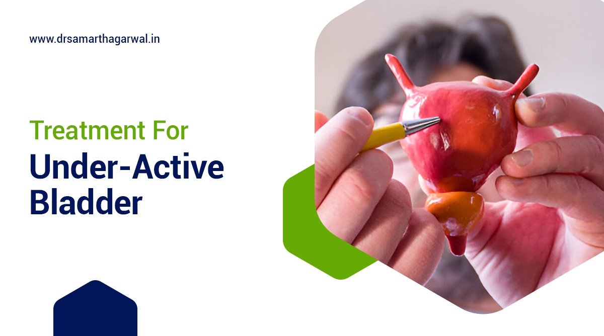
Glandular Hypospadias Surgery

What Is the Treatment for Glanular Hypospadias?

Glanular hypospadias is a type of congenital condition where the urethral opening is in the head of the penis. The treatment for glandular hypospadias typically involves surgery, which is usually performed between 6-12 months of age. The surgery involves straightening the penis, reconstructing the urethra, and performing a circumcision. Some types of glandular hypospadias may require more than one surgery. After surgery, the child may be prescribed antibiotics to prevent infection and an antispasmodic medication to suppress muscle spasms. 

What Are the Complications of Glandular Hypospadias?

Complications of glandular hypospadias include urethrocutaneous fistula, urethral stricture, urethral diverticulum, and recurrent curvature. A urethrocutaneous fistula is a hole between the urethra and the outer skin of the penis, causing urine to exit the body at the wrong location.

A urethral stricture is a narrowing of the urethra caused by scarring, making urination difficult and leading to abnormal urine stream direction. A urethral diverticulum is a widening of the urethra near the repair area, causing urine to gather and potentially leading to urinary tract infections. Recurrent curvature is a re-curving of the penis after hypospadias repair.

  1. Opening location: Glandular hypospadias typically involves an opening near the head of the penis.
  2. Pyuria: This is a condition where there is pus in the urine causing a burning sensation while urinating. It is a common complication of hypospadias.
  3. Obstructive urethral strictures: Urethral strictures, or narrowing of the urethra, are more common in patients with hypospadias and can lead to urinary symptoms such as pain or inability to empty the bladder completely.
  4. Fistulas: Fistulas are common, painful channels that form between the skin and the urinary tract. They can develop after surgery and are open to the skin, which can cause urinary contamination and increase the risk of infection.
  5. Urethral irritation and continence issues: These are some of the most common complications of hypospadias repair. Patients may experience discomfort and pain in the urinary tract, and urinary tract infections are more common. Incontinence is not as common as it is in distal hypospadias repairs, but it is still possible.
  6. Phimosis: This is a condition where the foreskin, the skin that covers the head of the penis, is too tight. It can occur in some patients with hypospadias and may require circumcision.
  7. Anesthesia complications: Anesthesia is used during hypospadias surgery to help manage pain and reduce discomfort. Complications such as aspiration, respiratory depression, and cardiac arrest can occur during and after anesthesia.
  8. Postoperative bleeding: Bleeding can occur after hypospadias surgery, and it is usually noticed in the first few days. It may require additional intervention such as blood transfusions in severe cases.
  9. Trauma and scrotal wounds: These can occur if the surgery is not performed properly, and they can be painful and can require wound healing and infection prevention measures.
  10. Penile relocation: In some rare cases, the hypospadias is so severe that the penis needs to be realigned and positioned differently. This may lead to additional complications such as penile retraction, intolerance to sexual activity, and decreased sexual function.

What Are the Three Types of Hypospadias?

  1. There are three main types of hypospadias, each depending on the location of the urethral opening:
    • Glanular Hypospadias: The opening is on the head of the penis.
    • Subcoronal Hypospadias: The opening is just below the head of the penis.
    • Distal Hypospadias: The opening is located midway between the head and the middle of the shaft.
  2. More severe cases of hypospadias can be classified as Midshaft, Penoscrotal, or Perineal Hypospadias.
    • Midshaft Hypospadias: The opening is halfway down the side of the penis.
    • Penoscrotal Hypospadias: The opening is where the shaft meets the scrotum.
    • Perineal Hypospadias: The opening is along the center of the divided scrotum.

Which Type of Hypospadias Needs Surgery?

The types of hypospadias that need surgery are glandular, coronal, penile, and perineal.

Hypospadias is a congenital condition where the urethral opening is not located at the tip of the penis. The type and severity of hypospadias determine whether surgery is necessary. In most cases, hypospadias requires surgery to correct the defect and restore normal urinary function.

There are several types of hypospadias, categorized based on the location of the urethral opening. The types that typically require surgery are:

  • Glanular (balanic) hypospadias: Thethral opening is located in the head of the penis, but not at the tip.
  • Coronal hypospadias: The urethral opening is located at the base of the glans penis.
  • Penile hypospadias: The urethral opening is located along the shaft of the penis.
  • Perineal hypospadias: The urethral opening is located on the scrotum or perineum.

Surgery is usually recommended for hypospadias to correct the defect and prevent potential complications, such as urinary tract infections, sexual dysfunction, and psychological distress. The type and complexity of the surgery vary depending on the severity and location of the defect.

In some cases, mild hypospadias may not require surgery, but this decision is made on a case-by-case basis by a healthcare provider or urologist. The goal of surgery is to create a normal urinary stream, improve sexual function, and enhance the overall quality of life for individuals with hypospadias.

What Is the Difference Between Glandular and Coronal Hypospadias?

The table below provides a detailed comparison of Glandular Hypospadias and Coronal Hypadias, two types of hypospadias, highlighting their differences in location, type, effect on urination, surgical correction, incidence, severity, and symptoms.

Glandular HypospadiasCoronal Hypospadias
Opening of the urethra is located on the head the penis, but not at the tip.Opening of the urethra is located just below the head of the penis.
Milder form of hypospadiasMore severe form of hypospadias
Does not cause misdirected urinary stream or curvature of the penisCan cause misdirected urinary stream and curvature the penis
Most common form of hypospadiasLess common form of hypospadias
Includes distal hypospadiasIncludes subcoronal and midshaft hypadias
Classified as anterior or minor hypospadiasClassified as midshaft hypospadias
Less severe curvature of the penisMore severe curvature of the penis

What Is the Success Rate of Hypospadias Surgery? 

The success rate of Hypospadias Surgery is above 90% as per many recent studies.

According to AlTaweel, Waleed M., et al’s 2017 study ‘Hypospadias repair during adulthood: series’, the overall success rate was 95% with no difference between primary and secondary repair.

According to Uygur, M. Cemil, et al’s 2002 ‘Factors affecting outcome of one-stage anterior hypospadias repair: analysis of cases’, one-stage anterior hypospadias repair was successful in 95% of, with a complication rate of 18%. According to Diao, B., et’s 2006 study ‘Anterior hypadias: one-stage repair. Report of 40 cases, 40 cases of anterior hypospadias were successfully repaired using a one-stage procedure.

How Long Does Hypospadias Surgery Take to Heal?

Hypospadias surgery typically takes around 6 weeks to fully heal. Mild cases may take 60-120 minutes to repair, while more severe cases can take 3 hours or more. After surgery, a urinary catheter is usually needed for 5-14 days, and full healing takes around 6 weeks.

How Long Does Hypospadias Surgery Take? 

Hypospadias surgery can take anywhere from 60 minutes to three hours, depending on the severity of the condition. Mild cases may take around 60-120 minutes, while more severe forms can take longer.

43

If you are concerned about Hypospadias surgery then please consult with the top urologist surgeon in Siliguri, Dr. Samarth Agarwal today!

At What Stage Does Kidney Cancer Spread

Kidney cancer, also known as renal cell carcinoma, is a type of cancer that forms in the lining of the tubules in the kidney. Early-stage kidney cancer often has no symptoms, but as the condition progression can lead to a range of signs. This article discusses the stage at which kidney cancer begins to spread and the potential impacts on a person’s health.

The various stages of kidney cancer represent the extent of the disease and guide treatment decisions. The primary staging system for kidney cancer consists of the TNM classification (Tumor, Node, Metastasis) and the stage grouping (I-IV). The stage indicates the size of the tumor, the presence of cancer cells in nearby lymph nodes, and the spread to distant organs.

At What Stage Does Kidney Cancer Spread?

Kidney cancer spreads in stages, with stage I being the earliest and stage IV the most advanced.

The following details explain the stages of kidney cancer:

  1. Stage I: Cancer is confined to the kidney and is less than 7 cm in size. No cancerous cells are present in the nearby lymph nodes or other organs.
  2. Stage II: The tumor is larger than 7 cm but still confined to the kidney, or it has grown into major veins near the kidney. There are no cancerous cells found in the nearby lymph nodes or other organs.
  3. Stage III: Cancer has spread to the nearby lymph nodes (N1) but has not reached distant lymph nodes or other organs (M0). The tumor may also be any size and have grown into the fatty tissue that surrounds the kidney.
  4. Stage IV: Cancer has spread to distant lymph nodes (N2), to other organs such as the lungs or bones (M1a), or both (M1b). The size of the primary tumor does not influence the stage IV designation in this case.

Understanding these stages helps in planning effective treatment strategies for kidney cancer. It is crucial to consult a healthcare provider for a comprehensive evaluation and customized treatment plan. Early detection, as with all cancers, leads to better treatment outcomes.

What are the treatment options for Kidney Cancer?

Learn about the diverse array of treatment options available for kidney cancer in this comprehensive list. From surgical interventions aimed at removing cancerous tissue to advanced therapies targeting specific abnormalities within cancer cells, explore the various approaches to managing kidney cancer at different stages.

Gain insights into prognosis and treatment considerations, empowering patients and caregivers with valuable information for informed decision-making.

  1. Surgery:
    • Surgeons aim to remove as much kidney cancer as possible.
    • Partial nephrectomy preserves healthy kidney tissue by removing only the tumor.
    • Radical nephrectomy involves removing the entire kidney and surrounding tissues if necessary.
  2. Nonsurgical Treatments:
    • Cryoablation freezes cancer cells using a special needle.
    • Radiofrequency ablation heats cancer cells through a probe, destroying them.
  3. Advanced and Recurrent Kidney Cancer Treatments:
    • Surgery: Removal of as much cancer as possible, even if complete removal isn’t feasible.
    • Targeted Therapy: Focuses on specific abnormalities within cancer cells to induce cell death.
    • Immunotherapy: Utilizes the body’s immune system to fight cancer by interfering with cancer cells’ ability to hide.
    • Radiation Therapy: Kills cancer cells with high-energy beams, often used to control or reduce symptoms in advanced cases.
    • Clinical Trials: Offer innovative treatments, assessing safety, effectiveness, prevention, or detection methods.
  4. Prognosis and Treatment Considerations:
    • Prognosis depends on the cancer stage at diagnosis.
    • For early-stage disease, prognosis is typically excellent with the possibility of cure.
    • Later-stage patients have access to various treatments, potentially extending life significantly.
    • Treatment decisions consider factors like overall health, cancer type and stage, spread, and patient preferences.

What are the signs kidney cancer has spread?

The following are some common symptoms and potential indicators of kidney cancer, including suspicious physical findings and less noticeable signs. These may include visible or undetectable blood in the urine, kidney lumps or masses, flank pain, fatigue, poor appetite, low-grade fever, a general feeling of malaise, high blood pressure, anemia, high calcium levels, bone pain, and metastasis to other organs.

  1. Blood in your urine (hematuria): One of the common signs of kidney cancer, it is the presence of blood in your urine, which may be visible or only detectable with a urine test.
  2. Kidney lump or mass: A lump or mass in your kidney area can be an indication of a kidney tumor, which could be cancerous.
  3. Flank pain: Persistent pain in the lower back or side near the kidneys might be a symptom of kidney cancer, often referred to as flank pain.
  4. Fatigue: Feeling tired, weak, or lacking energy is a frequent symptom associated with kidney cancer.
  5. Poor appetite: A decreased appetite and weight loss are also common symptoms, which might be related to feeling unwell or the presence of a growing tumor.
  6. Low-grade fever: Kidney cancer can sometimes cause a low-grade fever, as an indirect sign of the body’s inflammatory response.
  7. General feeling of being unwell: A vague sensation of not feeling well is common among patients with kidney cancer.
  8. High blood pressure (hypertension): While the relationship is not always direct, having high blood pressure can increase the risk factor for kidney cancer.
  9. Anemia: A condition characterized by a lower-than-normal number of red blood cells can develop due to kidney cancer, leading to symptoms like fatigue and weakness.
  10. High calcium levels (hypercalcemia): In some cases, kidney cancer can produce a hormone that causes an increase in calcium levels in your body.

When kidney cancer progresses, it may metastasize, leading to more advanced symptoms such as:

  1. Bone pain: Cancer that has spread to the bones can cause various painful symptoms, including bone pain.
  2. Metastasis: Kidney cancer cells can spread to other organs, such as the liver, lungs, and brain, leading to visible or possible symptoms related to those organs.

How long can you live with kidney cancer that has spread?

Kidney cancer survival rates depend on various factors such as the stage at diagnosis, overall health, and the effectiveness of treatment.

According to the American Cancer Society, the 5-year relative survival rate for stage 4 kidney cancer is 17%. This percentage indicates the number of people surviving for at least five years after the initial diagnosis.

Medical News Today cites an average kidney cancer growth rate of 0.3 centimeters (cm) per year; however, growth patterns can vary significantly among individuals.

Other factors affecting the outlook for patients with advanced kidney cancer include the tumor’s type and grade, as well as the presence of specific genetic mutations.

Test results, such as imaging scans or biopsies, help determine the stage, type, and grade of the cancer. Staging ranges from I (localized, 93% 5-year survival) to IV (distant metastasis, 17% 5-year survival), according to the American Cancer Society.

Treatment options for advanced kidney cancer may include targeted therapies, immunotherapy, hormone therapy, or palliative care to maintain quality of life. As cancer treatment improves, the outlook for patients with advanced kidney cancer also evolves.

For instance, a study published in the Journal of Clinical Oncology revealed that the median overall survival for patients with metastatic clear cell renal cell carcinoma who received sotorasib was 11.3 months. This was an improvement compared to the median overall survival of approximately 8.4 months for the same patient population without sotorasib treatment.

Where is the first place kidney cancer spreads to?

Kidney cancer spreads most commonly to lymph nodes, lungs, and other parts of the body like the liver, brain, ovaries, testicles, and bones. The risk of metastasis varies based on factors like tumor stage, location, and histology. Clear cell renal cell carcinoma, for example, often spreads to the lungs, liver, or bones.

Is stage 3 kidney cancer curable?

Stage 3 kidney cancer has a high survival rate, with approximately 75% of diagnosed individuals living for 5 years or more after diagnosis, according to Cancer Research UK and Healthline. Some stage III cancers can be cured with surgery alone, with 38-70% of patients achieving remission, states Tahoe Forest Cancer Center. The National Kidney Foundation reports that most people with early stage cancer (stages 1, 2, and 3) can be cured with surgery.

What part of the body is most affected by kidney cancer?

Kidney cancer mainly affects the kidneys, with most cases being renal cell carcinoma. However, if left untreated, it can spread to other parts of the body such as the bones, lungs, or brain.

Can you recover from kidney cancer?

Most people with early stage kidney cancer (stages 1, 2, and 3) can be cured with surgery, such as a radical nephrectomy, where the entire kidney is removed. The chances for recovery are good, with a five-year survival rate for stage 1 kidney cancer being 81%. Recovery time varies, ranging from 2-7 days in a hospital setting to 6-12 weeks for full recovery. However, in advanced stages, treatment may not be able to cure the cancer, but it can control it for a time and reduce symptoms. Long-term follow-up and management of side effects are essential for kidney cancer survivors.

Dr Samarth Agarwal receiving recognition

If you are worried about this deadly disease then contact with Dr. Samarth Agarwal for a consultation.

Can You Do Urethral Stricture Treatment with Medicine?

Urethral stricture treatment with medicine is a therapeutic approach that focuses on using medications to manage and relieve symptoms associated with urethral strictures. This treatment option is often used in conjunction with other approaches, such as dilation and ureterotomy, to provide optimal outcomes.

The symptoms of urethral stricture disease include painful urination, urinary frequency, urinary incontinence, and straining during urination. If left untreated, urethral strictures can lead to significant morbidity and decreased quality of life. Urethral strictures can be caused by injuries, infections, or surgical that damage the urethra, leading to scarring and narrowing of the urethra.

According to a 2021 study published in the Journal of Urology, the use of local therapy in preventing urethral strictures may carry clinical potential to improve urethral stricture treatment outcomes. Treatment options for urethral strictures range from minimally invasive procedures to surgical interventions, and medications play a crucial role in managing symptoms and preventing complications.

What is The Best medicine for urethral stricture?

The best medicine for urethral stricture is not specified, as treatment typically involves surgical or procedural interventions.

The primary mode of treatment is to make the urethra wider using a medical instrument called a dilator or through surgical options such as open urethroplasty. Urethral dilatation or internal urethrotomy may be tried, especially for short strictures. Under general anesthesia, the urethra is widened using a series of progressively larger dilators or a urethrotome.

In some cases, antibiotics may be prescribed if there is an underlying infection. Self-catheterization might be a choice for those diagnosed with a short stricture. However, medications are not a primary treatment for urethral stricture, and surgical or procedural interventions are often necessary to effectively treat the condition.

It is essential to work with a urologist to determine the best course of treatment, as delayed or inadequate treatment can lead to complications. Prevention of urethral stricture involves avoiding injury to the pelvic area and taking care to avoid infection. The outlook for urethral stricture is generally positive, and with proper treatment, it is a treatable condition.

Urethral stricture Treatments

Urethral stricture Treatments

How to treat urethral stricture at home?

To treat urethral stricture at home, consume prob-rich foods like yogurt, fermented cheese, kefir, and kimchi, which promote good bacteria growth in the gut and help fight urinary tract inflammation and infection.

How to prevent urethral stricture recurrence?

The ways to prevent urethral stricture recurrence are by avoiding injury to the urethra and pelvis, being careful with self-catheterization, using lubricating jelly liberally, using the smallest catheter needed for the shortest time, and avoiding sexually transmitted infections.

Urethral stricture recurrence can be prevented by adopting certain precautions and lifestyles. Firstly, it is essential to avoid injury to the urethra and pelvis, which can lead to urethral stricture disease. Self-catheterization should be performed with care, using lubricating jelly liberally to minimize the risk of injury Additionally, using the smallest possible cath needed for the shortest time can reduce the likelihood of urethral stricture recurrence.

Furthermore, avoiding sexually transmitted infections, such as gonorrhea, which was once the most common cause of strictures, can help prevent urethral stricture recurrence. The use of antibiotics has helped to prevent this.

In terms of treatments, urethroplasty is considered the most effective approach to prevent urethral stricture recurrence. This surgical reconstruction of the urethra can help to minimize the risk of recurrence. Additionally, some tips, such as maintaining a healthy diet, encouraging the growth of good bacteria through probiotics, and keeping the urinary tract healthy, can also help to prevent urethral stricture recurrence.

Penile Urethral Stricture

Penile Urethral Stricture – Causes, Coping Strategies 

Does urethral stricture heal itself?

Urethral stricture does not heal itself.

Urethral stricture is a condition where the urethra becomes narrow, causing difficulty and discomfort during urination. The most common causes of urethral stricture are chronic inflammation or injury, which can lead to scar tissue formation. This scar tissue causes the urethra to become, resulting in symptoms such as difficulty starting to urinate, a weak or slow stream, and straining to urinate.

Urethral stricture requires medical attention and treatment. In some cases, the condition may need immediate attention. If left untreated, urethral stricture leads to complications such as urinary retention, infections, and kidney damage.

Treatment for urethral stricture involves surgery, such as urethroplasty, to remove the scar tissue and widen the urethra. In some cases, treatment also involves dilating the urethra, a specialized instrument. The goal of treatment is to relieve symptoms, improve urinary flow, and prevent complications.

While treatment for urethral stricture is generally successful, the condition can recur, requiring multiple procedures. It is essential to work with a healthcare provider to manage symptoms and prevent complications.

In some cases, urethral stricture may be associated with other underlying conditions, such as prostate cancer, radiation therapy, or neurologic disorders. In these cases, treatment may involve addressing the underlying condition in addition to managing the urethral stricture.

Can urethral stricture be cured naturally? 

While urethral stricture has a generally positive outcome and is treatable, it can happen again, requiring multiple procedures. However, certain natural remedies like probiotics, yoga, and homeopathic medicines can help alleviate symptoms and promote healing.

How to dilate the urethra at home?

You can dilate the urethra at home by washing your hands, opening the dilator packet, inserting the dilator into the urethra, moving it up and down three or four times, and then removing it.

Dilating the urethra at home is a process for individuals who have undergone a urethral stricture repair surgery or have conditions such as hypospadias, circumcision, or Balanitis Xerotica Obliterans (O) that cause narrowing of the urethra. Self-dilatation helps to prevent the narrowing from recurring and improves urine flow Here’s a step-by-step guide on how to dilate the urethra at home. First, wash your hands with soap and water to maintain hygiene. Next, open the dilator packet by peeling the tabs apart. You may need to add water or a cold lubricating gel to the dilator, depending on the type used. Ensure you do not squeeze your penis as this can squash the urethra.

Insert the dilator into the urethra to the length advised by your doctor or nurse. You may need to push the catheter a bit to get it to go in. Once inserted, move the dilator up and down the urethra three or four times to help widen the urethra and improve urine flow. After completing the dilatation process, remove the dilator and dispose of it in your rubbish bin.

It is essential to follow the plan advised by your doctor or nurse and perform self-dilatation regularly to prevent the narrowing from recurring. If you do not stick to the plan, the narrowing may not get better and may even get worse. In some cases, even though you stick to the plan, the narrowing still may not get better, and you may need to undergo a small operation to stretch the urethra.

Urethral Stricture Disease Symptoms

Urethral Stricture Disease Symptoms

How can I get a permanent solution without surgery for urethral stricture? 

A permanent cure for urethral stricture can be achieved with REGROW by Regrow Biosciences, which is a one-time treatment that uses patients’ own stem cells.

Which exercise is best for urethral stricture?

The best exercises for urethral stricture are Vajrasana (Thunderbolt Pose Ardha Kurmasana (Half Tortoise Pose), and Kegel exercises, which strengthen the pelvic floor muscles, improve blood circulation, and provide relief from urethral strictures.

What causes the narrowing of the urethra?

Narrowing of the urethra is often caused by injury, inflammation, or scar tissue, which can result from surgery, infection, catheterization, straddle injury, or trauma to the urethra or pelvis.

Dr Samarth Agarwal receiving recognition

Contact Dr. Samarth Agarwal if you have any questions or concerns about your Urinary health!
Meet the top Urologist in Siliguri

BCG and Interferon for Bladder Cancer 

BCG (Bacillus Calmette-Guérin) and interferon therapy is a treatment approach used to combat bladder cancer. This form of intravesical therapy involves instilling the BCG vaccine into the bladder to stimulate the immune system to fight cancer cells.

BCG is a type of vaccine that has been for many years to prevent tuberculosis, but it has also been found to be effective in treating bladder cancer. The symptoms of bladder cancer include blood in the urine, painful urination, and frequent urination. If left untreated, bladder cancer can lead to serious complications, such as metastasis to other organs and even death.

According to a 2015 research paper published by the National Institutes of Health (NIH), adding interferon to BCG is an effective therapy for both treatment and salvage therapy in patients with bladder cancer. Interferon is a type of protein that helps to stimulate the immune system to fight cancer cells. When used in combination with BCG, interferon has been shown to enhance the immune response and increase the effectiveness of treatment.

Furthermore, a 2019 paper published in Springer by J.J. Duplisea emphasized that the combination of interferon α and low dose B has markedly superior efficacy and lower toxicity than standard dose BCG therapy for high-risk non-muscle-in bladder cancer.

What is interferon for bladder cancer?

Interferon is a type of protein that mediates the host immune response in the human body. In the context of bladder cancer, interferon plays a significant role in inducing a non-specific cellular and humoral immune response towards tumor cells. This immune response helps in reducing the recurrence and progression rates of superficial bladder cancer.

The use of interferon in bladder cancer treatment is particularly appealing due to its minimal side effects and very low dropout rate compared to other treatment methods like BCG (Bacillus Calmette-Guerin). Current research has focused on the use of interferon in combination with immunotherapeutic and cytotoxic drugs to enhance its effectiveness.

Interferon-α, in particular, has been extensively studied and has shown promise in reducing the recurrence and progression rates of superficial bladder cancer. Its antitumor effect relies on T cells and macrophages within the tumor microenvironment. Interferon-α2B, a subtype of interferon-α, has been found to enhance Th1 cytokine responses in bladder cancer, which could potentially improve the efficacy of treatment.

In some cases, bladder cancer can become resistant to BCG treatment, making it an inherently resistant disease state. In such cases, interferon-based gene therapy has shown potential as an alternative treatment option.

However, it’s important to note that while interferon therapy holds promise, it is not a cure for bladder cancer and its effectiveness may vary from patient to patient. For optimal results, it is often used in combination with other treatments and in a risk-stratified clinical framework. As with any medical treatment, the decision to use interferon for bladder cancer should be made in consultation with a healthcare provider.

What are the side effects of BCG interferon treatment?

BCG interferon treatment for bladder cancer is associated with minimal side effects compared to BCG alone. Here are some of the reported side effects:

  1. Fever and Flu-like symptoms: Patients may experience fever, chills, and flu-like symptoms after treatment. These symptoms are usually mild and temporary.
  2. Bladder irritation: Patients may experience bladder irritation, such as a burning sensation during urination, frequent urination, or the urge to urinate immediately after filling the bladder.
  3. Allergic reactions: Some patients may have allergic reactions to BCG, such as hives, rash, or difficulty breathing.
  4. Blood in urine: BCG treatment may cause blood in the urine, which is usually temporary.
  5. Lower back pain: Some patients may experience lower back pain after treatment.
  6. Rare but serious side effects: In rare cases, BCG treatment can cause severe side effects such as sepsis, which is a life-threatening infection.

It’s important to note that these side effects may not occur in all patients, and the severity can vary. Patients should discuss any concerns with their healthcare provider.

In addition, the combination of BCG and interferon may enhance the immune response, potentially increasing the effectiveness of the treatment. However, more research is needed to fully understand the benefits and risks of this combination therapy.

How successful is BCG treatment for bladder cancer?

Depending on the bladder cancer, BCG can have a success rate of up to 83%.

According to a study “Immunoprophylactic intravesical application of bacillus Calmette-Guerin after transurethral resection of superficial bladder cancer.” published in Croat Med J in April 2003 by Dr. Librenjak et al, the long-term follow-up of intravesical bacillus Calmette-Guerin (BCG) therapy in superficial transitional cell carcinoma (TCC) of the bladder indicated a success rate of 79% in terms of disease progression-free survival and a 83% rate of overall survival. The median progression-free survival and overall survival times were 162 months and 209 months, respectively.

According to Jiang, Song, and Redelman-Sidi, Gil’s 2022 study ‘Bacillus Calmette-Guérin (BCG) Treatment Bladder Cancer’, Bacillus Calmette-Guérin (BCG) is the most successful microbial immunotherapy of cancer, and intravesical treatment withCG is recommended for most patients with high-risk-muscle invasive bladder cancer (NMIBC). In patients with NMIBC, treatment with BCG is associated with a reduced risk of bladder cancer recurrence.

Is there a better treatment for bladder cancer than BCG?

In certain cases, intravesical chemotherapy may serve as a better treatment option for bladder cancer when BCG (Bacillus Calmette-Guérin) treatment is ineffective or unavailable.

Gemcitabine, mitomycin, valrubicin, epirubicin, and docetaxel are commonly used drugs for intravesical chemotherapy. A study presented in March 2023 confirmed that the combination of gemcitabine and docetaxel, employed after surgery for high-risk non-muscle-invasive bladder cancer, is an effective and safer alternative to BCG (2022 National Cancer Institute publication).

For high-risk patients who fail BCG, radical cystectomy often stands as the recommended treatment (as per a 2015 article from the National Institutes of Health, NIH). However, several patients may be unfit or reluctant to undergo this procedure, necessitating alternative treatment plans.

Intravesical chemotherapy offers a viable approach in such situations, with specific drug combinations potentially delivering impressive results. It is crucial that healthcare providers carefully consider patients’ high-risk features when electing between BCG therapy and intravesical chemotherapy based on individual needs and responses to treatment.

What is the alternative to BCG treatment for bladder cancer?

There is a potential alternative to traditional BCG treatment in patients with bladder cancer that has shown promise in reducing the chances of recurrence and progression. One such alternative is the use of interferons, specifically interferon-α2B.

Intravesical interferon, when utilized in combination with immunotherapeutic and cytotoxic drugs, acts as a non-specific immunostimulatory agent toward tumor cells that can minimize side effects and achieve a very low dropout rate compared to the BCG treatment.

Moreover, current research has specifically focused on using interferon for patients with non-muscle-invasive bladder cancer to reduce the rates of recurrence and progressive disease. Interferon-α2B is recommended as an adjuvant therapy for intermediate-risk and high-risk NMIBC since it enhances Th1 (T-helper 1) immune-cell cytokine responses in a bladder environment, thus potentially facilitating improved outcomes. Some studies comparing BCG therapy with the interferon-alpha-2B approach have comparable recurrence rates.

However, it is important to note that the timing of treatment with interferons may impact the success; it has been suggested that the administration of salvage intravesical therapy should not be offered if a patient fails BCG induction therapy within six months. Salvage intravesical treatment with interferon can be considered alongside other treatments, such as chemotherapy or device-assisted therapy like intravesical immunostimulants, for suitable candidates who prefer bladder preservation or have severe underlying medical comorbidities. Furthermore, research on the implementation of interferon in bladder cancer treatment is still ongoing as there are no standardized criteria nor a set regimen established as of yet.

In cases where cancer progresses significantly, alternative treatment options (such as surgical procedures like radiotherapy and chemotherapy) may be presented by the medical team to ensure bladder cancer eradication along with appropriate patient follow-ups depending on the chosen treatment plan. Before making a decision, it is necessary to discuss treatment options with healthcare specialists and weigh the advantages and disadvantages comprehensively to ensure the best plan for each patient.

What is the survival rate for BCG therapy? 

The survival rate of BCG therapy spans between 66% to 86%.

According to Pirzada, Muhammad T., et al.’s 2017 study “Non-Muscle-Invasive Bladder Cancer,” the survival rate for BCG induction therapy in high-risk bladder cancer patients at Shaukat Khanum Memorial Cancer Hospital & Research Centre in Pakistan was evaluated using a retrospective cohort study. The main outcome measures were the three-year disease-free survival and progression-free survival. The data included 68 high-risk (Ta and T1 with G3 or high-grade subtype) bladder cancer patients who underwent transurethral resection followed by six-weekly intravesical BCG instillation. The study found that the three-year disease-free survival rate was 66.2% and the progression-free survival rate was 86.8%. The study also noted that the tumor extent was the only factor significantly influencing disease-free survival independently. The results showed that induction BCG alone could be a viable option for high-risk non-muscle-invasive bladder cancer patients in terms of effective disease-free and progression-free survival rates.

According to a 2018 study published in Clinical Interventions in Urology by Bazarbashi S and a team from the Department of Urology at King Saud Medical City, the survival rate for BCG therapy in their patient population was compared with previously published data. The study included a review of medical records from patients diagnosed with nonmuscle invasive bladder cancer (NMIBC) and treated with transurethral resection (TUR) followed by intravesical BCG. The collected data revealed that at 5 years post-treatment, the relapse-free survival rate was 61.3% (95% confidence interval (CI) 44.2–78.4%). This was in line with the international data for similar treatments. The study titled “Intravesical Bacillus Calmette–Guerin (BCG) as Adjuvant Therapy of Non-Muscle-Invasive Bladder Cancer: A Five-Year Prospective Study” offered insights into the long-term effectiveness of BCG therapy for preventing the recurrence of NMIBC and highlighted the ongoing efforts to improve outcomes for patients who undergo this treatment.

How many rounds of BCG do you need for bladder cancer?

Patients with bladder cancer requiring intravesical BCG treatment typically undergo a course of weekly treatments for six weeks. The specific number of BCG instillations may vary depending on the healthcare provider’s recommendations and the patient’s response to treatment.

According to a review published in the Journal of Urology, the standard BCG treatment regimen consists of 6 weekly instillations. In cases where the response to treatment is inadequate, the course may be repeated. For high-risk non-muscle invasive bladder cancer, some clinicians may recommend maintenance therapy with BCG at longer intervals.

Furthermore, a study in the European Urology Oncology journal found that initiating BCG within 6 weeks of TURBT (transurethral resection of bladder tumor) was associated with a lower rate of cancer recurrence and progression.

In some instances, the number of BCG instillations may be reduced due to adverse effects or the patient’s overall health status. However, the primary goal is to achieve a sufficient therapeutic response to prevent or delay cancer recurrence.

What are the long-term side effects of BCG treatment?

Long-term side effects of BCG treatment for bladder cancer in the US often include lower urinary tract symptoms (LUTS). The most common LUTS associated with BCG treatment are frequency, urgency, nocturia, and dysuria (burning sensation while urinating).

BCG treatment acts by boosting the immune system, and thus it may provoke irritation in the bladder wall, leading to the symptoms mentioned. Notably, these side effects usually alleviate over time or can be controlled with medication.

Studies have shown that up to 90% of patients may experience some short-term side effects after undergoing BCG treatment for superficial bladder cancer. It is essential to recognize that the emergence of these side effects following BCG instillation reflects an active response by the immune system targeting the cancerous cells in the bladder.

When it comes to the combination of interferon and BCG treatment, the literature reveals that it significantly enhances Th1 cytokine responses in the bladder. Interferon α2b (IFN-α2b), administered in combination with BCG for bladder cancer treatment, can raise the chance of developing granuloma formation, which is a fibrotic, chronic response in the bladder tissue caused by the immune response triggered by BCG treatment [4, 5]. If these granulomas grow or extend deeper into the bladder wall over time, they might potentially impede normal bladder functioning and cause LUTS.

In cases where these symptoms persist for a longer period or become intolerable, patients may be candidates for various management strategies such as different dosing regimens or the addition of alternative therapies to control the symptoms. Regular follow-ups with healthcare professionals are essential in monitoring the progression and resolution of side effects to optimize treatment management

What is the most common complication of BCG?

The most common complication of BCG (Bacillus Calmette-Guérin) treatment for bladder cancer in the United States is a disseminated infection, which can range from infection of an organ to severe sepsis. Other systemic adverse events include malaise, fever, and lung infection.

BCG treatment involves the administration of immunotherapy drugs directly into the bladder through a catheter. This treatment is the most effective for early-stage bladder cancer, particularly high-risk non-muscle invasive bladder cancer (NMIBC). The treatment targets cancer cells inside the bladder without affecting the rest of the body.

Intravesical BCG acts as an immune modulator, eliciting a TH1-type response in the bladder. However, it can lead to complications such as fever, lung infection, and in rare cases, disseminated infection. Disseminated infection is the most serious complication and can lead to severe sepsis.

The likelihood of developing a disseminated infection increases with the number of BCG treatments. The risk is approximately 8% for all patients receiving BCG treatment. The most common organs affected by disseminated infection are the lungs, followed by the liver, bones, and soft tissues.

In some cases, aggressive bladder cancer can develop after BCG treatment. If this happens, cystectomy (surgery to remove the bladder) is usually recommended.

To minimize the risk of complications, it is essential to follow the healthcare provider’s guidelines before and after BCG treatment. These guidelines may include avoiding caffeinated beverages and diuretics for a certain period before the treatment and monitoring for side effects such as fever and signs of infection.

What is next if BCG treatments don’t work?

According to Zlotta, Alexandre R., et al.’s 2009 study ‘Management of BCG failures in non-muscle-invasive bladder cancer’, up to 40% of patients with non-muscle-invasive bladder cancer (NMIBC) will fail intravesical BCG therapy. The choice of an appropriate course of action following the failure of intravesical bacillus Calmette-Guérin (BCG) treatment is a controversial management issue for both patients with NMIBC and their physicians.

Dr Samarth Agarwal urologist Siliguri

Contact Dr. Samarth Agarwal if you have any questions or concerns about your bladder cancer treatment.

What Are Male Urinary Incontinence Treatment Options?

The symptoms of urinary incontinence can include a frequent or urgent need to urinate, a feeling of urgency or discomfort, and a feeling of not being able to control urination. Treatment options for urinary incontinence can vary depending on the underlying cause and severity of the condition.

These can include pelvic floor muscle exercises, medications such as antimuscarinics or calcium channel blockers, and surgical procedures such as urinary catheters or the insertion of a sphincter cuff.

What are the treatment options for male incontinence?

The management of male urinary incontinence generally follows a stepwise approach, which includes:

  1. Lifestyle modifications: Addressing lifestyle factors such as smoking, alcohol consumption, and weight may improve urinary incontinence outcomes.
  2. Conservative treatments: a. Pelvic floor physical therapy (PFPT): PFPT aims to strengthen pelvic floor muscles and improve bladder control. PFPT has shown beneficial effects for both stress and urge incontinence. b. Bladder training: Bladder training aims to increase the volume of urine that can be eliminated at one time, reducing urinary urgency and leakage. c. Male slings: Specifically designed for male urinary incontinence, male slings work by correcting urinary leaks during defecation or stress maneuvers.
  3. Invasive treatments: a. Prostatectomy: Prostatectomy removes the prostate gland, which can be effective in alleviating urinary incontinence caused by an enlarged prostate. b. Artificial urinary sphincter (AUS): An AUS is a surgically implanted device that works by creating a urinary reservoir and relieving pressure on the bladder by allowing urine to flow directly from the reservoir to the urethra at the appropriate time. AUS has shown effective outcomes for both men and women with urinary incontinence. c. Transurethral stricture disease (TUSD): TUSD can be treated with endoscopic dilation or repeat surgery if initial management fails.

In some cases, medications may be necessary to manage symptoms, such as anticholinergics for urge incontinence, alpha blockers for bladder spasms, or calcium channel antagonists for patients with SUI.

To facilitate the care and management of male urinary incontinence, primary care physicians and urologists should collaborate, with urologists providing specialized evaluation and management of the underlying condition, while primary care physicians focus on long-term follow-up, monitoring, and ongoing management.

You might also like: Aqua Therapy for Enlarged Prostate

How can I permanently fix incontinence?

Incontinence can not be fixed permanently. While surgery may be an effective treatment option for some individuals with incontinence, it is not always a cure. 

How to increase urine flow in males?

To improve urine flow in males, consider implementing the following strategies based on credible health sources:

  1. Maintain an active lifestyle: Regular physical activity, such as walking, jogging, and swimming, can help contract the muscles involved in urination and reduce urinary problems.
  2. Practice pelvic floor exercises, also known as Kegels, to strengthen your pelvic muscles and aid in controlling your urine flow.
  3. Adopt a healthy lifestyle, minimizing drinks like coffee, caffeinated beverages, and alcohol, and making changes to your diet to improve overall urinary health.
  4. Consider medication options, such as alpha blockers or beta-3 agonists, to relax the muscles and improve urine flow.
  5. Apply heat to your lower abdomen to soothe your bladder and promote easier urination.

What are urinary incontinence treatments for elderly males?

Here are 5 urinary incontinence treatments for elderly males:

  • Pelvic floor muscle exercises
  • Medications such as anticholinergics
  • Benign prostatic hyperplasia treatments
  • Slings or artificial urinary sphincters
  • Surgery such as a male sling or pessaries

According to a 1997 study published in the journal “Gerontology” by P.Iqbal and C.M. Castleden: Urinary incontinence is common in the elderly, affecting 6-8% of people over 64 years in the community and up to 31% in hospital and long-term care. The authors established that incontinent patients can be diagnosed clinically, and improving symptoms is likely in the majority. 

Treatment of urinary incontinence requires general and specific measures. General measures include moderation of fluid intake, treatment of aggravating conditions, and drugs. Specific measures are pelvic floor exercises, vaginal cones, interferential therapy, oestrogens for stress incontinence, bladder retraining, anticholinergic drugs for urge incontinence, and alpha-blockers and 5-alpha reductase inhibitors for overflow incontinence due to prostatic hyperplasia.

You might also like: Ketamine Bladder Syndrome Symptoms

When Should You Seek Treatment for Urinary Incontinence?

If you experience frequent urination, leaking urine, or difficulty controlling your bladder, it’s essential to consult a healthcare professional for an evaluation. Here are some guidelines:

  1. If you leak urine frequently and it interferes with your daily activities, causing embarrassment or limiting your social interactions, it is time to see a doctor.
  2. If you are experiencing urinary incontinence along with other symptoms, such as fever, pain in the lower back or side, or difficulty emptying your bladder, seek medical attention right away.
  3. Based on your medical history, your healthcare provider may recommend various diagnostic tests, such as a bladder catheterization or urodynamic testing, to determine the cause and severity of your incontinence.
  4. Depending on the diagnosis, treatment options may include dietary modifications, pelvic floor exercises, medications, or surgical intervention.
  5. In some cases, lifestyle changes, such as simpler fluid intake patterns or modification of medications, can reduce symptoms and improve your quality of life.
  6. In certain instances, a healthcare professional may suggest a specialist evaluation by a urologist or a geriatrician for further evaluation and management.

How Successful Is Treatment for Urinary Incontinence?

Urinary incontinence is a common health problem that affects millions of people globally, and its treatment is crucial as it can significantly impact one’s quality of life. Various treatment options exist for urinary incontinence, including behavioral and medical treatments, as well as surgical interventions. According to recent studies, about 80% of people with urinary incontinence can improve or even be cured with effective treatments.

Behavioral treatments such as bladder training, fluid and dietary modifications, and pelvic floor exercises can help individuals manage urinary incontinence. Antimuscarinics, estrogen, and β-blockers are commonly prescribed medications to treat urinary incontinence. Clinical trials have shown promising results for these treatments, with success rates ranging from 40% to 80%.

According to a 2023 study published in the International Journal of Urology, researchers evaluated the effectiveness of various therapies for urinary incontinence. They highlighted the importance of subjective and objective criteria in assessing the quality-of-life impact of incontinence therapy. Behavioral regimens, when combined with pharmacologic therapy, are likely to extend the duration of response. They also mentioned the presence of a significant placebo effect in up to 30% to 40% of patients and emphasized the necessity of demonstrating that a drug or treatment effect exceeds the placebo effect. Key instruments for evaluating incontinence therapy outcomes include the Bristol Female Lower Urinary Tract Symptom IIQ Incontinence Impact Questionnaire, LUTS Lower urinary tract symptoms, OAB Short Form-36 Item Health Survey, SUI Stress urinary incontinence, UDI Urogenital Distress Inventory, and UUI Urinary urgency incontinence.

You might also like: Home Treatment for BPH

According to Riemsma, Rob, et al’s 2017 systematic review ‘Can incontinence be cured? A systematic review of cure rates’, the present study aimed to assess cure rates and the number of people still dependent on containment strategies following treatment for urinary (UI) or fecal incontinence (FI) in individuals with UI, FI, disabled or neurological problems or diseases, or elderly or cognitively impaired. The searches retrieved 14,036 records, which were reduced to 127 papers reporting on 98 individual studies after title and abstract screening. The majority of studies were from Europe and focused on surgical interventions for SUI, pharmacological treatments for UUI, and sacral neuromodulation for FI.

It was reported that surgical interventions for women with SUI had a median cure rate of 82.3% (IQR, 72–89.5%), while men treated with male slings showed a median cure rate of 53% (IQR, 48–54%). Antimuscarinics showed a median cure rate of 49% (IQR, 35.6–58%) for women with UUI. Pelvic floor muscle training (PFMT) did not demonstrate high cure rates, but was still recommended due to its secondary benefits. For FI, sacral neuromodulation showed a median cure rate of 38.6% (IQR, 35.6–40.6%), and no studies assessed the success of containment strategies. There was a lack of data for individuals with disabilities.

Where to Go for Urinary Incontinence Treatment

According to Moore, Katie C., and Lucas, Malcolm G.’s 2010 review “Management of male urinary incontinence,” the majority of male urinary incontinence is secondary to sphincter weakness following prostate surgery. With an aging population and increasing number of prostate surgeries, the incidence of male incontinence is rising. This review focuses on the management of post-prostatectomy incontinence.

Evaluation of the incontinent male includes a thorough history, examination, and assessment of impact on daily activities. Bladder dysfunction, rather than detrusor overactivity, is more common and contributes to incontinence in 60% of men. Urodynamics are crucial to identify the cause of leakage and assess other parameters affecting success rate of future intervention.

Dr Samarth Agarwal urologist Siliguri

If you have any Incontinence issues, you can contact one of the most popular urologists in Siliguri.

What Is The Best Treatment for Hydrocele

Hydrocele treatment involves addressing the abnormal accumulation of fluid around the testicle, which can cause discomfort, pain, and embarrassment. The treatment approach depends on the underlying cause and severity of the condition.

The symptoms of hydrocele include a painless swelling in the scrotum, heaviness or discomfort in the scrotum, and a feeling of pressure or tenderness in the affected area. Hydrocele can be caused by a variety of factors, including injury, infection, or inflammation of the epididymis, a congenital defect, or a blockage in the lymphatic system.

In a 2010 paper published in PubMed, M. Cimador and colleagues emphasized that if hydrocele occurs after varicocelectomy, initial management include observation with or without hydrocele aspiration, and large persistent hydroceles are usually treated with surgical repair

Treatment options for hydrocele range from watchful waiting for mild cases to surgical repair for more severe cases. Other treatment approaches include sclerotherapy, which involves injecting a sclerosant into the hydrocele under ultrasound guidance. In some cases, surgery may be necessary to correct an underlying defect or remove the hydrocele.

What are the treatment options for a hydrocele?

Here are the main treatment options for a hydrocele and a brief description of each:

  1. Observation:
    • For small, asymptomatic hydroceles, the healthcare provider may recommend simply monitoring the condition over time.
    • This approach is often taken for infants and young children, as hydroceles can sometimes resolve on their own as the child grows.
    • Regular check-ups are necessary to ensure the hydrocele is not increasing in size or causing any discomfort.
  2. Aspiration:
    • This procedure involves using a needle to drain the fluid from the hydrocele sac.
    • It is a relatively simple and minimally invasive option, but the hydrocele may recur, and repeated aspirations may be necessary.
    • Aspiration is often used as a temporary measure or for individuals who are not good candidates for surgery.
  3. Sclerotherapy:
    • In this procedure, a sclerosing agent is injected into the hydrocele sac to cause inflammation and scarring, which helps prevent the accumulation of fluid.
    • Sclerotherapy is less invasive than surgery and can be an effective option for some patients, but it may also have a higher recurrence rate compared to surgical treatment.
    • The healthcare provider will discuss the potential risks and benefits of this approach with the patient.
  4. Surgical Repair (Hydrocelectomy):
    • Surgical repair, or hydrocelectomy, is the most definitive treatment for a hydrocele.
    • During the procedure, the surgeon will make an incision and remove the hydrocele sac, allowing the fluid to drain and preventing future accumulation.
    • Hydrocelectomy is generally considered a safe and effective treatment, with a low risk of recurrence, but it does involve a more invasive procedure and a longer recovery time compared to other options.

The healthcare provider will consider factors such as the size and location of the hydrocele, the patient’s age and overall health, and the individual’s preferences when recommending the most appropriate treatment option.

You might also like: Hydrocele Operation Side Effects

Is hydrocele curable without surgery?

 Hydroceles can be curable without surgery, specifically when addressing a non-communicating hydrocele. This condition, characterized by fluid accumulation in the scrotum, generally does not allow fluid to flow back into the abdomen, distinguishing it from a communicating hydrocele related to an inguinal hernia. Treatment methods such as aspiration and sclerotherapy emerge as non-surgical options. During aspiration, a doctor extracts fluid from the scrotum, and sclerotherapy involves the injection of a sclerosing agent to prevent fluid recurrence. However, non-communicating hydroceles may sometimes resolve spontaneously, especially in infants, where the tunica vaginalis naturally seals shut.

Can hydrocele heal naturally?

 In infants hydroceles often resolve without intervention within the first year of life. Communicating hydroceles, characterized by fluid passage between the abdomen and scrotum, pose a greater likelihood of requiring surgical repair due to their nature.

The Department of Urology frequently notes that fluid accumulation around the testicle, a hallmark of hydroceles, arises due to various causes ranging from congenital factors to injuries, all influencing the necessity and timing for potential surgery. Typically, non-communicating hydroceles surrounding the testicle might diminish on their own as the body reabsorbs the fluid.

Surgery to repair a hydrocele becomes a consideration when it causes discomfort, is associated with scrotal pain, or leads to chronic pain, thus impacting quality of life. Doctors may advise surgery based on assessments of hydrocele causes, potential for hydrocele recurrence, or when the hydrocele leads to swelling in the scrotum that does not decrease over time.

Hydrocele surgery aims to prevent recurrence of hydrocele and alleviate symptoms such as fluid in the scrotum, which if left unaddressed, could require further surgical interventions.

In adult cases, the appearance of a hydrocele causes concern regarding underlying testicular conditions or injuries, which a urologist must evaluate to determine the appropriate course. While some adult hydroceles might diminish slightly over time, persistent or enlarging hydroceles typically necessitate surgery to prevent complications like discomfort, infection, or in rare cases, testicular atrophy.

Therefore, while natural healing is plausible, especially in pediatrics, individuals experiencing hydrocele symptoms should consult a healthcare provider for an accurate diagnosis and to discuss the potential need for surgery. 

Which exercise is best for hydrocele?

Gomukhasana or the cow pose could be a beneficial exercise that provides temporary relief to hydrocele symptoms.

What is the best treatment for hydrocele at home?

Applying Epsom salt baths twice or thrice a week can effectively drain the fluid and alleviate pain, soreness, anderness of hydrocele, enhancing blood circulation in the scrotum region.

Are hydroceles dangerous?

Hydroceles can lead to complications such infection, pyocele, or hematocele. While hydroceles themselves are not harmful, they can be associated with underlying conditions that can affect fertility, such as an inguinal hernia or varicocele. Additionally, some studies have shown that in the presence of certain associated pathological findings, hydroceles can lead to infertility.

Types of Hydrocele

The two main types of hydroceles are communicating and non-communicating hydroceles. Communicating hydroceles occur when the processus vaginalis, a pouch of peritoneum that accompanies the testicle during its descent into the scrotum, remains open and communicates with the abdominal cavity.

This allows fluid to flow between the scrotum and abdomen. In contrast, non-communicating hydroceles occur when the processus vaginalis closes, and the fluid is isolated from the abdomen. Non-communicating hydroceles are the most common type and are often asymptomatic.

Congenital hydroceles, which occur in infants, can be further divided into communicating and non-communicating types.

Primary and secondary hydroceles are also distinguished, with primary hydroceles occurring when the processus vaginalis fuses at term or within 1-2 years of birth, and secondary hydroceles resulting from an underlying pathological condition.

You might also like: Best Treatment for Interstitial Cystitis

Dr Samarth Agarwal receiving recognition

If you have any symptoms of urology problems, then consult with Dr. Samarth Agarwal. One of the prominent top 10 urologist in Siliguri.

Infected Kidney Stone Treatment

Infected kidney stones, also known as infection stones, are a type of kidney stone that forms as a result of urinary tract infections (UT). The primary goal of treatment is to eliminate the stone and eradicate the underlying infection. Prompt treatment is crucial to prevent complications and improve patient outcomes.

The symptoms of infected kidney stones include severe pain, nausea, vomiting, fever, and chills. The causes of infected kidney stones are typically linked to urinary tract infections, with bacteria such as Proteus mirabilis being a common culprit. The benefits of prompt treatment include reducing the risk of complications, alleviating symptoms, and improving quality of life. According to a 2015 paper published in a medical journal by T Marien, the mainstay of treatment of infection stones is complete stone removal, with kidney stones that obstruct the urinary tract and cause obstructive pyelonephritis being the primary target.

The effectiveness of treatment depends on various factors, including the size and location of the stone, the severity of the infection, and the promptness of medical attention. In general, prompt treatment can lead to improved outcomes and reduced morbidity.

How to Treat a Kidney Stone Infection?

To effectively treat a kidney stone infection, it’s essential to prioritize prompt medical attention to eliminate the stone and eradicate the underlying infection.

Here are the key treatments for a kidney stone infection:

  • Complete stone removal through surgical procedures or lithotripsy.
  • Antibiotics to combat the underlying urinary tract infection.
  • Pain management through medication or other interventions.
  • Increasing fluid intake to help pass the stone.
  • Medications to reduce the risk of stone or to manage underlying medical conditions.
  • Behavioral changes, such as dietary modifications, to reduce the risk of future stone formation.
  • In severe cases, hospitalization may be required to manage complications or ensure proper treatment.

Remember to consult a urologist for personalized guidance on treating a kidney stone infection.

Do You Need Surgery for an Infected Kidney Stone?

You may need surgery for an infected kidney stone if it is causing severe symptoms, blocking the flow of urine, or causing infection.

Surgery for kidney stones is usually reserved for stones that are too large to pass on their own, cause severe symptoms, or block the flow of urine. The type of surgery depends on the location, size, and composition of the, as well as the individual’s overall health. In some cases, shock wave lithotpsy, a non-invasive procedure, may be used to break up smaller stones. Surgery is necessary if the stone is too large or lodged in the ureter, causing severe pain bleeding, or infection.

The surgical procedure may involve open surgery, laparoscopic surgery, or percutaneous nephrolithotomy, depending on the complexity of the case. In some instances a nephrostomy tube may be inserted to drain the kidney and relieve any blockage. Hospitals with experienced urologists and advanced surgical equipment may also offer robotic-assisted surgery for more complex cases. Your doctor will discuss the best course of treatment with you based on your individual situation.

How Long Does It Take to Recover from Kidney Stone Infection?

The recovery time for kidney stone infection treatment varies depending on the procedure used and the individual’s overall health.

Recovery typically takes around six weeks to allow for internal healing. Most patients will feel better within a few days, but it is essential to avoid lifting, pushing, or pulling objects until the doctor gives the all-clear. For those undergoing surgical procedures like ureteroscopy or percutaneous nephrolithotomy, the recovery time may be longer. Hospital stays usually last one or two nights, and full recovery can take up to six weeks. However, many patients can resume their normal activities within a week.

The size of the kidney stone also plays a significant role in the recovery process. Smaller stones (less than 4 mm) usually pass on their own about 31 days, while larger stones may take longer or require medical intervention. In some cases, large stones may need to be surgically removed.

During the recovery period, patients may experience some discomfort or pain, which can be managed with medication. It’s essential to follow the doctor’s instructions and attend follow-up appointments to ensure a smooth recovery.

Is a Kidney Stone Infection Bad?

Yes, a kidney stone infection poses significant health risks. Kidney stones, particularly when infected, can lead to severe complications including kidney damage. The National Institute of Diabetes and Digestive and Kidney Diseases indicates that obstruction caused by kidney stones can result in kidney infection, which in severe cases, may progress to kidney failure if untreated.

The predominant type of kidney stone, calcium oxalate stones, forms when calcium combines with oxalate in the urine. Insufficient fluid intake can increase the concentration of minerals, making it easier for these stones to form. Uric acid stones, another type, emerge from a high intake of protein foods and insufficient hydration. Cystine stones result from a rare genetic disorder, and struvite stones often develop after urinary system infections.

Patients with a history of kidney stones experience an elevated risk for developing more stones. Factors that increase the likelihood of stone formation include diets high in salt and protein but low in calcium, obesity, certain medical conditions such as diabetes and digestive diseases, and specific supplements and medications.

The treatment for kidney stones varies depending on the size and type of stone but may involve increased water intake, medication, or surgical procedures to remove or break down the stones.

Prevention strategies include consuming enough fluids to produce at least 2 liters of urine per day, limiting salt and high-protein food intake, and maintaining a balanced diet with an appropriate amount of calcium.

What Is the Treatment Procedure for Kidney Stone Infection?

The treatment procedure for kidney stone infection involves a combination of medical therapy and surgical interventions depending on the size, location, and type of stone.

Kidney stone treatment typically begins with medical therapy, which focuses on managing symptoms, preventing further stone formation, and helping the body pass the stone naturally. In this approach, a healthcare professional may recommend drinking plenty of fluids, taking pain relief medication, and using anti-sickness medicine to alleviate symptoms. Additionally, alpha-blockers may be prescribed to help the stone pass more easily. In some cases, a healthcare professional may also recommend a non-steroidal anti-inflammatory drug (NSAID) to help with pain management.

For larger stones or those that are causing urinary tract blockages, surgical interventions may be necessary. One common surgical procedure is percutaneous nephrolithotomy (PCNL), where a small incision is made in the back and a nephroscope is inserted through the incision to remove the stone. Another option is extracorporeal shock wave lithotripsy (ESWL), which uses shock waves to break the stone into smaller pieces that can then be passed out of the body. Ureteroscopy is another minimally invasive procedure that involves using a ureteroscope to remove the stone from the ureter. In some cases, open surgery may be necessary, especially for larger stones that are causing significant blockages.

In all cases, it is essential to work closely with a healthcare professional to determine the most appropriate treatment approach based on the individual’s specific needs and medical history.

Below is a table displaying treatment options for infected kidney stone:

Treatment ProcedureDescriptionUsed ForHospital StayRecovery Time
Percutaneous Nephrolithotomy (PCNL)Small incision in, nephroscope inserted to remove stoneLarge stones, kidney stones that obstruct urine flow1-2 nights1-2 weeks
Ureteroscopy with Holmium Laser LithotripsySmall scope inserted through urethra to break up stoneSmall to medium-sized stones in ureter1 night1-2 weeks
Shock Wave Lithotripsy (SWL)Non-invasive, uses shock waves to break up stoneSmall to medium-sized stones in kidney or ureterOutpatient1-2 days
Open SurgeryTraditional surgery to remove stoneLarge or complex stones, failed SWL or ureteroscopy2-5 nights4-6 weeks
Medication (e.g., allopurinol)To reduce uric acid levels and prevent future stonesUric acid stones, prevention of recurrent stonesN/AN/A

Note: The hospital stay and recovery time may vary depending on individual circumstances and the specific treatment procedure used.

Can Kidney Stones Go Away on Their Own?

Yes, kidney stones can go away on their own. Kidney stones are aggregations of mineral-based crystals that develop within the urinary system. These stones can form when substances in the urine, such as minerals, accumulate and solidify.

While they can be very painful, most stones will pass out of the body in the pee on their own, especially smaller stones that are less than 4 millimeters in size. In fact, around 80% of kidney stones can pass on their own. 

The amount of time it takes for you to pass a kidney stone depends on its size and location. Small stones usually take an average of 31 days to pass, while larger stones may take longer or require medical treatment. However, even small stones can take weeks to months to pass. It’s essential to follow up with a healthcare provider if you don’t pass the stone within four to six weeks. 

If the stone is large or causing a blockage, medical intervention may be necessary. A doctor may recommend pain relief medication, anti-sickness medicine, and alpha-blockers to help the stone pass more easily. In some cases, surgical interventions like percutaneous nephrolithotomy (PCNL), extracorporeal shock wave lithotripsy (ESWL), or ureteroscopy may be necessary to remove the stone.

Can I Live a Normal Life with Kidney Stones?

Yes, you can live a normal life with kidney stones. Managing this condition involves understanding the type of kidney stone, such as calcium stones or calcium phosphate stones, which are the most common types of stones in the kidney. Treatment for kidney stones aims to relieve symptoms, remove the kidney stone, and prevent recurrence.

The diagnosis and acute management of kidney stones require medical evaluation to determine the exact nature and size of the stone. Small kidney stones often pass through the urinary tract on their own, causing minimal symptoms. However, a large kidney stone may block the flow of urine and necessitate medical intervention to break or remove the stone.

Drinking enough fluids considerably reduces the risk for kidney stones, as dehydration is a key factor that can increase your risk. Furthermore, specific lifestyle changes and medical treatments can help prevent the recurrence of kidney stones, addressing the recurrence of kidney stones effectively.

The causes of kidney stones vary, yet a significant factor includes a diet high in salt and not drinking enough fluids, which can lead to the formation of stones in adults. Kidney stones’ symptoms include severe pain, blood in the urine, and nausea, which require prompt diagnosis and acute management for relief.

To reduce the increased risk of kidney stones, modifications in diet and hydration levels are essential, along with medical therapies that can alter the environment in which stones form. Thus, with appropriate diagnosis, treatment, and preventive strategies, individuals with kidney stones can lead normal, healthy lives.

Can You Prevent Kidney Stones?

Preventing kidney stones requires a combination of dietary changes, lifestyle modifications, and medical interventions. Drinking plenty of water is essential to prevent kidney stone formation, as it helps to dilute the concentration of minerals in the urine.

The recommended daily fluid intake is 2-3 quarts to ensure that you urinate frequently and prevent the formation of stones. Additionally, a dietitian can help you make necessary changes to your diet and lifestyle to prevent kidney stones. 

Eating a balanced diet that is low in salt, sugar, and animal protein can help reduce the risk of kidney stone formation. It is also important to limit foods high in oxalate, such asets, chocolate, and spinach, as they can increase the risk of calcium oxalate stones. Maintaining healthy weight and staying physically active can also help reduce the risk of kidney stone formation.

Furthermore, managing underlying medical conditions, such as high blood pressure, diabetes, and obesity, can also help prevent kidney stones. If you have a family history of kidney stones, it is essential to consult with a professional to develop a personalized prevention plan.

Dr Samarth Agarwal receiving recognition

Contact Dr. Samarth Agarwal if you have any questions or concerns about your Urinary health!
Meet the top Urologist in Siliguri

Hydrocele Operation Side Effects

A hydrocele is a medical condition in which fluid accumulates in the scrotal sac surrounding the testicle, causing the scrotum to swell. An operation to remove the hydrocele is commonly used, but it can cause side effects.

The purpose of hydrocelectomy is to remove the hydrocele and alleviate symptoms such as pain, discomfort, and limited mobility in the affected testicle. Hydrocelectomy is also known to help improve self-esteem and quality of life in men experiencing hydrocele.

Symptoms of hydrocele may include persistent swelling or lump in the scrotum, discomfort or pain in the scrotum, and difficulty moving or even speaking due to the tightening of the scrotal sac. Hydrocele surgery is typically recommended when other methods of treatment, such as sclerotherapy or hydrocephalostomy, are not successful.

Some common side effects of hydrocelectomy surgery include infection, bleeding, wound dehiscence, and respiratory depression. Estimated recovery time for hydrocelectomy surgery can range from a few days to a few weeks, and patients are advised to include adequate rest, exercise, and wound care in their recovery. Patients should consider discussing potential complications and side effects with their surgeon before deciding on surgery.

What Are the Side Effects of Hydrocele Surgery? 

Here are the side effects of hydrocele surgery! If you are having any side effects after the hydrocele surgery, consult with your urologist to get all the answers.

Side EffectDescription
Pain and DiscomfortPain and discomfort around the surgical site are common side effects after hydrocele surgery. This can vary from mild to severe and may persist for several days to weeks post-surgery. Pain medication prescribed by the doctor can help manage this discomfort.
SwellingSwelling in the scrotum and surrounding areas is typical after hydrocele surgery. It may take some time for the swelling to subside completely, and the scrotum may appear larger than usual immediately after the procedure. Using ice packs and wearing supportive underwear can help reduce swelling.
BruisingBruising around the scrotum and groin area is common following hydrocele surgery. This is typically due to the manipulation of tissues during the procedure. Bruising usually resolves on its own over time, but it may take a few weeks for it to completely disappear.
InfectionInfection at the surgical site is a potential risk with any surgical procedure, including hydrocele surgery. Signs of infection may include increased redness, swelling, warmth, pain, or discharge from the incision site. Antibiotics may be prescribed to prevent or treat infections.
BleedingSome bleeding from the surgical site is normal after hydrocele surgery. However, excessive bleeding or persistent bleeding should be reported to the doctor immediately. It may require medical attention to prevent complications such as hematoma formation.
Scrotal HematomaA scrotal hematoma is a collection of blood within the scrotum, often occurring as a result of bleeding during or after surgery. While small hematomas may resolve on their own, larger ones may require drainage or other interventions by a healthcare professional.
Fluid Build-up (Seroma)Seroma formation, which is an accumulation of fluid in the scrotum, is a possible side effect of hydrocele surgery. This can cause swelling and discomfort. In some cases, the fluid may need to be drained by a healthcare provider to relieve symptoms.
Nerve DamageNerve damage in the surrounding area, such as the scrotum or groin, may occur during hydrocele surgery. This can lead to numbness, tingling, or changes in sensation in the affected area. Nerve damage is typically temporary but may persist in rare cases.
Hydrocele RecurrenceDespite successful surgery, there is a risk of hydrocele recurrence, where fluid re-accumulates in the scrotum. This may necessitate additional treatment or surgical intervention. The likelihood of recurrence varies depending on factors such as surgical technique and underlying causes.
Adverse Reaction to AnesthesiaAdverse reactions to anesthesia, such as nausea, vomiting, dizziness, or allergic reactions, are possible during or after hydrocele surgery. An anesthesiologist will monitor the patient closely to minimize these risks and provide appropriate treatment if any complications arise.

It’s essential for individuals undergoing hydrocele surgery to discuss potential side effects and complications with their healthcare provider beforehand. Additionally, they should follow post-operative instructions carefully and seek medical attention if they experience any concerning symptoms or complications after surgery.

Is a Hydrocele Harmful?

Hydroceles are generally not harmful and may go away on their own without treatment. However, if symptoms and discomfort persist or the hydrocele does not heal on its own, surgery may be necessary to relieve symptoms. Some prognosis for hydrocele treatment also say that large hydrocele may cause discomfort while walking or sitting and an open hydrocele may cause a bump in the scrotum in children. Hydroceles are generally not associated with long-term health problems. It’s recommended to consult a professional for proper diagnosis and treatment.

What Is the Best Age for Hydrocele Surgery?

The best age for hydrocele surgery is usually around 2-3 years old. Surgery is usually recommended if the condition persists past this age. However, for formerly premature infants younger than 60 weeks’ postconceptual age, surgery should be scheduled as soon as possible with 24-hour postoperative care. It’s also important to note that for a congenital hydrocele that is decreasing in size, surgery may be deferred until the child is older. However, for a hydrocele that is stable, surgery may be recommended at a later age. It is best to consult with a healthcare provider for proper diagnosis and treatment.

Which Surgery Is Best for Hydrocele?

A hydrocelectomy is recommended for the treatment of hydrocele in children and adults. A hydrocelectomy is a surgical procedure that involves removing or repairing a hydrocele which is a fluid-filled sac surrounding the testicle. The procedure usually takes less than an hour to complete, and the risks include healing issues, infection, swelling, bruising, and scarring.

What Happens if Hydrocele Is Left Untreated?

Hydrocele will not cause immediate harm, but leaving it untreated can result in complications after a certain period. A snapping hydrocele, scarring, and infection are some of the possible complications that can occur if not treated. However, symptoms such as pain, scrotal puffiness, and discomfort may occur. A hydrocelfectomy or hydrocelectomy may be performed to remove the hydrocele if it does not resolve on its own.

Is It Okay to Live with A Hydrocele?

Yes, a hydrocele is usually not dangerous, and it is usually only treated when it causes discomfort or embarrassment, or it gets so large that it threatens the blood supply of the testicle.

How Does Recovery Look Like After Hydrocelectomy?

Recovery after a hydrocelectomy, a surgery to remove a fluid-filled sac around the testicle, typically involves a few days of rest followed by gradual return to normal activities. In the first 1-2 days, expect swelling and discomfort in the scrotum, which can be managed with ice packs and pain medication. You should be able to resume light activities within 2 days, but strenuous exercise and heavy lifting are best avoided for 2-4 weeks to allow for proper healing.

Swelling can persist for several weeks, gradually subsiding as the scrotum returns to its normal size. Using a supportive jockstrap can help minimize discomfort during this time.  Most men can return to work or school within 4-7 days after surgery. It’s important to maintain good hygiene and keep the incision clean and dry to prevent infection. Be sure to follow your doctor’s specific instructions regarding showering, bathing, and wound care.

You might also like: Most Common Urology Procedure and Surgeries

What Is the Success Rate of Hydrocele Surgery?

The success rate of Hydrocele surgery depends on the surgery method applied. While hydrocelectomy is a common and known surgery, methods like sclerotherapy has high success rate as well.

In a 2013 study published in the Journal of Urology, Dr. John Francis et al. investigated the effectiveness of hydrocele aspiration and sclerotherapy with doxycycline. Their findings suggest that this minimally invasive procedure offers a safe and successful option for treating hydroceles. They reported an 84% success rate in resolving hydroceles with a single aspiration and sclerotherapy treatment using doxycycline. This success rate is comparable to surgical intervention (hydrocelectomy) but avoids potential surgical complications and hospital stays.

In a 2021 study by Korkes et al. compared surgery (Jaboulay technique) to sclerotherapy for treating hydroceles. They found that sclerotherapy has a high success rate (92.8%) with minimal complications, a shorter hospital stay (4 hours) and faster recovery time compared to surgery. Additionally, sclerotherapy was significantly less expensive compared to surgery. This study suggests that sclerotherapy may be a preferable treatment option for idiopathic hydrocele over traditional surgery. 

You might also like: Urethral stricture Treatments

How Is a Hydrocelectomy Performed?

Preparation for Surgery: Before the hydrocelectomy procedure, the patient will typically undergo a pre-operative assessment to ensure they are fit for surgery. This may involve blood tests, imaging studies, and a review of their medical history. The patient will also receive instructions on fasting and medication management prior to the surgery.

Anesthesia Administration: Hydrocelectomy is usually performed under general anesthesia, which means the patient will be unconscious throughout the procedure. In some cases, local anesthesia with sedation may be used instead. The choice of anesthesia depends on factors such as the patient’s overall health and preferences, as well as the surgeon’s recommendation.

Incision and Access: Once the anesthesia has taken effect, the surgeon will make an incision in the scrotum or lower abdomen, depending on the type of hydrocele and the preferred surgical approach. For a traditional hydrocelectomy, the incision is typically made in the scrotum. However, for large hydroceles or cases where additional procedures (such as hernia repair) are needed, an incision in the lower abdomen may be preferred.

Fluid Drainage and Sac Excision: After gaining access to the hydrocele sac, the surgeon will carefully drain the fluid from the sac using a needle or suction device. Once the fluid is drained, the sac lining (tunica vaginalis) may be partially or completely excised to prevent fluid accumulation from recurring. The surgeon will take care to preserve surrounding structures such as blood vessels, nerves, and the spermatic cord.

Closure and Recovery: After addressing the hydrocele, the surgeon will close the incision using sutures or surgical staples. In some cases, a drainage tube may be placed temporarily to prevent fluid accumulation and facilitate healing. The patient will then be taken to a recovery area where they will be monitored closely as they wake up from anesthesia. Pain medication and instructions for post-operative care will be provided to ensure a smooth recovery process.

Potential Complications: While hydrocelectomy is generally considered a safe procedure, complications such as infection, bleeding, scrotal hematoma, nerve injury, and recurrence of the hydrocele are possible. Patients should follow their surgeon’s recommendations for post-operative care and report any unusual symptoms or complications promptly for appropriate management. Regular follow-up appointments will be scheduled to monitor recovery progress and address any concerns.

You might also like: Best Treatment for Peyronie’s Disease

Who Should Consider Hydrocelectomy?

Individuals with a moderate or severe hydrocele should consider hydrocelectomy. This surgical procedure targets the fluid sac that causes swelling in the scrotum, effectively offering relief. Those experiencing discomfort from a communicating hydrocele, where the sac is connected to the abdominal cavity, thereby increasing the risk of hernias, are prime candidates. 

Hydrocelectomy, performed under general anesthesia, ensures a relatively safe intervention with low complication rates, contradicting the misconception that complications are common. Post-surgery, patients are often prescribed pain medicine to manage discomfort and are scheduled for a follow-up appointment to monitor recovery, facilitating a smooth return to work or school. Thus, hydrocelectomy serves as a preemptive solution that helps prevent further health issues stemming from the hydrocele.

43

Consult with Dr. Samarth Agarwal if you have any urinary disease symptoms. Dr. Agarwal can easily be reached at all the locations mentioned here.

Best Treatment for Peyronie’s Disease

Peyronie’s disease is a chronic and painful condition that is characterized by fibrotic stiffening of the penis. The management of this disease involves a variety of options including surgical and non-surgical treatments.

The treatment selection based on the severity of the disease, the overall health of the patient, and their preferences plays a crucial role in achieving the best outcomes. Here are a range of treatment options for Peyronie’s disease in the most recent literature, focusing on various surgical and non-surgical treatments. 

Among the new surgical methods, the major developments are angioplasty of the penis for the elimination of the plaque and revascularization of the penis through a subdermal approach, as well as improvements in various surgical techniques for penis reconstruction and culmination surgery. 

On the other hand, radiation therapy, siltendra, vitamin E, shockwave and anti-inflammatory treatments are effective tools in treating the non-surgical aspects of Peyronie’s disease.

According to a 2023 research paper published in the International Journal of Environmental Research and Public Health by Dr. Hasan Beyari, the following key information was found:

Peyronie’s disease (PD) is a condition that affects up to 9.6% of men, and penile deformity, not the plaque, is the main complaint of a PD patient. For selected cases, surgical treatment should focus on functional penile length restoration, as patients with PD are likely to have experienced a decrease in penile length due to the curvature. The geometrical technique is the most precise procedure for lengthening the short side, thereby recovering the length lost to scarring. Maximum length restoration was shown to be possible and limited by the length of the dissected neurovascular bundle. The preoperative evaluation includes a complete sexual and medical history, assessment of penile deformity, rigidity, vascular status, and arterial anomalies, as well as patient and partner satisfaction.

The preoperative evaluation should also establish the presence and severity of associated erectile dysfunction (ED), as the diameter and length of intracavernosal vessels may influence the surgical indications and outcomes. The presence of arterial anomalies, such as arterial insufficiency or veno-occlusive dysfunction, may require additional treatment, such as vascular reconstructive procedures or penile prosthesis implantation.

Once penile length and function have been assessed, the patient’s referral for surgery can be determined. A systematic approach to penile curvature correction, utilizing geometrical principles and proper patient selection, can lead to optimal outcomes for the patients.

What Are the Best Treatments for Peyronie’s Disease

The best treatments for Peyronie’s disease depend on the severity of your condition and your individual goals. There are generally two main approaches: nonsurgical and surgical.

Nonsurgical options are typically recommended during the early stages of Peyronie’s disease. These can include:

Medications

There are two main types of medications used for Peyronie’s disease: oral medications and injectable medications.

Oral medications, like pentoxifylline, have been used to improve blood flow and potentially reduce curvature, but research shows mixed results on their effectiveness.

Injectable medications target the scar tissue itself. Collagenase injections, like Xiaflex, are an FDA-approved option that can help break down the plaque. Other injectable medications, such as verapamil, are still being studied to determine their long-term benefits.

It’s important to note that medications are generally considered most effective in the early stages of Peyronie’s disease. Your urologist can help you decide if medications are the right course of treatment for you.

Injections

Collagenase clostridium histolyticum (Xiaflex) is the FDA-approved medicine for the treatment of Peyronie’s disease. Verapamil is another medicine that can be given as an injection and works by disrupting scar tissue and preventing further scar tissue from forming. There is no single gold standard injectable therapy for Peyronie’s disease and even the only FDA-approved injectable agent has shown mixed results. Xiaflex has been associated with serious injury to the penis in some cases. It’s important to note that there are different clinical trial results and resources for Peyronie’s disease treatment.

Traction therapy

Penile traction therapy emerges as a significant nonsurgical treatment that may aid in mitigating the penile curvature associated with this condition. This therapy involves the application of a device that exerts a stretching force on the penis, aiming to reduce curvature by remodeling the scar tissue.

Penile traction therapy finds its utility primarily in the chronic phase of Peyronie’s disease when the condition stabilizes. Health care professionals prescribe this therapy to patients as it may help straighten the penis and enhance erectile function without the need for invasive procedures.

Additionally, early treatment using penile traction devices can prevent further curvature progression and, in some cases, reduce the existing curve or bend in the penis.

The goal of combining penile traction with other nonsurgical or surgical interventions is to enhance penile straightness and function, reduce pain, and address any associated erectile dysfunction.

Surgery

If nonsurgical options aren’t effective or your Peyronie’s disease is causing significant sexual problems, surgery may be an option. There are different surgical procedures available, depending on the specific characteristics of your condition. These procedures aim to either straighten the penis by shortening the healthy tissue or by placing a patch (graft) on the plaque to allow for a more natural bend. In some cases, if Peyronie’s disease is accompanied by erectile dysfunction, a penile prosthesis implant may be recommended. This implant helps achieve and maintain an erection during intercourse.

According to Levine, Laurence A. and Larsen, Stephen M.’s 2013 article ‘Surgery for Peyronie’s disease’ published in the Asian Journal of Andrology, Peyronie’s disease (PD) is a fibrotic wound-healing disorder of the tunica albuginea that causes penile deformity and sexual dysfunction. Despite various non-surgical treatments, surgery remains the gold standard for correction of this condition. The authors discuss the preoperative evaluation, surgical algorithm, graft materials, and postoperative management of PD. They review the outcomes for tunical shortening, tunical lengthening, and penile prosthesis placement for penile straightening. 

It’s important to discuss all these treatment options with a urologist to determine the most suitable approach for your specific needs.

How long does it take to cure Peyronie’s disease? 

The duration required to cure Peyronie’s disease varies, primarily depending on the treatment approach and the disease stage. Early treatment often accelerates recovery, potentially reducing the timeline. Collagenase clostridium histolyticum, specifically approved for this condition, directly targets and breaks down the scar tissue formed during the acute phase, facilitating a quicker resolution. 

Penile traction therapy, another method, aims to gradually diminish curvature and mitigate scar tissue’s impact, further contributing to a comprehensive recovery process. When these treatments commence during the acute phase, before scar tissue solidifies, the chance of full recovery improves significantly. 

Therefore, while the exact timeframe can range from a few months to a year or more, engaging in early treatment with the right interventions like collagenase clostridium histolyticum and penile traction therapy can effectively reduce pain and restore functionality more swiftly.

You might also like: Complications of Hypospadias Surgery

Is it OK to live with Peyronie’s disease?

Living with Peyronie’s disease is feasible, especially since treatments can straighten the penis, and in some cases, the condition even goes away on its own. Various surgical methods, such as plication, graft, or penile implant, may be used to correct the curve or bend if the condition is getting worse or significantly affects erectile function. Non-surgical options aim to reduce pain and may prevent the disease from progressing. If the disease and erectile dysfunction coexist, interventions focus on enhancing the erect state and overall sexual health. Therefore, with appropriate management, the impact of Peyronie’s disease on one’s life can be minimized, making it entirely possible to live with the condition without severe compromise to quality of life.

How can I treat Peyronie’s disease at home?

Treating Peyronie’s disease at home focuses on managing symptoms during its acute phase and preventing progression. Oral medications, such as verapamil, may reduce plaque size and ease penile curvature. These drugs target the collagenase enzyme, breaking down scar tissue on the side of the penis. Another treatment, interferon injections, can decrease curvature and reduce pain by altering scar tissue properties.

For those seeking non-pharmacological options, penile traction devices aim to straighten the penis and may help restore length lost to Peyronie’s disease. Traction therapy, applied for several hours daily, gradually corrects penile curvature by stretching the penile tissue. This method proves most effective in the disease’s chronic phase, preventing further curvature and potentially improving erectile function.

Erectile dysfunction, a common Peyronie’s complication, benefits from specific oral medications that enhance erection quality, allowing men with Peyronie’s to maintain sexual activity. Importantly, these treatment options require patience and consistency, as improvements take time to manifest.

In cases where Peyronie’s disease progresses or penile curvature severely impedes sexual function, consulting a healthcare provider is essential. Professional interventions might include verapamil injections to break down plaque directly, or more invasive options like surgery to remove scar tissue or implant devices that straighten the penis. 

However, such measures are considered only after evaluating the disease’s stability and the patient’s overall health. In summary, while at-home treatments offer significant benefits in managing Peyronie’s disease symptoms and progression, collaboration with healthcare professionals ensures the most suitable and effective approach. 

You might also like: Aqua Therapy for Enlarged Prostate

What happens if Peyronie’s is left untreated?

If Peyronie’s disease goes untreated, the curvature of the erect penis may not improve and can get worse. The natural course of the condition means that, without treatment, some men will experience an increase in discomfort and a decrease in sexual function. Physicians may recommend early treatment to reduce pain and improve function, though some cases may stabilize or slightly improve without intervention. Nonsurgical options, like injections, aim to alleviate the condition’s symptoms without the need for surgery. However, for those with severe or worsening conditions, surgery might become necessary. 

Procedures such as plication, grafting, or the insertion of a penile implant may be used to correct the curvature and restore sexual function. These types of surgery are considered when Peyronie’s disease significantly affects the quality of life and nonsurgical methods are insufficient in managing the condition. Without timely and appropriate treatment, individuals may face persistent symptoms and the potential exacerbation of penile deformity, underscoring the need for treatment evaluation in early stages.

Can you regain length after Peyronie’s? 

Regaining length after Peyronie’s disease is possible through certain medical interventions. Scar tissue, which causes the penis to bend, can be addressed with methods aimed to straighten the penis. Initially, treatment may focus on nonsurgical options if the disease does not progress or goes away without treatment. However, when Peyronie’s disease persists or gets worse, causing significant curvature or pain, more direct methods may be used.

Penile traction devices and injectable treatments have shown to improve the condition by stretching the connective tissue inside the penis, potentially reducing the curvature and restoring some length. For more severe cases, surgical options such as plication, which involves removing or pinching a piece of the penile tissue to straighten the penis, or grafting, where scar tissue is replaced with a tissue called a graft, are effective. Additionally, a penile implant may be considered, especially in men who also experience erectile dysfunction.

A significant percent of men with Peyronie’s benefit from these treatments, although the choice of treatment depends on the severity of the condition and individual patient factors. It is crucial for individuals to seek medical advice early, as the success of treatments varies, and early intervention may prevent the condition from getting worse or reduce pain. Connective tissue disorders like Peyronie’s can often improve with targeted treatment, restoring both function and appearance to a degree that improves quality of life.

You might also like: Neurogenic Bladder Overactive Bladder Causes, Symptoms, Management

Is Peyronie’s Disease Permanent?

While Peyronie’s disease can result in curvature or pain in the penis, the symptoms may improve or even disappear naturally in some cases, especially if not severe. 

Treatment options for Peyronie’s disease depend on the severity of the symptoms and how long the condition has been present. In many cases, treatment may not be necessary for a mild case of Peyronie’s disease. However, if symptoms are severe and causing significant distress, medications, physical therapy, or even surgery may be recommended.

Best Treatment for Interstitial Cystitis

Interstitial cystitis (IC) is a chronic bladder condition characterized by bladder pain, urinary urgency, and frequency. Treatment for interstitial cystitis aims to alleviate symptoms and improve the quality of life for affected individuals. Various therapeutic options are available, including dietary and lifestyle interventions, oral medications, intravesical instillations, and in some cases, surgery. 

What Are Some Treatment Options for Interstitial Cystitis?

Interstitial cystitis (IC) is a chronic bladder condition characterized by pelvic pain, pressure, and sometimes painful urination. The treatment for interstitial cystitis typically involves a combination of approaches, as there is no single cure for the condition. Here are some common treatments for interstitial cystitis:

  1. Lifestyle modifications:
    • Dietary changes: Avoiding foods and beverages that can irritate the bladder, such as caffeine, alcohol, spicy foods, and acidic foods.
    • Stress management: Techniques like relaxation exercises, meditation, or counseling to reduce stress levels, which can exacerbate symptoms.
  2. Oral medications:
    • Pentosan polysulfate sodium (Elmiron): This oral medication is believed to help repair the bladder lining and reduce inflammation.
    • Tricyclic antidepressants (e.g., amitriptyline, imipramine): These medications can help relieve pain and reduce urinary frequency.
    • Anti-inflammatory drugs (e.g., ibuprofen, naproxen): These may help reduce inflammation and pain in some cases.
  3. Bladder instillations:
    • Dimethyl sulfoxide (DMSO): A liquid solution is instilled directly into the bladder through a catheter to help reduce inflammation and pain.
    • Heparin or lidocaine: These solutions can also be instilled into the bladder to provide temporary relief from symptoms.
  4. Bladder distention:
    • This procedure involves stretching the bladder under anesthesia, which can disrupt pain pathways and provide relief for some patients.
  5. Electrical nerve stimulation:
    • Techniques like transcutaneous electrical nerve stimulation (TENS) or sacral nerve stimulation can help modulate pain signals.
  6. Bladder hydrodistention with fulguration (burn) or resection:
    • In severe cases, a surgical procedure may be performed to remove or burn away painful or inflamed areas of the bladder lining.
  7. Complementary and alternative therapies:
    • Acupuncture, physical therapy, and certain supplements (e.g., calcium glycerophosphate) may be helpful for some individuals.

It’s important to note that treatment for interstitial cystitis is often tailored to the individual, and it may take some trial and error to find the most effective approach. Regular follow-up with a healthcare provider is recommended to monitor symptoms and adjust the treatment plan as needed.

Symptoms of Interstitial Cystitis

The main symptoms of interstitial cystitis (IC) include:

  1. Chronic pelvic pain or pressure:
    • Pain or discomfort in the pelvic area, lower abdomen, or around the bladder.
    • The pain may increase as the bladder fills and decrease after urination.
  2. Painful urination (dysuria):
    • Burning sensation or discomfort during urination.
  3. Frequent and urgent need to urinate (urinary frequency and urgency):
    • The feeling of needing to urinate frequently, even after just emptying the bladder.
    • Sudden, strong urges to urinate that are difficult to control.
  4. Incomplete bladder emptying:
    • The sensation of not being able to fully empty the bladder after urination.
  5. Waking up frequently at night to urinate (nocturia):
    • Disrupted sleep due to the need to urinate multiple times during the night.

Other potential symptoms of interstitial cystitis may include:

  1. Pain during sexual intercourse (dyspareunia).
  2. Discomfort or pain in the lower back or upper thighs.
  3. Increased discomfort or flare-ups during menstrual periods.
  4. Bladder or pelvic pain that worsens with certain foods, beverages, or activities.

The symptoms of interstitial cystitis can vary in severity and may come and go in cycles. Some individuals experience periods of remission with few or no symptoms, followed by flare-ups of more severe symptoms.

It’s important to note that the symptoms of interstitial cystitis can mimic those of other conditions, such as urinary tract infections or bladder cancer. Therefore, proper diagnosis by a healthcare professional is crucial.

Aqua Therapy for Enlarged Prostate

You might also like: Aqua Therapy for Enlarged Prostate

What causes interstitial cystitis?

Interstitial cystitis (IC) is a disorder characterized by an overly sensitive bladder, leading to symptoms such as pain in the pelvic area and an urgent need to urinate frequently, often up to 60 times a day. The pain associated with IC can manifest in various areas, including the lower abdomen, urethra, or vagina, making daily activities challenging and impacting social functioning.

Despite extensive research, the exact cause of IC remains unknown. However, potential factors contributing to its development include allergies, vascular diseases, autoimmune conditions, defects in the bladder wall lining, and the presence of abnormal substances in the urine. 

Additionally, unusual types of infections that are not detected through standard tests may also play a role in IC. It is essential to note that IC is a chronic condition with no definitive cure, and while some patients may experience periods of remission, symptoms can persist or worsen over time. Therefore, managing IC often requires long-term treatment strategies aimed at alleviating symptoms and improving quality of life.

What Disease Is Associated with Interstitial Cystitis?

Interstitial cystitis (IC) is a chronic bladder condition that is often associated with other diseases and disorders, particularly those involving chronic pain or autoimmune processes. Some diseases and conditions that have been linked to interstitial cystitis include:

  1. Fibromyalgia: A chronic musculoskeletal pain disorder characterized by widespread pain, fatigue, and sleep disturbances. Fibromyalgia and interstitial cystitis frequently co-occur, and both conditions involve central nervous system sensitization and pain amplification.
  2. Irritable bowel syndrome (IBS): A functional gastrointestinal disorder characterized by abdominal pain, bloating, and altered bowel habits. IBS and interstitial cystitis share some overlapping symptoms and may have common underlying mechanisms.
  3. Chronic fatigue syndrome: A complex disorder characterized by profound fatigue, sleep disturbances, and cognitive impairment. Interstitial cystitis and chronic fatigue syndrome may share common pathways related to immune dysregulation and central sensitization.
  4. Endometriosis: A condition in which the endometrial tissue (the lining of the uterus) grows outside the uterus, causing pelvic pain and other symptoms. Endometriosis and interstitial cystitis are both associated with pelvic pain and may have shared underlying inflammatory processes.
  5. Vulvodynia: Chronic pain or discomfort in the vulvar area. Interstitial cystitis and vulvodynia can co-occur, and both conditions involve sensitization of pelvic pain pathways.
  6. Sjögren’s syndrome: An autoimmune disorder characterized by dry eyes and dry mouth, as well as potential involvement of other organs. Sjögren’s syndrome has been associated with an increased risk of developing interstitial cystitis.
  7. Systemic lupus erythematosus (SLE): An autoimmune disease that can affect various organs and systems, including the kidneys and bladder. SLE has been linked to an increased risk of interstitial cystitis.

It’s important to note that the association between interstitial cystitis and these conditions does not necessarily mean a causal relationship. However, understanding these associations can help healthcare professionals better manage and treat patients with interstitial cystitis, particularly those with overlapping conditions or comorbidities.

Obstructing Kidney Stone Treatment

You might also like: Obstructing Kidney Stone Treatment

Does Interstitial Cystitis Ever Go Away?

In some patients, the symptoms gradually improve and even disappear. Pain management, including other drugs and non-drug therapies, can significantly enhance the quality of life.

What Is the Fastest Way to Get Rid of Interstitial Cystitis?

Take medicines to reduce symptoms at the first sign of a flare-up. Drink extra water to dilute urine. Apply an ice pack to the pelvic area for relief. Try pelvic floor physical therapy to alleviate tightness in muscles.

What Does a Urologist Do for Interstitial Cystitis?

A urologist plays a crucial role in diagnosing and treating interstitial cystitis, focusing on relieving symptoms and improving the patient’s quality of life. They employ various approaches such as cystoscopy to examine the bladder, bladder stretching procedures, and prescribing medications to manage pain and inflammation.

Additionally, urologists may recommend behavioral therapies and lifestyle modifications tailored to alleviate discomfort and improve bladder function. Collaboration between the patient and the urologist is essential to tailor treatment plans effectively and address individual needs.

Meet Dr Samarth Agarwal

If you are having any symptoms of interstitial cystitis then do not wait consult with renown urologist of Siliguru, Dr. Samarth Agarwal at the earliest.

Urethral stricture Treatments

Urethral stricture treatments encompass various interventions aimed at alleviating the narrowing of the urethra, a condition that obstructs urine flow. One common approach involves urethral dilation, a procedure to widen the stricture.

This treatment is necessary to restore normal urine flow, preventing complications such as urinary retention and kidney damage. Research by Heyns et al. (1998) highlights the efficacy of repeated dilation for male urethral strictures, emphasizing the importance of this therapeutic modality.

Urethrotomy, another treatment option, involves making incisions in the stricture to relieve the blockage. While less invasive than some alternatives, urethrotomy may require repetition due to the risk of stricture recurrence. Despite this limitation, it offers benefits such as improved urine flow and reduced risk of urinary tract infections.

In cases where dilation or urethrotomy proves insufficient, urethroplasty emerges as a more definitive solution. This surgical procedure involves reconstructing the urethra, often using tissue grafts to widen the narrowed segment permanently. Urethroplasty provides long-term relief from urethral strictures and is associated with favorable outcomes in terms of symptom resolution and quality of life.

How do you treat a urethral stricture?

Treating a ureteral stricture involves addressing the narrowing of the urethra to restore normal urine flow. Urethral stricture, a narrowing caused by scar tissue, disrupts urine flow from the bladder through the urethra. Mayo Clinic suggests urethroplasty as a definitive treatment for urethral stricture, particularly effective in cases of a short stricture. This surgical procedure entails removing the scar tissue and reconstructing the affected segment of the urethra, thereby restoring its normal diameter.

Surgical Therapy

Surgical options such as urethral dilation, internal urethrotomy, permanent urethral stents, open reconstruction, primary repair, free graft repair, buccal mucosal graft, and pedicled skin flaps are available for treating urethral strictures, aiming to alleviate narrowing and restore normal urinary flow.

Urethral dilation

This procedure involves widening the narrowed urethra using progressively larger dilators, aiming to relieve obstruction and improve urine flow in patients with urethral strictures.

Open Reconstruction

Open reconstruction involves surgical techniques such as urethroplasty, where the narrowed portion of the urethra is surgically removed and replaced or repaired using various tissue grafts or flaps to reconstruct the urethral lumen.

Internal urethrotomy

In internal urethrotomy, the stricture is incised using endoscopic equipment inserted through the urethra, allowing for the release of scar tissue and widening of the narrowed area.

Primary repair

Primary repair involves directly addressing the urethral stricture through surgical techniques without the need for additional grafts or flaps, aiming to restore normal urethral function.

Buccal mucosal graft 

In buccal mucosal grafting, tissue from the inner lining of the cheek (buccal mucosa) is harvested and used to repair or augment the narrowed urethra, offering a viable option for urethral reconstruction.

Permanent urethral stents

Permanent urethral stents are devices placed within the urethra to keep it open, providing support and preventing re-narrowing after procedures such as dilation or urethrotomy.

Free graft repair

Free graft repair involves using tissue grafts taken from other parts of the body to reconstruct or replace the narrowed portion of the urethra, improving urinary flow.

Pedicled skin flaps

Pedicled skin flaps involve transferring skin and tissue with an intact blood supply from nearby areas to reconstruct the urethra, providing a durable solution for urethral strictures.

How do you treat a stricture without surgery?

 The primary method of treating a urethral stricture without surgery involves the use of a catheter or dilatation technique. A catheter, a flexible tube inserted into the urethra, facilitates urine flow from the bladder. Dilatation involves gradually widening the urethra at the stricture site to improve urine flow. Urethral stricture, a narrowing of the urethra that impedes the flow of urine from the bladder, can arise from infection, inflammation, or injury. Symptoms of urethral stricture include a decreased urine stream, pain during urination, and recurrent urinary tract infections.

Which exercise is best for urethral stricture?

In the realm of urology, identifying the optimal exercise for addressing urethral stricture disease necessitates a comprehensive understanding of its origins, whether they be bulbar urethral strictures, posterior urethral issues, or complications arising from hypospadias surgery. This condition, characterized by the narrowing of the urethra which carries urine from the bladder, significantly impacts the ability to urinate effectively. Exercises centered around pelvic floor strengthening may indirectly support the health of the urethra by improving urinary flow and facilitating more complete bladder emptying, thereby potentially mitigating the severity of symptoms associated with urethral stricture.

Diagnostic procedures such as retrograde urethrogram and cystoscopy play a pivotal role in the accurate diagnosis of urethral stricture disease, thereby guiding treatment options. While exercise itself may not directly remedy the structural narrowing, it can contribute to overall pelvic health, possibly preventing the exacerbation of recurrent urethral strictures. Notably, catheter placement is a conventional approach to manage acute symptoms by ensuring the urethra remains open for urine passage. However, in cases of penile urethral strictures or those related to the prostate, surgical intervention may be requisite to restore normal function.

You might like: Most Common Urology Procedure and Surgeries

What is the best medicine for urethral strictures?

What are the causes of urethral stricture?

Urethral stricture involves the narrowing of the urethra, impeding the flow of urine from the bladder. This condition primarily stems from the formation of scar tissue in the urethra, a consequence of injury, infection, or surgical procedures. Scar tissue formation can occur anywhere along the urethra but is notably prevalent in the bulbar urethral strictures near the base of the penis and the anterior urethral strictures toward the tip.

The primary causes of scar tissue include trauma to the penis, complications from medical procedures such as urinary catheter insertion or internal urethrotomy, and infections of the urethra. Diagnosis of urethral stricture often involves techniques such as retrograde urethrography and cystoscopy, which offer visual and contrast imaging to confirm the presence and extent of the narrowing.

You might also like: Common Symptoms of Urinary Problems

 Treatment for male urethral strictures varies, ranging from urethral dilatation, which involves stretching the narrow area, to more definitive surgical interventions. Internal urethrotomy entails cutting the stricture with a special instrument, whereas urethroplasty may involve the removal of the strictured segment and reconstruction, potentially incorporating a buccal mucosal graft.

For recurrent urethral strictures, more complex procedures, including the implantation of an endourethral prosthesis, may be considered. Urologists play a crucial role in managing this condition, often starting with less invasive treatments like urethral catheterization to relieve immediate symptoms before progressing to surgical solutions based on the stricture’s severity and recurrence. Prevention focuses on minimizing risk factors such as avoiding injury to the penis and prompt treatment of urethral infections to reduce the development of scar tissue.

What are the symptoms of urethral stricture?

Urethral stricture manifests as a narrowing of the urethra due to scar tissue, resulting from previous trauma, infection, or surgery. This condition commonly compromises the ability to urinate, leading to symptoms such as decreased urine flow, straining to urinate, incomplete bladder emptying, and recurrent urinary tract infections (UTIs).

A definitive diagnosis of urethral stricture often involves a cystoscopy, where a urologist examines the urethra and bladder for narrow areas, and a retrograde urethrogram, which highlights the length and location of the stricture through X-ray imaging.

 Treatment options for urethral stricture disease include urethral dilation, where the narrow section is mechanically widened; internal urethrotomy, involving a precise cut into the stricture via an endoscope; and urethroplasty, a reconstructive surgery where the stricture is removed and the gap bridged with tissue grafts, such as a buccal mucosal graft.

For recurrent urethral strictures, especially bulbar urethral strictures, an urethroplasty offers a higher success rate compared to repeated urethrotomies or dilations. In some instances, an endourethral prosthesis might be considered for treating recurrent stricture disease, yet this approach remains less common. 

How Is Urethral Stricture Diagnosed?

 Diagnosing urethral stricture involves identifying the narrowing within the urethra, the tube that carries urine from the bladder out of the body, caused primarily by scar tissue. Key diagnostic methods include a retrograde urethrogram and cystoscopy.

A retrograde urethrogram employs radiographic imaging to visualize the urethra by injecting contrast material, highlighting narrow segments and the length of the stricture. Cystoscopy allows direct visualization of the urethral segment by inserting a thin camera through the urethra.

What are the risk factors for urethral stricture?

The risk factors for urethral stricture primarily involve conditions or actions that lead to the development of scar tissue within the urethra. Such scar tissue narrows the urethral lumen, making it difficult for individuals to urinate.

Urethral injury stands as a significant risk factor, often resulting from trauma or prior medical procedures. Incising the stricture during surgery can inadvertently contribute to recurrent urethral strictures.

Infections, especially sexually transmitted ones, can provoke inflammation that results in membranous urethral strictures. Lichen sclerosus, a chronic skin condition affecting the genital area, notably increases the risk of developing bulbar urethral strictures.

Moreover, interventions involving an indwelling urethral catheter, particularly when used long-term, can induce stricture formation due to continuous irritation. Prostate surgery, a common procedure amongst older men to address prostate issues, might inadvertently lead to male urethral stricture disease.

Finally, conditions that lead to chronic inflammation or repeated urethral trauma, such as rough catheter insertions or pelvic fractures, substantially elevate the likelihood of stricture formation. 

FAQ

Can urethral stricture be cured naturally?

Yoga serves as a natural remedy for curing urethral stricture. Regular yoga practice can significantly improve the condition by maintaining overall digestive and urinary tract health. Certain yoga poses are particularly beneficial for managing urethral stricture, offering a holistic approach to treatment.

What happens if stricture is left untreated?

If left untreated, a urethral stricture can result in various complications. These include urinary retention, which can lead to an enlarged bladder and kidney problems. Furthermore, there’s a risk of urinary tract infections, testicular infections, kidney stones, and damage to the bladder and kidneys due to obstructed urine flow.

What is the permanent solution for urethral stricture?

Urethroplasty is often considered a permanent solution for urethral stricture. During this procedure, the narrowed portion of the urethra is surgically removed, and scar tissue is excised. Additionally, new tissue, such as a graft from the mouth (buccal mucosal graft) or a flap of skin, may be added to reconstruct and reshape the urethra, ensuring a long-term remedy for the condition.

26afdc6f original

If you are looking for an Urologist in Siliguri then look no further than consulting Dr. Samarth Agarwal. You can also contact your Urologist directly here.

Steam Therapy for Enlarged Prostate

Steam therapy, also known as water vapor thermal therapy, is a minimally invasive treatment option for individuals suffering from an enlarged prostate, a condition known as benign prostatic hyperplasia (BPH). This therapy involves using steam to ablate prostatic tissue, providing relief from symptoms associated with BPH such as urinary frequency, urgency, and nocturia.

The purpose of steam therapy for an enlarged prostate is to alleviate symptoms and improve the quality of life for individuals affected by BPH. By targeting and reducing excess prostatic tissue through the application of steam, this therapy aims to restore normal urinary function and reduce bothersome symptoms commonly experienced by patients with BPH.

Research studies, such as one conducted by KT McVary et al., have demonstrated the benefits and success rates of steam therapy for enlarged prostate. These studies have shown that steam therapy is effective in relieving lower urinary tract symptoms associated with BPH, with minimal risk of adverse effects and complications. Additionally, steam therapy has been found to preserve sexual function, making it an attractive treatment option for men concerned about potential side effects on sexual health.

More on: Benign Prostate Hyperplasia Treatment

What Is the Rezūm Water Vapor Therapy or Steam Treatment for Enlarged Prostates?

Steam therapy, also known as water vapor thermal therapy or Rezūm therapy, is a minimally invasive treatment for enlarged prostates, medically termed as benign prostatic hyperplasia (BPH). This therapy involves the use of steam to ablate excess prostate tissue, thereby reducing the size of the prostate and alleviating associated symptoms such as urinary frequency, urgency, weak urine stream, and incomplete bladder emptying.

Rezūm therapy has gained prominence as an effective alternative to traditional treatment options like medications and surgical procedures such as transurethral resection of the prostate (TURP) or laser prostatectomy. Unlike these procedures, Rezūm therapy offers several advantages, including a quick outpatient procedure with minimal downtime, preservation of sexual function, and long-lasting relief from BPH symptoms.

Benefits of Rezūm therapy include its efficacy in treating small to average-size prostates, minimal risk of side effects, and the ability to produce long-lasting results. It is particularly suitable for patients who are unwilling or unable to take BPH medications or undergo traditional surgical interventions. Additionally, Rezūm therapy does not impact erectile function and rarely affects ejaculation, making it a favorable option for many individuals with BPH.

More on: BPH Benign prostatic hyperplasia symptoms

How does the steam treatment for enlarged prostates work?

Steam treatment for benign prostatic hyperplasia (BPH), involves the use of steam to reduce the size of the prostate gland and alleviate associated symptoms. This minimally invasive therapy, exemplified by procedures like Rezūm, employs a specialized needle inserted into the urethra, generating a steam ball within the prostate that effectively eliminates excess tissue responsible for BPH symptoms. The procedure, typically conducted in a clinic under local anesthesia, offers a low risk of side effects and allows patients to resume regular activities shortly after. Throughout the process, steam selectively targets prostate tissue, leading to its destruction without affecting surrounding areas, thus providing long-lasting relief from BPH symptoms. This approach presents a valuable alternative for individuals seeking effective treatment without the need for prolonged hospital stays or extensive recovery periods.

What to expect with steam therapy/ rezūm procedure? 

Anticipate relief from nasal congestion, sinus pressure, and potential improvement in respiratory discomfort, primarily caused by cold, sinus infections, or related respiratory issues. Steam inhalation, often referred to as steam therapy, involves breathing in warm, moist air to help loosen mucus, soothe inflamed nasal passages, and alleviate symptoms associated with respiratory ailments like colds or sinus infections. While it won’t cure the underlying condition, it can offer comfort and aid in symptom management. However, caution is advised to prevent burns or adverse effects, and it’s essential to follow proper techniques and safety measures during steam therapy sessions.

What are the side effects of steam treatment for prostate problems?

The side effects of steam treatment for prostate problems include painful or frequent urination, blood in the urine or semen, decrease in ejaculatory volume, urinary tract infection (UTI), inability to urinate or completely empty the bladder, urgent need to urinate, inability to ejaculate, urinary incontinence, inflammation of the epididymis, worsening erectile dysfunction, pain or discomfort with ejaculation, pelvic or penile pain or discomfort, poor or splayed urine stream, inflammation of the prostate gland, scarring and narrowing of the urethra. In rare cases, narrowing of the bladder neck, bladder stone, and severe infection may occur. These side effects are generally manageable and temporary, with most patients experiencing symptom relief within a few weeks post-treatment. However, it is crucial for patients to discuss the benefits and risks of steam therapy with their healthcare provider before undergoing the procedure.

FAQ

Does heat shrink the prostate?

Heat treatments like steam therapy and transurethral microwave thermotherapy (TUMT) are effective in reducing the size of the prostate by destroying excess tissue lining the organ. While they alleviate symptoms, they do not directly shrink the prostate itself.

How much does Rezum procedure cost?

The cost of Rezum procedure varies depending on factors like insurance coverage and healthcare provider. Without insurance, it can range from approximately $3,600 to Rs 4 lakh. Prices may differ based on geographical location and individual healthcare plans. It’s advisable to consult with healthcare professionals for specific pricing details.

Dr.Samarth Agarwal

From steam therapy to TURP, if you are having problems with your prostate then you can always get in touch with Dr. Samarth Agarwal.

Kidney stone treatment laser: Know All About

Kidney stone treatment laser, also known as laser lithotripsy, is a minimally invasive procedure used to break down kidney stones into smaller fragments, facilitating their passage through the urinary tract. By directing laser energy onto the stones, urologists can effectively pulverize them without the need for surgical incisions. Laser lithotripsy is renowned for its high success rates and reduced risk of complications compared to traditional surgical methods.

Causes of kidney stones vary, but commonly include factors such as dehydration, dietary habits, and underlying medical conditions like hyperparathyroidism or urinary tract infections. Additionally, certain lifestyle factors such as obesity and a sedentary lifestyle may contribute to stone formation.

In terms of effectiveness, laser lithotripsy boasts impressive outcomes, with research indicating its efficacy in fragmenting kidney stones of various compositions. A study published in 2020 in the International Journal of Environmental Research and Public Health highlighted the advantages of thulium fiber laser technology, suggesting it as a promising option for ureteroscopy and kidney stone treatment.

What is Laser Kidney Stone Surgery?

Laser Kidney Stone Surgery, known medically as laser lithotripsy, employs a laser to break kidney stones into smaller pieces that can pass from the kidney to the bladder more comfortably. This procedure serves as a treatment for kidney stones that cannot pass through the urinary tract on their own.

Typically performed by a urologist, the process involves the insertion of a ureteroscope through the urethra and bladder into the ureter or kidney, where the stone resides. Laser lithotripsy is a minimally invasive option, requiring no incisions and often performed under general anesthesia to ensure the comfort of the patient.

During the procedure, a laser fiber introduces a laser beam directly to the stone, effectively breaking it into small pieces or stone fragments that can be removed or left to exit the body through urination. A stent is placed temporarily to aid the flow of urine from the kidney to the bladder, assisting in the healing process and preventing blockages. This treatment resolves symptoms and complications associated with kidney stones, such as urinary tract infections and persistent pain.

How do People Prepare For Laser Kidney Stone Surgery?

People preparing for laser kidney stone surgery typically undergo a series of steps under the guidance of the department of urology. Initially, the medical team assesses the stones’ size in the kidney to determine the most effective treatment method, such as laser lithotripsy or percutaneous laser treatment. Following the assessment, doctors might prescribe medication to manage pain and facilitate the procedure. The surgery, recognized for being minimally invasive, often uses a holmium laser to break the stones into smaller pieces, enhancing the ability to remove stones efficiently.

Before the procedure, patients receive general anesthesia, ensuring they remain unconscious and pain-free throughout. A key aspect of the surgery is the placement of a stent from the bladder to the kidney, which allows the kidney to drain properly and facilitates the passage of stone fragments. This preparation phase might also include instructions on fasting and adjustments to current medications.

The actual process of kidney stone removal involves inserting a laser fiber through the urinary tract to the stone’s location, where the laser’s energy breaks the stones into passable pieces. This method, highly effective for treating both kidney and ureteral stones, ensures that pieces of the stone can later be passed through urination.

 Post-surgery, the medical team will continue to monitor the patient’s recovery in an outpatient setting, providing necessary pain medication and recommendations for hydration to aid in passing the stone fragments. The use of shock wave lithotripsy or ureteroscopy might be advised for any remaining stone fragments, ensuring complete stone removal.

Finally, a follow-up appointment checks the effectiveness of the kidney stone surgery and the patient’s overall health, removing the stent if it was placed and evaluating the need for any further treatment. 

How does laser stone surgery work?

 Laser stone surgery employs a high-intensity laser, often a holmium laser, to fragment the stone in the kidney or ureter. Surgeons perform this treatment under general anesthesia to ensure the patient remains comfortable and still.

The process begins with the insertion of a ureteroscope into the urethra, progressing through the bladder and into the ureter or kidney, allowing direct visualization of the stone. The laser then precisely targets the stone, effectively breaking it into small, passable fragments. In certain cases, a small basket attached to the ureteroscope removes stones or stone fragments to ensure clear passage of urine.

To facilitate healing and prevent ureteral obstruction, a stent is placed temporarily within the ureter. This stent ensures the uninterrupted flow of urine from the kidney to the bladder, mitigating the risk of urinary tract infections and promoting the expulsion of stone fragments.

Laser lithotripsy represents a significant advance in urology, offering a less invasive alternative to percutaneous and shock wave lithotripsy procedures. Post-surgery, physicians may prescribe medications to alleviate discomfort and prevent infection, ensuring a swift recovery. Patients typically undergo this procedure as outpatients, allowing them to return home the same day. Through laser lithotripsy, individuals suffering from painful kidney stones receive efficient stone treatment, experiencing relief and the ability to urinate without pain or obstruction. 

Who are the best candidates for the kidney stone removal process?

The best candidates for the kidney stone removal process are individuals who have larger or stubborn stones in their kidney that fail to pass on their own. Particularly, those who experience severe pain, recurrent infections, or obstruction in the urinary tract due to the stones are ideal patients. This type of surgery, known as laser lithotripsy, uses a laser to break up the kidney stones into tiny pieces that can then exit the body in the urine. Unlike major surgery, this procedure is minimally invasive, utilizing a small lighted instrument, termed ureteroscopy, to access the stones.

This method is effective in breaking down stones of varying sizes, including those considered small stones. 

Candidates for this surgery typically have good kidney function, a factor crucial for recovery and the overall success of the procedure. Extracorporeal shock wave lithotripsy (ESWL) is another option, but laser lithotripsy often becomes the preferred choice when ESWL is ineffective or when stones are found in positions that ESWL cannot adequately address.

After the procedure, patients can expect to return to normal activities shortly, guided by the advice of their healthcare provider. The surgery aims not only to remove current stones but also to minimize the risk of future occurrences by eliminating all pieces of stone, thus maintaining the health and function of the kidneys. 

What is the success rate of kidney stone laser? 

Kidney stone removal by laser is highly successful as per many studies like below.

According to Uygun, Ibrahim et al.’s 2012 study “Efficacy and safety of endoscopic laser lithotripsy for urinary stone treatment in children,” endoscopic holmium: yttrium aluminum garnet (YAG) laser lithotripsy has proven to be effective and safe in treating urinary stones in children. Over a period of six years, the study reviewed 120 laser lithotripsy procedures performed on 111 children with stones located in various parts of the urinary tract. The results indicate a high success rate, with complete stone clearance achieved in 91.9% of patients. Additionally, the study found no major complications during or after the procedures, confirming the efficacy and safety of holmium laser lithotripsy for treating urinary stone locations in children of all ages.

In a 2018 paper published in the International Brazilian Journal of Urology by Prattley, Sarah et al., the outcomes of ureteroscopy for the treatment of stone disease in elderly patients (≥70 years) were assessed over a period of five years. The study aimed to evaluate the feasibility and outcomes of ureteroscopy in this demographic, which is often underreported in the literature. The findings shed light on the efficacy and safety of ureteroscopy as a treatment modality for urolithiasis in elderly patients, providing valuable insights for clinical practice and decision-making.

What are the advantages of laser lithotripsy?

This procedure holds several advantages. Firstly, being minimally invasive, it reduces the chances of complications compared to traditional surgery. This aspect ensures that the procedure remains an outpatient one, meaning patients can return home on the day of their procedure.

Secondly, the use of a stent, which is placed during the procedure to facilitate the stone’s passage, is typically removed in the office, simplifying post-procedure care.

Another benefit is that laser lithotripsy specifically designed to break up stones, such as utilizing the pulse using the Moses effect, precisely targets stones while minimizing damage to surrounding tissues. This precision reduces the risk of blood in the urine or pain in the penis following the procedure.

Moreover, since the laser is passed through the urethra into the bladder, there’s no need for incisions, contributing to a quicker recovery. If for any reason a patient needs to reschedule the procedure, the process is straightforward, ensuring treatment is accessible when needed. 

What are the side effects of laser treatment for kidney stones?

Laser lithotripsy, a urology procedure utilized to break up kidney stones, employs a focused laser beam directly targeting stones in your kidney or ureter, rendering them into smaller stone fragments that can pass more easily through the urinary tract.

The primary side effects of this treatment involve discomfort or pain necessitating pain medication, primarily due to the passage of stone fragments and the irritation they cause within the urinary tract. In some instances, the procedure requires the placement of a stent within the ureter to ensure the fragments’ smooth passage, which may introduce its own set of complications such as increased urge to urinate or bladder discomfort. 

Additionally, although laser lithotripsy effectively minimizes the size of kidney stones, its action can occasionally lead to urinary tract infections or bleeding, especially if the stone fragments do not clear completely or cause damage to the surrounding tissue during their passage. Despite these potential side effects, laser lithotripsy presents a significant advantage over traditional ESWL (Extracorporeal Shock Wave Lithotripsy) by offering a more targeted approach that reduces the risk of injury to other tissues and enables the treatment of stones that ESWL might not effectively address. 

How long does it take to recover from laser kidney stone surgery?

Recovery time from laser kidney stone surgery varies. External shock wave lithotripsy or ureteroscopy with laser lithotripsy typically allow for normal activities within two to three days. Stone fragments usually pass within 24 hours, though large stones may take weeks. Pain and nausea post-surgery can persist for 4 to 8 weeks, but most people resume regular activities within a week.

FAQ

Is laser treatment for kidney stones safe?

Laser treatment for kidney stones is generally safe and effective. It involves using a laser to break down stones into smaller fragments, which can then be passed out of the body naturally. While precautions are necessary to avoid adverse events, such as careful patient selection and technique, laser lithotripsy is considered safer compared to other methods, with lower complication rates and quicker recovery times.

Is laser lithotripsy painful? 

During laser lithotripsy, patients may receive anesthesia to ensure they don’t feel pain. The procedure involves inserting tools through the urethra into the ureter and possibly the kidney without causing discomfort. However, after the procedure, patients may experience a burning sensation while urinating or mild to moderate pain in the flank or bladder area, which is typically manageable with medication.

What is the recovery time for kidney stone laser treatment?

Recovery time for kidney stone laser treatment depends on the procedure. Generally, patients can resume normal activities within one to two days after laser lithotripsy. This minimally invasive procedure breaks down stones into tiny pieces, allowing for faster recovery compared to other methods. Some may require a week for full recovery, but most experience significant improvement within a couple of days, with minimal discomfort.

How much pain is normal after laser lithotripsy?

After laser lithotripsy, patients may experience various levels of discomfort. Commonly reported sensations include a burning feeling during urination and mild to moderate pain in the flank or bladder area. Some individuals may also notice blood in their urine, which is generally expected and resolves over time. Severe pain that persists or heavy bleeding warrants immediate medical attention, while mild to moderate discomfort typically subsides as the body heals.

Treatment for Overactive Bladder in Males

 Treatment for overactive bladder in males often involves a combination of therapeutic approaches aimed at alleviating symptoms and improving quality of life. Overactive bladder (OAB) is a condition characterized by a sudden and uncontrollable urge to urinate, often accompanied by frequency and nocturia. Behavioral interventions, such as bladder training and pelvic floor exercises, are commonly recommended as initial treatments for managing OAB symptoms in men. These approaches aim to enhance bladder control and reduce urinary urgency and frequency through structured routines and exercises. Additionally, medical therapies, including pharmacological treatments, may be prescribed to alleviate OAB symptoms in men.

Symptoms of overactive bladder in males typically manifest as a sudden and urgent need to urinate, often resulting in frequent trips to the bathroom throughout the day and night. Other common symptoms include urinary incontinence, nocturia (waking up multiple times during the night to urinate), and a feeling of incomplete emptying of the bladder after urination. These symptoms can significantly impact daily activities and quality of life, leading to frustration and embarrassment for affected individuals.

The causes of overactive bladder in males can vary and may include factors such as age-related changes in bladder function, neurological conditions, prostate problems, urinary tract infections, and certain medications. Research studies, such as those conducted by Kaplan et al., have highlighted the effectiveness of medical therapies, such as solifenacin and mirabegron, in providing symptom relief for men with overactive bladder.

What is the first line of treatment for overactive bladder in males?

The first line of treatment for overactive bladder in males typically involves behavioral therapies, which aim to manage symptoms without causing side effects. These therapies often include pelvic floor muscle exercises, known as Kegel exercises, to strengthen the muscles supporting the bladder and improve bladder control. Additionally, lifestyle modifications such as limiting fluid intake and avoiding bladder irritants may help alleviate symptoms.

Behavioral therapies are considered effective initial interventions for overactive bladder in males, as they address the underlying mechanisms contributing to urinary urgency and frequency. These therapies focus on retraining the bladder and improving urinary habits through structured routines and exercises. By incorporating behavioral modifications, such as scheduled voiding and bladder training, men with overactive bladder can experience improved bladder function and reduced symptom severity.

In conjunction with behavioral therapies, medical management may also be recommended to further control overactive bladder symptoms. Medications that relax the bladder, such as anticholinergic agents like oxybutynin and tolterodine, are commonly prescribed to reduce episodes of urinary urgency and urge incontinence. Additionally, newer medications like mirabegron may be utilized to help relieve symptoms by targeting bladder function.

What are the treatment options for male overactive bladder (OAB)?

Treatment options for male overactive bladder (OAB) include lifestyle modifications, such as fluid management and scheduled voiding, medication like antimuscarinics and beta-3 adrenergic agonists, and in some cases, minimally invasive procedures like botox injections or neuromodulation.

According to Burgio, Kathryn L. et al’s 2020 study ‘Effectiveness of Combined Behavioral and Drug Therapy for Overactive Bladder Symptoms in Men’, overactive bladder symptoms affect between 12% and 17% of community-dwelling adults and increase in prevalence with age in both men and women. The study demonstrates that combining behavioral and drug therapies significantly improves outcomes compared to each therapy alone, with fewer adverse effects reported in the behavioral therapy group.

Behavioral Therapy

Behavioral therapy is a preferred method for managing overactive bladder in males, offering effective symptom relief with minimal side effects.

Overactive bladder (OAB) symptoms, such as urgency, frequent urination, urge incontinence, and nocturia, can significantly impact men’s quality of life. While drug therapies like α-adrenergic receptor antagonists and antimuscarinic agents are commonly used, behavioral therapies have emerged as valuable alternatives or complementary approaches.

In a paper titled “Behavioral therapy for overactive bladder” published in Urology in May 2000, the author Mattiasson A. discusses the effectiveness of behavioral therapy for overactive bladder (OAB). The paper highlights the lack of consensus in the literature regarding the definition and optimal delivery mode of behavioral therapy for OAB. However, it emphasizes that behavioral therapy is possibly the “best” single treatment for urinary incontinence when viewed from a risk:benefit analysis. 

Additionally, patients receiving combined therapy often report higher levels of satisfaction and fewer adverse effects compared to those undergoing drug therapy alone. Notably, behavioral therapy may allow for the use of lower medication doses, reducing the risk of side effects like dry mouth and constipation. Despite the efficacy of combined therapy, many clinicians do not routinely integrate behavioral components into standard OAB treatment protocols.

However, optimizing treatment for OAB symptoms in men may involve incorporating behavioral therapy as a fundamental aspect of care. By addressing both the physiological and behavioral aspects of OAB, healthcare providers can tailor treatment plans to better meet individual patient needs, potentially improving treatment outcomes and enhancing quality of life for men living with OAB.

Bladder training

If you have male bladder overactive bladder (OAB), you could do bladder training to improve your bladder control and management. Bladder training involves learning to reduce urination frequency, increasing urine holding capacity, and managing the urge to urinate, all of which can be effective in managing OAB symptoms.

ladder training involves consciously focusing on urination and urine holding capacity to improve bladder control. The process typically involves the following steps:

  1. Reducing urination frequency by emptying your bladder less frequently, which will potentially reduce the amount of urine you produce and ultimately reduce urinary urgency.
  2. Increasing urine holding capacity by using contracts and breathing techniques to train your bladder to hold more urine for extended periods, gradually increasing the time between urinations.
  3. Taking steps to manage the urge to urinate, including using relaxation techniques, deep breathing, distraction, or tension methods.

These strategies can help individuals with OAB manage their symptoms and regain control over their bladder. Over time, the individual can work to gradually reduce urinary urgency and frequency, and ultimately sustain bladder control for extended periods.

Pelvic floor muscle therapy (PFMT)

Pelvic floor muscle therapy (PFMT) is a specialized form of physical therapy aimed at managing overactive bladder (OAB) symptoms in males. Through coordinated exercises, such as Kegels, PFMT helps strengthen and control the pelvic floor muscles responsible for regulating urination. PFMT involves assessing and targeting individual muscle needs, potentially aiding in improving conscious bladder control and reducing urinary urgency, frequency, and incontinence. Various exercises, including Kegels, abdominal workouts, glute exercises, and posture strengthening, are tailored to address specific muscle weaknesses and improve overall pelvic floor function.

Additionally, biofeedback techniques can assist individuals in correctly identifying and engaging pelvic floor muscles. Research suggests that PFMT is effective in alleviating symptoms associated with OAB, offering a conservative yet promising treatment approach for male patients. Furthermore, studies highlight the potential of PFMT to complement other OAB treatment modalities and even prevent bladder symptoms.

Overall, PFMT holds promise as a non-invasive, evidence-based intervention for managing male overactive bladder, emphasizing the importance of targeted exercises in enhancing pelvic floor muscle function and urinary control.

Biofeedback-assisted therapy

Biofeedback-assisted therapy for male overactive bladder involves utilizing biofeedback, a type of assisted behavioral therapy, to help individuals gain awareness and control over their bladder function and associated muscles. This therapy, often used as a first-line defense, can significantly improve bladder control, with studies showing reductions in overactivity by as much as 94%. Additionally, other treatment options such as bladder Botox injections may be recommended for those with overactive bladders.

Electrical stimulation

Electrical stimulation therapy for male overactive bladder involves using a mild electric current to treat the condition and alleviate the strong urge to urinate. This therapy aims to provide better control over the bladder muscles, particularly when other treatments like medication and lifestyle changes haven’t been effective.

The therapy can be administered through various methods, including sacral nerve stimulation (SNS), transcutaneous electrical nerve stimulation (TENS), and eCoin Tibial Neurostimulator. SNS requires surgery to implant a pacemaker-like device near the sacral nerve roots, while TENS involves placing thin wires either inside the vagina or in the anal area to deliver electrical pulses to strengthen bladder muscles. Additionally, eCoin Tibial Neurostimulator is a battery-powered device implanted near the ankle to stimulate the tibial nerve.

While electrical stimulation therapy may offer benefits in controlling overactive bladder symptoms, it’s essential to discuss with healthcare providers about the most suitable treatment option, potential side effects, and alternative options if the therapy proves ineffective. Further research and consultation with medical professionals can provide insights into personalized treatment plans for managing male overactive bladder effectively.

Pharmacologic Therapy

Pharmacologic therapy for male overactive bladder typically involves the use of medications that relax the bladder muscle, such as Fesoterodine and Mirabegron. These drugs help alleviate symptoms like urinary urgency, frequency, and incontinence by inhibiting abnormal contractions of the bladder detrusor muscle stimulated by certain muscarinic receptors.

In addition to pharmacotherapy, behavioral therapies like bladder training are often recommended as the first-line approach for managing overactive bladder in males, as they can effectively reduce symptoms without significant side effects. Alternative treatments such as acupuncture and biofeedback may also be considered. However, it’s essential to note that complementary or alternative therapies have not been proven to treat overactive bladder effectively.

For those seeking medical intervention, it’s crucial to consult with a healthcare provider to determine the most suitable treatment plan, considering factors such as individual symptoms, medical history, and medication tolerance. Lifestyle modifications, including managing fluid intake and using absorbent pads, can complement pharmacological interventions to improve overall bladder control.

While pharmacotherapy plays a vital role in managing overactive bladder, it’s essential to recognize potential sex differences in pharmacokinetics and pharmacodynamics, which may influence treatment outcomes. Further research is needed to better understand these differences and tailor treatment strategies accordingly.

Anticholinergics

Anticholinergic medications, such as oxybutynin, tolterodine, darifenacin, solifenacin, trospium, and fesoterodine, are commonly prescribed for male overactive bladder. These drugs work by blocking the action of acetylcholine, reducing bladder contractions and the urgency to urinate. While they may take several weeks to show full effect, they can alleviate symptoms of overactive bladder, though common side effects like dry mouth and constipation should be monitored. It’s essential for healthcare providers to consider individual patient factors and potential drug interactions when prescribing anticholinergics for male overactive bladder, ensuring the most effective and safest treatment approach.

Beta3-receptor agonists

Beta3-receptor agonists, such as mirabegron, function by selectively binding to beta-3 adrenergic receptors in the bladder, leading to relaxation of the bladder during the storage phase of micturition. This mechanism involves the activation of adenylyl cyclase and the subsequent formation of cyclic adenosine monophosphate (cAMP), resulting in detrusor muscle relaxation. 

Clinical studies have shown the efficacy and safety of beta3-agonists like Mirabegron in the treatment of overactive bladder (OAB) in both males and females. Trials have demonstrated reductions in the mean number of micturitions per 24 hours and improvements in secondary endpoints such as volume voided per micturition and urgency episodes. 

Moreover, beta3-agonists have been found to provide better efficacy than placebo and similar efficacy to anticholinergics, with a favorable safety profile and tolerability. While mirabegron has been studied more extensively in female populations, recent research has shown its effectiveness and safety in males with lower urinary tract symptoms (LUTS) secondary to benign prostatic hyperplasia (BPH).

Overall, beta3-agonists represent a valuable addition to the pharmacological treatment of OAB, offering an alternative to anticholinergics and potential combination therapy options for individuals who do not respond adequately to monotherapy.

Botulinum toxins

Botulinum toxin A (BoNT-A) injections have emerged as a promising treatment for overactive bladder (OAB) and urinary urgency incontinence (UUI) in males. By blocking the release of acetylcholine at the synaptic junction, BoNT-A induces flaccid paralysis, effectively inhibiting detrusor muscle contractions and reducing urgency and incontinence episodes.

Botulinum toxin injections into the bladder have shown efficacy in reducing urgency and urinary incontinence in males with OAB. Research suggests that BoNT-A temporarily inhibits detrusor muscle contraction by blocking acetylcholine release from nerve endings, offering a minimally invasive alternative for severe OAB symptoms. However, further studies are needed to comprehensively evaluate the risk-benefit profile of BoNT-A in the male population, as limited evidence currently exists regarding its efficacy and safety for male OAB.

What is the best treatment for overactive bladder in men?

Behavioral therapies, such as bladder training and pelvic floor exercises, are usually recommended as the first line of treatment due to their effectiveness and minimal side effects. Additionally, medications like anticholinergics or beta-3 adrenergic agonists may be prescribed to help relax the bladder muscles and reduce symptoms.

What are the Causes of overactive bladder in men?

The causes of overactive bladder in men can vary, with factors such as an enlarged prostate, urinary tract infections, hormonal changes during menopause, and neurological conditions like Parkinson’s disease or strokes contributing to its development. Additionally, lifestyle factors such as excessive caffeine or alcohol consumption and not emptying the bladder completely can exacerbate symptoms. It’s important to address these underlying causes through lifestyle modifications, medical treatments, and, in some cases, surgical interventions to effectively manage overactive bladder and improve quality of life. Consultation with a healthcare provider is crucial for accurate diagnosis and personalized treatment planning.

Natural Treatment for Overactive Bladder for Males?

There are only few natural treatments for OB for males that could work. Here are they:

Consider herbal extracts like Gosha-jinki-gan, Ganoderma lucidum, pumpkin seed extract, and saw palmetto to alleviate symptoms.

Also According to Chughtai, Bilal et al.’s 2013 study “Use of Herbal Supplements for Overactive Bladder” published by the National Institutes of Health, several herbal treatments, including gosha-jinki-gan, hachi-mi-jio-gan, buchu, cornsilk, cleavers, and horsetail, show promise for treating overactive bladder (OAB). However, definitive studies are lacking to prove their efficacy.

According to a study published in Hinyokika Kiyo in 2007 by Ogushi, Tetsuo, Gosha-jinki-gan (GJG), a traditional Chinese medicine, demonstrated potential effectiveness for urinary disturbance. The study administered GJG to elderly male patients with overactive bladder (OAB) symptoms for 6 weeks and assessed its efficacy and tolerability. After treatment, significant improvements were observed in various parameters, suggesting that Gosha-jinki-gan may be a new potential therapeutic agent for OAB in men without deterioration of voiding function.

Also explore lifestyle modifications such as pelvic floor exercises, Kegel exercises, and dietary changes including magnesium-rich foods like bananas, kale, cashews, and pumpkin seeds.

What calms down an overactive bladder?

To calm down an overactive bladder, it’s recommended to limit foods and drinks that might irritate the bladder, such as caffeine, alcohol, tea, carbonated drinks, citrus juice and fruit, chocolate, spicy foods, and tomatoes. Cutting back on these items could help alleviate symptoms.

How common is an overactive bladder?

An overactive bladder (OAB) is a prevalent urologic condition, affecting approximately 33 million individuals annually in the United States alone. However, this figure may be underestimated due to underreporting, as many people feel embarrassed discussing their symptoms with healthcare providers. OAB can significantly disrupt daily life, leading to frequent urination, sudden urges to urinate, and even incontinence. While common causes include neurological disorders, diabetes, and urinary tract infections, lifestyle factors such as high fluid intake and hormonal changes can also contribute to OAB. Fortunately, various treatment options, including pelvic floor exercises, bladder training, medications, and surgical interventions, are available to manage OAB and improve patients’ quality of life.

Dr Samarth Agarwal urologist Siliguri

If you have any signs of OAB then you must consult with the top urologist Siliguri, Dr. Samarth Agarwal.

Most Common Urology Procedure and Surgeries

Urology, a branch of medicine concerned with the urinary system’s disorders and the male reproductive organs, involves various procedures and surgeries to diagnose and treat these conditions. From addressing kidney stones to treating benign prostatic hyperplasia (BPH), urologists employ both traditional and minimally invasive techniques to provide optimal care. These procedures, essential for maintaining urinary and reproductive health, encompass a wide range of treatments aimed at improving patients’ quality of life. This article will delve into the most common urology procedures and surgeries, highlighting the advancements in minimally invasive techniques that have revolutionized urological care.

12 Common Urology Procedures

1. Vasectomy

A vasectomy is a surgical procedure where the vas deferens, the thin tube that carries sperm from the testicles, is cut and sealed. This outpatient procedure is a popular form of permanent male contraception, preventing sperm from reaching the semen and leaving the body. The simplicity of the procedure, combined with its high efficacy rate, has made vasectomies a favored choice for many men. The minimally invasive nature of this surgery means that it can often be performed in a urology specialist’s office, making it a convenient option for those looking to prevent future pregnancies.

Despite its popularity, some men may reconsider their decision and opt for a vasectomy reversal. This procedure is more complex than the original vasectomy and involves reattaching the severed ends of the vas deferens to allow sperm to enter the semen again. While successful, it requires advanced microscopic surgery techniques and may not guarantee the return of fertility. Therefore, men are advised to thoroughly consider the permanency of a vasectomy before undergoing the procedure.

2. Vasectomy Reversal

Vasectomy reversal is a surgical technique aimed at restoring fertility in men who have undergone a vasectomy. This procedure reconnects the vas deferens, allowing sperm to mix with semen and enable pregnancy. The success rate of vasectomy reversal depends on the time lapse since the original vasectomy; the shorter the time, the higher the chances of successful pregnancy. Urologists specializing in male fertility can perform this complex surgery, typically under general anesthesia. Despite being more intricate than a vasectomy, many men opt for reversal to conceive naturally.

The process of reversing a vasectomy involves careful reconnection of the vas deferens using microscopic surgical methods. This requires the urologist to have considerable skill and experience in the field of reproductive urology. Post-surgery, patients are advised to rest and follow specific guidelines to ensure the best outcome. It’s essential for men to consult with their urology specialist extensively, discussing success rates and potential complications before proceeding with a vasectomy reversal.

3. Cystoscopy

Cystoscopy is a diagnostic tool employed by urologists to examine the bladder and urethra. It involves inserting a cystoscope—a thin, flexible tube equipped with a camera—through the urethra into the bladder. This allows the urologist to visually inspect these areas for any abnormalities, such as tumors, stones, or infections. Cystoscopy can be performed as an outpatient procedure and is crucial for accurately diagnosing various urinary system issues.

The procedure is relatively quick and, while it may cause some discomfort, is generally well-tolerated by patients. In some cases, cystoscopy can also be used for minor treatments, such as removing small bladder stones or taking tissue samples for biopsy. The versatility and minimal invasiveness of cystoscopy make it an invaluable tool in urological diagnostic practices. Patients requiring this procedure may request an appointment with a urology associate who can further explain the process and its benefits.

4. Prostate Procedures

Prostate procedures are common urology procedures focused on treating various prostate conditions, including benign prostatic hyperplasia (BPH) and prostate cancer. Transurethral resection of the prostate (TURP) is a widely used procedure to relieve symptoms of BPH by removing excess prostate tissue obstructing the urethra. This minimally invasive urological surgery involves using a resectoscope inserted through the urethra, eliminating the need for external incisions. TURP has been a gold standard for BPH treatment, offering significant relief from urinary symptoms.

For men with prostate cancer, a prostate biopsy may be performed to diagnose and evaluate the extent of cancer. This involves the use of a fine needle to extract small samples of prostate tissue for laboratory analysis. Advances in urologic diagnostic techniques have improved the precision of prostate biopsies, reducing discomfort and potential complications. Furthermore, minimally invasive robotic surgery has become increasingly prevalent in treating localized prostate cancer, offering patients faster recovery times and reduced risk of side effects.

5. Ureteroscopy

Ureteroscopy is a minimally invasive procedure used to diagnose and treat kidney stones and tumors in the ureter. A ureteroscope—a thin, flexible device—is passed through the urethra and bladder into the ureter. Urologists can then visually inspect the urinary tract, employ laser technology to fragment kidney stones, or even remove small stones without making any incisions. Ureteroscopy has become a preferred method for treating kidney stones, especially those located within the ureter, due to its effectiveness and minimal recovery time.

This procedure offers a high success rate in removing kidney stones and alleviating symptoms. Patients typically undergo ureteroscopy under general anesthesia and can often return home the same day, making it an optimal choice for those seeking a swift resolution to their urologic condition. Post-operative care is generally straightforward, with most individuals resuming normal activities shortly after. Ureteroscopy highlights the advancements in minimally invasive urological surgery, promoting faster healing and reducing the risks associated with open surgery.

6. Lithotripsy

Lithotripsy is a non-invasive procedure utilized to shatter kidney stones into smaller, passable pieces using shockwaves. This technique allows patients to avoid surgery, offering a convenient and effective treatment option for kidney stones. During the procedure, focused ultrasonic waves or shockwaves are directed at the stone, breaking it into pieces small enough to be passed through the urinary tract. Lithotripsy has gained popularity for its ability to treat kidney stones without the need for an incision or hospital stay, enabling patients to resume their daily activities quickly.

While lithotripsy is highly effective for stones less than 2 centimeters in diameter, larger stones may require additional treatments or alternative surgical procedures. The process is usually performed on an outpatient basis, and though it may cause some discomfort, pain management options are available to ensure patient comfort. The ease and efficacy of lithotripsy exemplify the advancements in minimally invasive urological treatments, making it a first-line therapy for many individuals suffering from kidney stones.

7. Orchiopexy

Orchiopexy is a surgical procedure performed to correct undescended testicles, a condition commonly referred to as cryptorchidism. This condition affects newborn males when one or both testicles fail to descend into the scrotum before birth. Orchiopexy involves surgically moving the undescended testicle(s) into the scrotum and securing them in place. This procedure is generally recommended to be performed during early childhood to prevent potential complications such as infertility or increased risk of testicular cancer. As an essential urologic condition treatment, orchiopexy reflects the importance of timely intervention in pediatric urology.

The surgery is typically performed under general anesthesia, with most patients returning home the same day. Post-operative care includes limited physical activity and regular follow-up appointments to ensure the testicles remain in the proper position. The success rate of orchiopexy is high, offering favorable long-term outcomes for affected children. By addressing cryptorchidism early, urologists play a crucial role in preserving future fertility and reducing the risk of other health issues associated with undescended testicles.

8. Penile Plication

Penile plication is a surgical procedure designed to correct penile curvature, a condition often resulting from Peyronie’s disease. The surgery involves shortening the longer side of the penis with sutures to straighten its overall structure. This approach helps restore a more natural appearance and function to the penis, improving sexual health and performance. Penile plication offers a less invasive alternative to more complex reconstructive surgeries, with most men experiencing satisfactory outcomes.

The benefit of penile plication lies in its simplicity and effectiveness, providing relief to men suffering from discomfort or erectile dysfunction due to penile curvature. The procedure typically requires only a brief hospital stay and has a relatively short recovery period. Men considering penile plication are advised to discuss their expectations and potential outcomes with their urologist, ensuring a comprehensive understanding of the procedure’s benefits and limitations. The success of penile plication in alleviating penile curvature symbolizes the progress in treating urological conditions affecting men’s sexual health.

9. Penile Implants & Prosthesis

Penile implants and prostheses represent advanced surgical options for men with severe erectile dysfunction (ED) unresponsive to conventional treatments. These devices are surgically inserted into the penis to provide rigidity and support, enabling satisfactory sexual intercourse. Penile implants come in two main types: inflatable and malleable. Inflatable implants allow for the control of erection timing and firmness, while malleable implants provide a permanently firm penis that can be positioned as desired. The decision between the two types depends on patient and surgeon preference, considering factors such as ease of use and natural appearance.

The procedure to implant these devices is considered a last resort for treating ED and is usually recommended after less invasive options have been exhausted. Recovery from penile implant surgery involves a period of healing and adjustment, with most men returning to sexual activity within a few months. Despite the invasiveness of the procedure, patient satisfaction rates are high, with many men reporting improved sexual function and quality of life. Penile implants underscore the breadth of urological surgery’s capabilities in restoring sexual health and confidence to men with ED.

10. Male circumcision

Male circumcision is a common urological procedure involving the surgical removal of the foreskin from the penis. Traditionally performed for religious or cultural reasons, circumcision is also recognized for its health benefits, including a reduced risk of urinary tract infections, certain sexually transmitted infections, and penile cancer. This procedure can be performed at any age, though it is most commonly done in newborns. Circumcision is usually an outpatient procedure, with recovery typically involving minimal discomfort.

While the decision to perform circumcision is often influenced by personal, cultural, or religious factors, medical considerations also play a vital role. The American Urological Association acknowledges the benefits of circumcision while recommending that the decision be made by parents or the patient, considering medical information and personal beliefs. The procedure is safely performed by trained professionals and has a low complication rate, making it a widely accepted practice in urology. Circumcision exemplifies a procedure with both traditional significance and modern medical relevance, showcasing the diversity of treatments within the field of urology.

11. UroLift

The UroLift system is a revolutionary minimally invasive treatment for benign prostatic hyperplasia (BPH), providing an alternative to traditional surgical procedures like TURP. It involves placing tiny implants that lift and hold the enlarged prostate tissue away from the urethra, thereby relieving urinary obstruction without cutting, heating, or removing prostate tissue. This unique approach allows for a quicker recovery time compared to conventional prostate surgeries and has been shown to effectively improve urinary symptoms due to BPH.

UroLift’s minimally invasive nature translates into significant benefits for patients, including reduced risk of sexual dysfunction and the possibility of returning to daily activities shortly after the procedure. It’s particularly appealing to men seeking a solution to BPH symptoms without the potential complications associated with more invasive surgeries. As advancements in urology continue, procedures like UroLift represent the field’s move towards innovative treatments that prioritize patient quality of life and recovery.

12. Transurethral incision of the prostate (TUIP)

Transurethral incision of the prostate (TUIP) is another surgical option for men with benign prostatic hyperplasia. Unlike TURP, TUIP involves making one or two small incisions in the prostate to relieve pressure on the urethra, without removing significant amounts of prostate tissue. This procedure is often recommended for men with smaller prostate enlargements. TUIP is less invasive than TURP, resulting in a potentially shorter hospital stay and quicker recovery.

The effectiveness of TUIP in improving urinary flow with fewer side effects has made it a preferred option for certain patients. As it preserves more of the prostate tissue, the risk of sexual dysfunction post-operation is considered lower compared to more extensive prostate surgeries. TUIP exemplifies the advancements in surgical techniques that offer patients options with varying degrees of invasiveness, catering to individual needs and conditions. The ongoing development of such procedures underscores the dynamic nature of urology, constantly evolving to improve patient care and outcomes.

You might be interested in this: Overactive Bladder Therapy Options

What are some different Procedures related to Urology

Prostate Procedures

Prostate procedures in urology encompass various diagnostic and treatment interventions aimed at addressing conditions such as benign prostatic hyperplasia (BPH) and prostate cancer. Common procedures include transurethral resection of the prostate (TURP), prostate biopsy, UroLift, transurethral needle ablation (TUNA), and prostate laser surgery. TURP, for instance, involves the removal of prostate tissue through the urethra using a resectoscope and is typically done under anesthesia. It’s crucial to discuss the potential outcomes, risks, and recovery with a urologist before undergoing any procedure to ensure personalized care and optimal results.

Kidney Procedures

Kidney procedures in urology encompass a variety of interventions, ranging from lithotripsy for kidney stones to nephrectomy for kidney cancer, often performed laparoscopically for quicker recovery. Common procedures include percutaneous nephrolithotomy (PCNL) for large stones, laparoscopic cyst ablation for cysts, and laparoscopic pyeloplasty for ureter-kidney issues. Additionally, treatments such as extracorporeal shock wave lithotripsy (ESWL) and nephrectomy with vein involvement are available, tailored to the severity and nature of the kidney condition. For comprehensive care, consultation with a urology specialist is recommended to explore suitable procedures aligned with individual health needs.

Bladder Procedures

Bladder Procedures in Urology encompass various diagnostic and therapeutic interventions aimed at addressing bladder-related conditions. These procedures include cystoscopy, which allows for a detailed examination of the bladder and urethra using a specialized instrument equipped with a camera and light.

Additionally, treatments like lithotripsy utilize shock waves or lasers to break down stones within the bladder, kidney, or ureter, facilitating their passage through the urinary system. Other procedures such as orchiopexy are performed to repair conditions like undescended testicles. The scope of urologic procedures extends to encompass diverse techniques, from minimally invasive surgeries like robotic procedures to traditional interventions such as cystectomy for bladder cancer. 

Collaboration between urologists and multidisciplinary teams is crucial in diagnosing and managing complex bladder conditions effectively, emphasizing a holistic approach to patient care. Specialized technologies like blue light cystoscopy aid in the detection and diagnosis of bladder tumors, enhancing diagnostic accuracy and treatment outcomes. Overall, urology procedures play a pivotal role in diagnosing, treating, and managing a wide range of bladder-related issues, contributing to improved patient health and well-being.

Penis, Scrotal & Infertility

Penis, scrotal, and infertility procedures in urology encompass a range of treatments including vasectomy, vasectomy reversal, hydrocelectomy, varicocelectomy, orchiectomy, penectomy, inflatable penile implant, testicular sperm extraction (TESE), microsurgical epididymal sperm aspiration (MESA), and circumcision, among others.

These procedures are performed to address conditions such as male infertility, hydrocele, varicocele, undescended testis, sperm production or delivery issues, and scrotal reconstruction following trauma or other medical interventions. Utilizing office-based andrology procedures under local anesthesia has emerged as a cost-effective and patient-preferred alternative, offering reduced healthcare costs and comparable outcomes compared to traditional operating room settings (Alom et al., 2017). Technical considerations for scrotal reconstruction involve skin grafting techniques such as split-thickness or full-thickness skin grafts, with outcomes relatively similar across approaches (Lucas et al., 2017). Additional treatments like penile plication address conditions like Peyronie’s disease, aiming to correct penile curvature for improved function. Scrotal ultrasound and semen analysis are diagnostic tools used in the evaluation of male infertility, helping to identify potential causes such as sperm abnormalities, testicular issues, or blockages in sperm delivery (Mayo Clinic, Cleveland Clinic). Overall, a comprehensive approach to addressing penis, scrotal, and infertility issues in urology involves a combination of diagnostic assessments, surgical interventions, and post-operative care tailored to individual patient needs.

32

Contact Dr. Samarth Agarwal if you have any questions or concerns about your Urinary health!
Meet the top Urologist in Siliguri

What do urological surgeons do?

Urological surgeons, also known as urologists, are specialized physicians trained to diagnose and treat disorders of the urinary system in both men and women, as well as the male reproductive system. They possess expertise in a wide range of conditions, including kidney stones, urinary tract infections, bladder disorders, prostate conditions, and male infertility. Urologists utilize both surgical and non-surgical means to address these issues, with a focus on minimally invasive techniques to reduce patient recovery time and discomfort.

From conducting prostate biopsies to performing complex surgeries like penile implants and TURP, urologists are at the forefront of medical advancements in their field. They are adept at employing state-of-the-art technologies, including robotic surgery, to enhance precision and outcomes in urological care. Additionally, urologists play a key role in preventative care, offering advice and treatment options to maintain urinary health and prevent future complications. Their comprehensive training and dedication to patient care make urologists invaluable in diagnosing and treating urologic conditions, improving patients’ quality of life.

FAQ 1
If you are wondering how a Urologist can help you, please see our Urologist FAQ here.

Home Treatment for BPH – Benign Prostatic Hyperplasia

Benign Prostatic Hyperplasia (BPH) is a common condition affecting older men, characterized by the enlargement of the prostate gland. Home treatment offers a viable approach to managing BPH symptoms without resorting to medical interventions. One notable aspect of home treatment involves addressing the symptoms directly, providing relief and improving the overall quality of life for individuals dealing with BPH.

Symptoms of BPH often manifest as frequent urination, especially at night, a sense of urgency, weak urinary stream, and difficulty initiating or maintaining urination. Home treatment aims to alleviate these symptoms, offering individuals a practical means of managing the impact of BPH on their daily lives.

Effectiveness is a crucial factor when considering home treatments for BPH. Research studies have shown promising results in using nutraceuticals and herbal remedies to address BPH symptoms. For instance, a 2019 paper published in the International Journal of Environmental Research and Public Health by AFG Cicero demonstrated the efficacy of polyphenols such as quercetin, equol, and curcumin in managing BPH symptoms through clinical studies

Can BPH be reversed naturally?

Benign Prostatic Hyperplasia (BPH), commonly known as an enlarged prostate, cannot be completely cured, but various natural remedies and lifestyle changes can help manage and alleviate its symptoms. Lifestyle adjustments such as regular exercise, maintaining a healthy diet, and staying hydrated, along with specific natural remedies like saw palmetto, soy, and pygeum, have shown promise in slowing or stopping prostate growth. However, consulting with a healthcare professional is crucial for tailored advice, as individual cases may vary. If experiencing BPH-like symptoms, prompt medical consultation is recommended for early detection and appropriate intervention.

Dr Samarth Agarwal urologist Siliguri

While not all home treatments for BPH might work for you, but guidance from Siliguri’s own Urologist Dr. Samarth Agarwal would. Book a consultation with Dr. Samarth Agarwal here.

Top Home treatment options for benign prostatic hyperplasia

Kegel exercises

Kegel exercises are simple workouts beneficial for urinary symptoms like frequency, urgency, leakage, and loss of control. While they strengthen pelvic floor muscles, unfortunately, they don’t treat the underlying issue of an enlarged prostate, a common condition causing similar symptoms in men with BPH.

Kegel exercises, though valuable for pelvic health, don’t directly address BPH. The prostate’s enlargement triggers urinary symptoms, prompting the bladder to compensate. Kegels may alleviate symptoms but aren’t a cure. It’s crucial for men with BPH to explore treatments focusing on prostate health, such as medication, surgery, or minimally invasive procedures. Regular evaluation, especially during mild symptoms, can prevent complications like bladder failure. Kegels offer benefits like improved blood flow, reduced urine leakage, and enhanced sexual function but fall short in treating the core issue of an enlarged prostate. Effective BPH management requires comprehensive approaches tailored to individual needs, balancing effectiveness, side effects, and costs.

Saw palmetto (Serenoa repens)

Saw palmetto, with its plant-based chemicals, is believed to impact testosterone levels, potentially reducing the enzyme promoting prostate cell growth and exhibiting anti-inflammatory effects on the prostate.

Saw palmetto, derived from Serenoa repens, is theorized to modulate testosterone levels, potentially hindering the growth-promoting enzyme in prostate cells. Its anti-inflammatory properties on the prostate suggest efficacy in addressing benign prostatic hyperplasia (BPH) symptoms. Research indicates potential benefits, such as alleviating urinary symptoms, but results are inconclusive. Combining saw palmetto with lycopene and selenium may enhance its anti-inflammatory activity. Notably, the herb’s historical use for urinary tract issues aligns with contemporary focus on BPH. Understanding its mechanism requires further exploration, emphasizing the need for comprehensive studies to ascertain its effectiveness in managing BPH.

Beta-sitosterol

Beta-sitosterol, found in fruits, vegetables, nuts, and seeds, may help reduce cholesterol and alleviate symptoms of an enlarged prostate (BPH), improving urinary health.

Beta-sitosterol, a plant sterol, exhibits potential benefits for benign prostatic hyperplasia (BPH). Studies suggest it might limit cholesterol absorption, reduce swelling in the prostate, and improve symptoms. While Kegel exercises focus on pelvic floor muscles and urinary symptoms, they do not address prostate size. For BPH, more aggressive interventions like Urolift, Aquablation, iTind, Rezum, and HoLEP are common. Medication, surgery, or minimally invasive treatments like prostatic artery embolization (PAE) can help reduce prostate size. Kegel exercises, though beneficial for pelvic muscles, are not a direct solution for BPH. Consultation with a healthcare professional is crucial to explore suitable treatment options for individual cases.

Pygeum africanum (Prunus africana)

Pygeum africanum, derived from the bark of the African prune tree, has demonstrated efficacy in treating benign prostatic hyperplasia (BPH), reducing symptoms such as poor urine flow and nighttime urination.

Pygeum, an herbal extract from Prunus africana, has shown promise in alleviating symptoms of BPH. Its therapeutic effects involve inhibiting growth factors like epidermal growth factor (EGF) and insulin-like growth factor 1 (IGF-I). Clinical studies suggest it reduces BPH symptoms, including nocturia, frequent urination, and inflammation. Notably, research indicates potential benefits in slowing prostate cell growth. Although effective in symptom relief, more recent studies explore its role in treating the condition itself. Additionally, Pygeum’s anti-inflammatory properties may help prostatitis. While traditional uses suggest malaria and stomachache treatment, these lack robust scientific support. Dosages typically range from 75-200 mg daily, showing a satisfactory safety profile. Further research is needed to fully assess its effects, but Pygeum presents a promising alternative for BPH treatment.

Rye grass pollen (Secale cereale)

Rye grass pollen extract, such as Cernilton, is utilized as a home remedy for BPH by potentially reducing prostate pain and swelling. However, its effectiveness may vary, and it might not address all symptoms associated with an enlarged prostate, such as changes in urine production or sexual issues. How does Rye grass pollen (Secale cereale) function as a home remedy for BPH? gl:in

Rye grass pollen extract, commercially known as Cernilton, is derived from rye grass and is employed as a home remedy for Benign Prostatic Hyperplasia (BPH). Research suggests that it may alleviate symptoms like prostate pain and swelling associated with an enlarged prostate. The extract’s mechanism involves decreasing inflammation, interfering with certain chemicals, and potentially impeding the growth of prostate cancer cells. However, it’s important to note that while it might mitigate certain aspects of BPH, it may not comprehensively address all symptoms, such as changes in urine production or sexual concerns. It’s advisable for individuals considering this remedy to consult with healthcare professionals for personalized guidance. Additionally, other interventions like medication, surgery, or minimally invasive treatments such as prostatic artery embolization (PAE) are mentioned as potential approaches for reducing prostate size and relieving symptoms associated with BPH, highlighting a multifaceted approach to managing this condition.

Stinging nettle 

Stinging nettle, rich in compounds like phytosterols, lignans, and polysaccharides, exerts anti-inflammatory and anti-tumor effects, relieving symptoms of benign prostatic hyperplasia (BPH). It compares favorably to medications like finasteride in slowing prostate growth.

Stinging nettle’s efficacy in BPH lies in its modulation of the immune system and relief of clinical symptoms. Clinical trials show a significant reduction in International Prostate Symptom Score (IPSS), serum PSA, and prostate size in BPH patients treated with nettle. Its safety, positive impact on symptoms, and patient acceptance make it a recommended herbal remedy for BPH. Surgical interventions like Urolift, Aquablation, iTind, Rezum, and HoLEP provide more aggressive options for severe cases. Regular evaluation and early intervention, especially when symptoms are mild, are crucial to prevent potential complications like bladder failure.

Lycopene

Lycopene, found in tomatoes and other sources, has shown potential in inhibiting benign prostate hyperplasia (BPH) progression, with improvements in symptom scores.

Research suggests that lycopene’s bioavailability, obtained through a healthy diet, contributes to its positive effects on prostate health. Clinical trials indicate reduced prostate-specific antigen (PSA) levels and inhibited BPH progression with lycopene supplementation. Additional studies explore the synergistic effects of lycopene and FruHis in managing BPH symptoms, emphasizing the importance of adequate dosage and patient factors such as age and BMI. While preventive effects on prostate cancer are debated, lycopene demonstrates promise as a natural remedy for an enlarged prostate, offering potential benefits in alleviating symptoms and supporting prostate health.

How to Choose the Right Urologist for You ?

Green tea

Green tea may benefit BPH by regulating DHT and PSA, promoting normal prostate size, and relieving urinary symptoms like frequent urination and nighttime urination.

Green tea, a widely consumed beverage, contains catechins with anti-inflammatory, anti-carcinogenic, and antioxidant properties. These catechins regulate DHT and PSA, contributing to a normal prostate size and volume. Green tea’s efficacy extends to relieving urinary symptoms associated with BPH, such as frequent urination and problems with urinary flow. The specific polyphenols in green tea, notably EGC, have shown promise in managing BPH alongside medications and other treatments. While research suggests potential benefits, it’s essential to continue prescribed treatments, maintain a balanced diet, exercise, and consult with a healthcare provider regularly. For men exploring holistic approaches to BPH, integrating green tea into their lifestyle, along with other interventions, may offer additional support.

Soy

Isoflavones in soy, such as genistein and daidzein, exhibit potential effects on benign prostatic hyperplasia (BPH). Research suggests that soy isoflavones may influence serum PSA levels and contribute to apoptotic activity in prostate tissues. While studies explore their impact on advanced prostate cancer (PCa), more research is needed. Incorporating soy into the diet might offer benefits, but individual responses vary. Understanding the phytoestrogenic nature of soy, particularly isoflavones, sheds light on its potential role in supporting prostate health. It’s essential to consider soy consumption as part of a holistic approach to managing BPH, consulting healthcare professionals for personalized advice.

Cranberry 

Cranberry supplements have shown promise in reducing symptoms of benign prostatic hyperplasia (BPH), including decreased nocturnal urination, reduced urgency, and improved urine flow.

Cranberry, particularly in supplement form, has demonstrated efficacy in addressing lower urinary tract symptoms associated with BPH. A study in the British Journal of Nutrition reported improvements after 6 months of cranberry supplementation, indicating reduced nighttime urination, diminished urgency, and enhanced urine flow. Additionally, research on cranberry powder in rats suggested potential benefits in attenuating BPH by inhibiting 5-alpha reductase and reducing prostate weight and dihydrotestosterone levels. While further human studies are warranted, these findings highlight cranberry’s potential as a natural remedy for symptoms related to prostate enlargement.

Overactive Bladder Therapy Options

Can an enlarged prostate heal itself?

While there isn’t a cure for Benign Prostatic Hyperplasia (BPH), natural remedies and lifestyle adjustments may help manage symptoms. Studies suggest substances like beta-sitosterol could alleviate urinary issues, including improving urine flow. Lifestyle changes, such as double voiding and exercise, might slow or halt prostate growth. Consultation with healthcare professionals is essential to determine suitable interventions for individual cases.

Obstructing Kidney Stone Treatment

Kidney stones are a prevalent condition that affects a significant number of individuals worldwide, leading to severe pain and discomfort. The formation of kidney stones can severely obstruct the flow of urine from the kidney to the bladder, causing a range of symptoms and increasing the risk of complications. This article explores the triggers for the formation of kidney stones, their symptoms, various treatment options, and effective prevention strategies. Furthermore, it probes into understanding the different types of kidney stones and how they influence treatment choices, the diagnosis process, and managing emergency situations in which kidney stones might precipitate.

What Triggers the Formation of Kidney Stones?

The formation of kidney stones is primarily influenced by dehydration, dietary choices, and genetic predisposition. Dehydration, by reducing the volume of urine, leads to higher concentrations of minerals which can precipitate and form stones.

Diets high in sodium, oxalate, and protein can increase the risk of stone formation by elevating the levels of stone-forming substances in the urine. Moreover, individuals with a family history of kidney stones are at a heightened risk, indicating a strong genetic component to stone susceptibility.

Other factors contributing to kidney stone development include certain medical conditions like hyperparathyroidism, which alters calcium metabolism, leading directly to the formation of calcium stones.

Several factors can trigger the formation of kidney stones, significantly impacting an individual’s risk profile. Chronic dehydration, dietary habits laden with high salt or protein intake, and a genetic predisposition are principal contributors. Dehydration decreases urine output, which results in highly concentrated urine where minerals can crystallize and form stones.

Excessive salt in the diet can increase calcium in the urine, while high protein intake can increase uric acid levels, both of which are known to contribute to kidney stone formation.

Additionally, obesity, certain medications, and medical conditions such as inflammatory bowel disease (IBD) and renal tubular acidosis can increase the likelihood of developing kidney stones, underlining the multifactorial origins of this condition.

Dr Samarth Agarwal image 8

If you have any Common Symptoms of Urinary Problems and want a professional evaluation then consult with Urologist Dr. Samarth Agawarwal in Siliguri

Symptoms of kidney stones

Kidney stones often manifest through various symptoms, the most notable being severe pain or renal colic. This pain typically starts in the flank or lower abdomen and can radiate to the groin area, varying in intensity. Other symptoms include hematuria (blood in the urine), frequent urination, urination in small amounts, nausea, vomiting, and fever if an infection is present. These symptoms occur as the stone moves from the kidney to the ureter, obstructing urine flow and causing inflammation and irritation in the urinary tract. The size of the stone and its exact location significantly influence the severity and type of symptoms experienced by the individual.

Symptoms associated with kidney stones are varied and can significantly impact an individual’s quality of life. Renal colic, characterized by intense, sharp pain in the back, belly, or groin, is a hallmark symptom. Additionally, sufferers may experience blood in the urine (hematuria), which can be visible or microscopic. Frequent urges to urinate, painful urination, urine that is cloudy or foul-smelling, and episodes of nausea and vomiting are other common manifestations. If the stone leads to a blockage causing urinary tract infection, symptoms might escalate to include fever and chills, highlighting the importance of prompt treatment to prevent further complications.

Obstructing Kidney Stone Treatment Options

When it comes to obstructing kidney stone treatment, the options vary depending on the size, type of stone, and severity of the obstruction. Small kidney stones may often pass through the urinary tract without the need for medical intervention, supported by increased water intake to facilitate stone passage.

  1. Shockwave lithotripsy (SWL): A non-invasive procedure that uses ultrasound to pinpoint the location of the kidney stone and sends shock waves to break it into smaller pieces, allowing it to pass through the urinary tract.
  2. Ureteroscopy: A procedure that involves passing a thin, flexible telescope called a ureteroscope through the urethra, bladder, and ureter to locate the stone. The stone is then either removed or broken into smaller pieces using laser energy.
  3. Percutaneous nephrolithotomy (PCNL): A procedure that involves making a small incision in the back and using a thin telescope called a nephroscope to locate and remove the kidney stone directly from the kidney.
  4. Medical therapy: This includes the use of alpha blockers, such as tamsulosin, to help pass the stone by relaxing the muscles in the ureter. However, this is an off-label use of the drug and its effectiveness remains controversial.
  5. Extracorporeal shock wave lithotripsy (ESWL): A non-invasive procedure that uses shock waves to break up the kidney stone into smaller pieces, which can then pass through the urinary tract.
  6. Percutaneous nephrolithotripsy (PCNL): A procedure that involves gaining access to the kidney stones through a small incision in the lower back and breaking them into fragments using ultrasound or laser.
  7. Pyelolithotomy: A procedure that involves the removal of a stone from within the renal pelvis or from the ureter, and can be done as an open or laparoscopic procedure.
  8. Medications: Oral alkalinization can be used to increase urine pH for uric stones, and hypercalciuria for calcium stones.
  9. Hydration: Drinking plenty of fluids, particularly water, can help flush out smaller kidney stones and prevent new stones from forming.
  10. Dietary changes: Limiting salt intake, avoiding fizzy drinks, and adding fresh lemon juice to water can help prevent kidney stones.
  11. Ureteral stents: A thin, flexible tube that may be left in the urinary tract to help urine flow or a stone to pass.

However, for larger stones causing significant obstruction or pain, more active treatment options are considered. These can include medications to relax the ureter and facilitate stone passage, extracorporeal shock wave lithotripsy (ESWL) to break up stones into smaller pieces, and ureteroscopy, where a small scope is used to remove the stone directly. In more severe cases, percutaneous nephrolithotomy, a surgical procedure to remove kidney stones, may be necessary.

Treatments for obstructing kidney stones are determined by factors like the stone’s size, composition, and location, as well as the patient’s health. Small stones may be treated with enhanced fluid intake and pain management. Alpha-blockers can help with larger stones. Extracorporeal shock wave lithotripsy (ESWL) uses shock waves to break up stones. Ureteroscopy involves inserting a scope to fragment or remove the stone. In severe cases, percutaneous nephrolithotomy, a surgical procedure, may be used to remove the stone directly.

Overactive Bladder treatment options

Preventing Recurrent Kidney Stones: Strategies That Work

Preventing the recurrence of kidney stones is crucial for individuals with a history of this condition. Dietary modifications, including increasing fluid intake to maintain dilute urine, reducing salt intake, and limiting foods high in oxalates (such as spinach and almonds) and animal proteins, can significantly decrease the risk of stone formation.

Regular exercise is also beneficial in managing body weight and reducing the risk of kidney stones. In some cases, doctors may prescribe medications that alter the composition of urine to make it less conducive to stone formation, particularly for those with a history of recurrent stones. Monitoring and adjusting calcium intake, while once advocated, is now approached with caution, as calcium plays a critical role in binding oxalates in the gut, potentially reducing stone risk.

To safeguard against the recurrence of kidney stones, adopting lifestyle and dietary changes is essential alongside monitoring by healthcare professionals.

Staying well-hydrated is paramount; individuals are encouraged to drink at least 8 glasses of water daily, as adequate hydration dilutes the substances in urine that lead to stone formation. A balanced diet low in salt and animal proteins and rich in fruits and vegetables helps in reducing the risk factors associated with kidney stones. For individuals with specific types of stones, such as uric acid stones, a reduction in purine-rich foods (like red meat and shellfish) may be recommended.

Furthermore, certain medications that adjust urinary pH levels or decrease calcium or oxalate levels in the urine can be effective in preventing stone recurrence, tailored to the individual’s unique medical history and stone composition. Engaging in regular physical activity and maintaining a healthy weight also contribute to lowering the likelihood of developing additional kidney stones, emphasizing a holistic approach to prevention.

When Should You Consult a Urologist in Siliguri ?

Understanding the Different Types of Kidney Stones

Calcium stones: The common culprit

To summarize, calcium stones are the most common type of kidney stones, consisting of calcium oxalate and calcium phosphate. Dietary factors, such as high oxalate intake and metabolic disorders, contribute to their formation. High sodium intake can exacerbate the risk by increasing calcium levels in urine. Prevention strategies focus on dietary adjustments, including reduced oxalate and salt intake, and maintaining adequate hydration to dilute urine concentration.

Uric acid stones and dietary influences

Uric acid stones are a common type of kidney stone, often resulting from a high-protein diet rich in purines, such as meat and fish. Gout and genetic factors can also increase the risk. Prevention and treatment include a low-purine diet, proper hydration, and in some cases, medications to reduce uric acid levels or adjust urine pH. Dietary habits, especially the consumption of meat, poultry, and fish, play a significant role in stone formation, emphasizing the importance of dietary measures in prevention.

Struvite and cystine stones: Causes and treatment nuances

Struvite stones are formed due to bacteria in the urinary tract elevating the pH of urine, leading to stone formation. These stones can grow large and cause significant obstruction. Treatment involves managing the underlying infection and may require surgical intervention. Cystine stones result from a genetic disorder causing excessive cystine excretion in the urine. Treatment includes high fluid intake, dietary adjustments, and medications to decrease cystine concentration or alter urinary pH.

Diagnosing a Kidney Stone: What You Need to Know

Diagnosing a kidney stone typically involves a combination of physical examination, review of symptoms, and diagnostic imaging tests. The intense pain associated with kidney stones often prompts individuals to seek medical attention, at which point a healthcare provider will assess symptoms such as pain location, urinary habits, and the presence of blood in the urine.

Imaging tests play a crucial role in diagnosis, with non-invasive options like ultrasound and CT scans being preferred for their accuracy in detecting the size, location, and number of stones present. In some cases, urinalysis, blood tests, and a detailed medical history are utilized to identify underlying conditions that may contribute to stone formation, guiding the approach to treatment and prevention.

The process of diagnosing kidney stones is comprehensive, aiming to accurately identify the presence and characteristics of stones for effective treatment planning.

Severe pain typically leads individuals to consult with a healthcare provider, who will inquire about specific symptoms, including the nature and duration of the pain, any changes in urinary patterns, and the presence of hematuria. 

Diagnostic imaging is pivotal in confirming the diagnosis and mapping out the stones; ultrasound and computed tomography (CT) scans are among the most reliable methods for this purpose. These imaging techniques can ascertain the stone’s size and location, crucial for determining the appropriate treatment path.

Supplemental diagnostic tools such as urinalysis can detect signs of infection or other abnormalities in the urine, while blood tests help uncover any biochemical imbalances that may indicate the stone’s composition or underlying metabolic causes, thereby informing targeted preventative and therapeutic strategies.

Emergency Situations: Kidney Stones Leading to Complications

Identifying a kidney infection:

Kidney stones can lead to significant complications, with kidney infection being among the most urgent. If a stone obstructs the flow of urine, it creates an environment conducive to bacterial growth, potentially resulting in an infection. Symptoms of a kidney infection include severe pain, fever, chills, nausea, and vomiting, which necessitate prompt medical intervention.

The diagnosis is typically confirmed through urinalysis to identify bacteria or pus in the urine and may require antibiotic treatment to address the infection and measures to remove or bypass the obstructing stone to restore urine flow and prevent further damage to the kidney.

A kidney infection, or pyelonephritis, is a serious complication of kidney stones and warrants immediate medical attention. When a stone causes a blockage, urine becomes trapped, creating an ideal environment for bacteria to multiply, leading to infection. Signs of a kidney infection are distinct and can rapidly escalate, including high fever, intense back or side pain, nausea, vomiting, and cloudy or foul-smelling urine.

Diagnosing a kidney infection involves a combination of clinical evaluation, urinalysis to detect the presence of bacteria or white blood cells indicating infection, and sometimes imaging tests to assess the extent of the obstruction. Treatment typically comprises antibiotics to combat the infection and, depending on the stone’s size and location, procedures to remove the stone or relieve the obstruction, emphasizing the critical need for addressing both the cause and the complications of the condition.

When a kidney stone causes urinary tract obstruction

A urinary tract obstruction caused by a kidney stone can lead to severe pain and potential kidney damage if not promptly addressed. The obstruction prevents urine from flowing freely from the kidney to the bladder, causing the urine to back up, leading to pain, swelling, and increased pressure within the kidney.

Symptoms may include intense pain, visible blood in the urine, and reduced urine output. Treatment focuses on relieving the obstruction, either through medical management to enable the stone to pass on its own or through procedural interventions such as ureteroscopy or shock wave lithotripsy to remove or break up the stone, ensuring the restoration of normal urine flow and preventing long-term damage to the kidney.

Urinary tract obstructions are acute complications arising from kidney stones, which, if unaddressed, can cause significant discomfort and escalate to renal damage. The obstruction hampers the normal flow of urine, resulting in accumulation and backflow towards the kidney, exacerbating pressure and swelling, and manifesting as severe pain, hematuria, and decreased urine volume. Immediate intervention aimed at removing the blockage is crucial to alleviate symptoms and avert permanent kidney damage.

Depending on the size and location of the stone, treatment may involve pharmacological methods to facilitate the stone’s passage or surgical techniques like ureteroscopy, which involves using a scope to extract or disintegrate the stone, and shock wave lithotripsy, which employs sound waves to break the stone into smaller, more manageable pieces. These measures are essential to resuming normal urine flow and safeguarding renal health.

The critical importance of acute management in preventing renal damage

Acute management of kidney stones is pivotal in preventing renal damage, which can result from prolonged obstruction or infection. Immediate treatment aims to alleviate the obstruction and address any infection to restore urine flow and reduce pressure on the kidney. Failure to promptly treat kidney stones can lead to complications like hydronephrosis (swelling of the kidney due to urine buildup), kidney infection, and in severe cases, permanent kidney damage.

Interventions may include pain management, medical therapy to facilitate stone passage, or surgical removal of the stone, underscoring the importance of early diagnosis and treatment to prevent the progression to irreversible kidney damage.

Effective acute management of kidney stones is crucial to mitigate the risk of renal damage, emphasizing the necessity of swift and appropriate treatment strategies. Prolonged urinary tract obstruction caused by stones can lead to increased pressure on the kidneys, impaired kidney function, and heightened risk of infection, each posing a serious threat to renal health.

Prompt intervention, tailored to the stone’s characteristics and the patient’s clinical profile, is essential to relieve obstruction, treat any concurrent infections, and prevent complications such as hydronephrosis, which if left unmanaged, can progress to irreversible kidney damage. Treatment modalities span from conservative management for smaller stones to more aggressive surgical options for larger or more stubborn stones, highlighting the importance of a proactive approach to care in preserving kidney function and preventing long-term damage.

FAQ

Can you pass an obstructing kidney stone?

Passing a kidney stone is possible, especially for small stones. Healthcare providers recommend increasing fluid intake, engaging in physical activity, and, in some cases, taking medication to relax the ureter. The ability to pass a stone depends on its size, location, and individual’s pain tolerance and kidney health. For larger stones causing obstruction or pain, medical or surgical intervention may be needed.

Benign Prostate Hyperplasia Treatment

Benign Prostatic Hyperplasia (BPH) treatment aims to address the symptoms associated with the enlargement of the prostate gland. One of the primary treatment options for BPH is transurethral resection of the prostate (TURP), a surgical procedure that effectively removes excess tissue and improves urinary flow. Pharmacologic treatments, including alpha-adrenergic blockers and 5-alpha reductase inhibitors, are also commonly employed to manage BPH symptoms.

In addressing the effectiveness of these treatments, research published in the International Journal of Urology emphasizes the complexity of BPH management. Surgical interventions like TURP have been proven effective, providing relief from emptying and retention disorders associated with BPH. Pharmacological approaches, such as alpha-adrenergic blockers and 5-alpha reductase inhibitors, play a crucial role in symptom relief.

Additionally, lifestyle modifications and smooth muscle relaxant alpha-blocker therapy are initial management strategies for BPH. These alpha-blockers often demonstrate a quick onset of action, contributing to their efficacy in addressing lower urinary tract symptoms associated with BPH.

What is benign prostatic hyperplasia?

Benign prostatic hyperplasia (BPH) is an age-related health issue, also known as an enlarged prostate, affecting the prostate gland located just below the bladder.

BPH, characterized by prostate enlargement, commonly occurs in men over 50. The prostate’s growth can lead to symptoms such as frequent urination, weak urine stream, and nighttime urgency. The exact cause is unclear, but hormonal changes and age-related factors contribute. Treatment options vary, including lifestyle changes, medications, minimally invasive procedures, and surgery, depending on symptom severity. While BPH doesn’t raise prostate cancer risk, complications like urinary retention can occur. Awareness of symptoms and appropriate medical consultation are crucial for effective management.

What causes benign prostatic hyperplasia?

Benign prostatic hyperplasia (BPH), marked by the enlargement of the prostate gland, commonly affects men as they age. The gland’s growth, not cancerous, occurs around the urethra and is associated with factors like aging, altered testicular cell composition, and testosterone levels. The prevalence of BPH increases with age, ranging from 8% to 60% by age 90. The exact cause of prostate enlargement remains unknown, with hormonal changes, especially from the testes, considered a primary factor.

Lifestyle elements like obesity and physical inactivity may influence BPH symptoms. Understanding the epidemiology of clinical BPH is crucial, as prevalence rises with age, and inflammatory markers may play a role in lower urinary tract symptoms. Although the exact cause remains unclear, a comprehensive strategy for prevention and control involves addressing age-related factors and hormonal changes associated with BPH development.

If you have any BPH Benign prostatic hyperplasia symptoms then you can consult with Dr.Samarth Agarwal, top Urologist in Siliguri.

BPH Benign prostatic hyperplasia symptoms

Treatment Options for BPH

Medicines for an enlarged prostate

The medical treatment for BPH involves a range of medications. Alpha-Blockers, such as tamsulosin and doxazosin, relax the smooth muscle of the bladder and prostate, making urination easier. 5-Alpha Reductase Inhibitors, like finasteride and dutasteride, shrink the prostate by preventing hormone-induced growth. Tadalafil (Cialis), known for erectile dysfunction, is also used for an enlarged prostate. Additionally, a combination of medications might be prescribed for optimal results. It’s crucial to consult with a healthcare professional to determine the most suitable medicine based on individual symptoms and health conditions.

Surgery and other treatments for an enlarged prostate

Surgical treatments for an enlarged prostate, such as Transurethral Resection of the Prostate (TURP), involve procedures like laser therapies, open prostatectomy, and minimally invasive techniques. TURP, a common surgery, utilizes a resectoscope to trim excess prostate tissue, providing relief from urinary symptoms. Alternative procedures like Holmium laser and prostatic urethral lift (PUL) implants offer viable options.

While complications may include bleeding or erectile dysfunction, the choice of surgery depends on factors like prostate size and individual health. Consultation with a doctor is crucial to assess risks and benefits tailored to each patient’s needs.

Transurethral resection of the prostate (TURP)

Transurethral resection of the prostate (TURP) is a common surgical procedure that involves removing part of the prostate gland to alleviate symptoms of an enlarged prostate.

TURP, a widely used procedure for benign prostate disease, entails the surgical removal of the middle part of the prostate through the urethra. This alleviates symptoms like difficulty urinating caused by an enlarged prostate. Alternative surgical methods, including open enucleative prostatectomy, laser TURP, transurethral incision of the prostate (TUIP), and UroLift®, offer choices based on individual cases. TURP’s effectiveness is notable for moderate to severe urinary issues, though potential risks like urinary tract infections and low sodium levels exist.

Newer techniques, such as the PLASMA system and holmium laser enucleation of the prostate (HoLEP), aim to provide comparable effectiveness with fewer side effects and shorter recovery times. Notably, prostate artery embolization (PAE) is emerging as a minimally invasive alternative, particularly for patients unsuitable for TURP due to factors like anesthesia intolerance or larger prostates. Understanding the risks and benefits, as well as considering individual health conditions, aids in determining the most suitable approach to address an enlarged prostate and associated urinary symptoms.

Transurethral incision of the prostate (TUIP)

Transurethral incision of the prostate (TUIP) is a minimally invasive procedure for treating urinary symptoms associated with an enlarged prostate, also known as benign prostatic hyperplasia (BPH).

TUIP involves using a resectoscope inserted through the penis to make one or two small cuts in the prostate, relieving symptoms like urgency, slow urination, and nighttime frequency. Typically preferred for smaller prostates in younger men concerned about fertility, TUIP offers advantages such as lower bleeding risk and minimal hospital stay.

While effective, potential complications include urinary tract infections and the need for re-treatment, and noticeable improvement may take several weeks. Consideration of alternatives and consultation with a healthcare professional is crucial in determining the most suitable treatment for BPH, tailored to individual circumstances and health conditions.

Transurethral microwave thermotherapy (TUMT)

Transurethral microwave thermotherapy (TUMT) is an outpatient procedure addressing urinary symptoms from an enlarged prostate (benign prostatic hyperplasia or BPH), involving a microwave antenna inserted through the penis to destroy excess prostate tissue, improving urine flow. 

TUMT is considered safe with minimal risks, offering advantages such as lower bleeding risk and no hospital stay. It may take weeks to months for noticeable improvement, and potential complications include urinary symptoms and infections. TUMT’s efficacy and safety are supported by studies, with considerations for patient eligibility based on health conditions and medication use. Despite being generally well-tolerated, re-treatment might be necessary for some individuals.

According to the study conducted by Daniel S Kellner et al. in 2004, titled “Efficacy of high-energy transurethral microwave thermotherapy in alleviating medically refractory urinary retention due to benign prostatic hyperplasia,” the researchers aimed to determine the effectiveness of high-energy transurethral microwave thermotherapy (HE-TUMT) in treating patients experiencing medically refractory complete urinary retention secondary to benign prostatic hyperplasia (BPH).

Between April 2000 and July 2003, 39 patients in urinary retention due to BPH underwent treatment with HE-TUMT. After the procedure, a Foley catheter was reinserted and removed at 3 weeks for a voiding trial. Patients unable to void were recatheterized, and voiding trials were repeated at 2-week intervals. Evaluation criteria included history and physical examination, prostate-specific antigen level, prostate volume, cystourethroscopy, International Prostate Symptom Score, quality of life score, peak uroflow, and postvoid residual.

The study reported that 32 out of 39 patients were able to void successfully after HE-TUMT, resulting in an overall success rate of 82%. Patients who achieved successful voiding after HE-TUMT had a mean of 1.6 voiding trials and required catheters for a mean period of 4.1 weeks. However, only 15% of the patients who were voiding successfully were able to discontinue their medication for BPH.

Laser therapy

Prostate laser surgery, including techniques like photoselective vaporization and holmium laser enucleation, effectively alleviates moderate to severe urinary symptoms caused by an enlarged prostate, known as benign prostatic hyperplasia (BPH).

Prostate laser surgery employs various methods, such as photoselective vaporization (PVP), holmium laser ablation (HoLAP), and holmium laser enucleation (HoLEP), to address BPH symptoms by removing excess prostate tissue.

The choice of laser surgery depends on factors like prostate size, patient health, and the availability of laser equipment. While these procedures generally lead to improved urinary flow, potential side effects include temporary difficulty urinating, urinary tract infection, bleeding, retrograde ejaculation, and, rarely, the need for retreatment. It’s crucial to discuss benefits and risks with your doctor before choosing a laser therapy approach.

Prostate lift

Prostatic urethral lift (PUL) is a minimally invasive procedure for managing benign prostatic hyperplasia (BPH) by lifting and compressing the enlarged prostate, providing relief from lower urinary tract symptoms (LUTS).

Prostatic urethral lift (PUL) is a promising minimally invasive procedure for managing BPH-associated LUTS. This procedure involves the placement of tiny implants in the prostate, lifting and compressing the enlarged tissue. PUL can be performed under various types of anesthesia and has shown improvements in LUTS while preserving erectile and ejaculatory function.

According to the study conducted by Jae Hung Jung, Balaji Reddy, Karen Ann McCutcheon, Michael Borofsky, Vikram Narayan, Myung Ha Kim, and Philipp Dahm, titled “Prostatic Urethral Lift for the Treatment of Lower Urinary Tract Symptoms in Men with Benign Prostatic Hyperplasia,” published in 2019, the prostatic urethral lift (PUL) offers a minimally invasive surgical approach for managing lower urinary tract symptoms (LUTS) in men with benign prostatic hyperplasia (BPH)1. This study aims to assess the effects of PUL in treating LUTS associated with BPH.

Combination therapies and novel procedures like PUL, with their associated benefits and potential side effects, contribute to the comprehensive approach to address prostate growth and large prostate symptoms.

Water vapor thermal therapy (WVTT)

Water vapor thermal therapy (WVTT) is a minimally invasive surgical procedure used to treat benign prostatic hyperplasia (BPH). Here are the key points about WVTT from the provided text:

  1. It involves using a few drops of heated water to treat the excess prostate tissue causing BPH symptoms.
  2. It is performed under sedation or local anesthesia, without the need for general anesthesia.
  3. Patients consider several attributes when choosing WVTT, including:
    • Treatment durability (avoiding need for retreatment)
    • Cost of the procedure
    • Preserving sexual function
    • Avoiding daily BPH medications
    • Avoiding more invasive surgeries like transurethral resection of the prostate (TURP)
    • Avoiding long wait times for other surgical BPH treatments
    • Quick recovery and return to normal activities

Robotic waterjet treatment

Robotic waterjet treatment, Aquablation therapy, utilizes a heat-free waterjet with robotic precision for long-lasting relief from benign prostatic hyperplasia (BPH).

Aquablation therapy is an advanced, minimally invasive procedure for BPH. Using a heat-free waterjet controlled by robotic technology, it precisely removes prostate tissue, offering relief without compromising sexual function. The procedure, performed through the urethra, treats the entire prostate regardless of size. It uniquely combines a cystoscope and ultrasound imaging for real-time visualization. Aquablation therapy boasts low rates of irreversible complications and has demonstrated safety and effectiveness in treating symptomatic BPH, maintaining sexual preservation. The efficacy is supported by trials, indicating its potential as a benchmark in robotic waterjet ablation therapy for refractory BPH.

Open or robot-assisted prostatectomy

Robotic-assisted simple prostatectomy is a preferred option for benign prostatic hyperplasia (BPH), offering advantages such as smaller incisions, reduced blood loss, and faster recovery compared to open surgery.

Robotic techniques, like the da Vinci Surgical System, enable precise removal of the enlarged prostate, resulting in smaller incisions, less blood loss, reduced pain, a shorter hospital stay (usually one to two days), and faster overall recovery. Temple Health has pioneered an improved robotic simple prostatectomy technique, becoming the highest-volume center in the US since 2014. Research supports the feasibility and effectiveness of robotic-assisted procedures for large prostates, providing similar functional outcomes to open approaches with advantages like reduced length of stay and blood loss.

Embolization 

Prostatic artery embolization (PAE) is an effective and safe treatment for symptomatic benign prostatic hyperplasia (BPH). PAE, performed by interventional radiologists, offers a lower risk of urinary incontinence and sexual side effects compared to more invasive surgical procedures like transurethral resection of the prostate (TURP). Typically suitable for those ineligible or uninterested in traditional surgery, PAE involves blocking prostatic arteries, leading to prostate shrinkage and symptom relief.

With over 95% of men experiencing symptomatic improvement, it’s considered a valuable option. Patient evaluation, understanding pelvic arterial anatomy, and meticulous embolization are crucial for optimal results. Possible risks include post-PAE syndrome, hematoma, and infection, emphasizing the importance of skilled practitioners. Emerging studies and meta-analyses support PAE’s efficacy, positioning it as a promising choice for managing BPH symptoms.

What is the safest treatment for BPH?

Laser therapy is a safe option for treating BPH, offering a lower risk of side effects than non-laser surgery. It becomes particularly suitable for individuals who can’t undergo other prostate procedures due to blood-thinning medications. The high-energy laser effectively destroys or removes overgrown prostate tissue, providing a minimally invasive alternative with reduced risks.

What is the first choice in treatment for BPH?

Alpha blockers stand as the primary choice for treating lower urinary tract symptoms (LUTS) caused by BPH. These drugs, including tamsulosin and alfuzosin, exhibit efficacy and safety proven through numerous randomized trials. Alpha blockers begin to work swiftly and are typically recommended as a first-line treatment, especially for men with mild to moderate symptoms. Their effectiveness makes them a key intervention to alleviate the symptoms associated with an enlarged prostate.

Overactive Bladder treatment options – OAB treatment

Understanding Overactive Bladder

Overactive bladder (OAB) is a prevalent condition affecting millions, significantly impacting the quality of life for both men and women. The syndrome is chronic and characterized by urinary urgency, frequency, and, at times, incontinence. OAB prevalence varies among demographic groups, with distinctions in age, gender, and race.

Can an overactive bladder ever be cured?

Overactive bladder (OAB) is a collection of symptoms, causing increased urgency and frequency of urination. Unfortunately, OAB doesn’t have a definitive cure. If left untreated, symptoms may worsen, leading to weakened bladder muscles and thinner pelvic floor tissues. Treatment involves behavioral changes, medications, and, in some cases, nerve stimulation.

Bladder retraining, exercises, and managing underlying conditions like diabetes are essential. While treatments can improve symptoms, complete disappearance might not always be achievable. Vaginal estrogen therapy post-menopause and medications like Mirabegron are among the approaches. 

Always contact your doctor to find out the best treatment plan for you.

Medical treatments for an Overactive Bladder

Medications

Anticholinergic Drugs for OAB 

Anticholinergic drugs, such as Oxybutynin, are commonly prescribed for Overactive Bladder (OAB). They work by relaxing bladder muscles and controlling spasms, reducing urine leaks. Fesoterodine, an extended-release medication, may be considered for those switching due to side effects. Side effects include dry mouth, constipation, drowsiness, and potential heart rate changes. Collaborate with your doctor to find the most suitable medication, considering potential interactions.

Hormones for OAB

Hormonal changes, especially estrogen deficiency, play a crucial role in overactive bladder (OAB) symptoms. Research suggests that estrogen therapy, specifically local estrogen, can alleviate OAB symptoms, showing effectiveness in both pre- and post-menopausal women.

Overactive bladder (OAB) is significantly influenced by hormonal factors, particularly estrogen levels. Studies, such as the one conducted using a national health database in Taiwan, demonstrate a high correlation between estrogen deficiency and female OAB symptoms, emphasizing the impact of hormonal changes on voiding function.

Beta-3 adrenergic drugs for OAB

Beta-3 adrenergic drugs, like Mirabegron, treat overactive bladder (OAB) symptoms by relaxing the detrusor smooth muscle, increasing bladder capacity. These drugs are indicated for urge urinary incontinence, urgency, and urinary frequency in adults.

Diuretics and antidiuretics

Diuretics and antidiuretics, such as desmopressin (DDAVP), are used for overactive bladder (OAB) treatment. Diuretics may be prescribed for nighttime urination issues, while antidiuretic drugs like mirabegron relax the bladder. These medications, along with antimuscarinics and antidepressants, aim to manage OAB symptoms.

Overactive bladder (OAB) presents symptoms like a sudden urge to urinate and frequent bathroom visits. Diuretics, including desmopressin, may be prescribed for nighttime urination. Antidiuretic drugs like mirabegron help relax the bladder. Antimuscarinics (e.g., oxybutynin) and antidepressants (e.g., duloxetine) are additional medication options. Research suggests a link between OAB symptoms and diuretic use in older adults, emphasizing the need for further studies to understand this association’s impact on health-related quality of life. Behavioral strategies, incontinence products, and surgical options are also considered in OAB management.

Antimuscarinics

Antimuscarinic drugs, including darifenacin, fesoterodine, oxybutynin, propiverine, solifenacin, tolterodine, and trospium, are commonly prescribed for Overactive Bladder (OAB).

They work by blocking muscarinic receptors, specifically M2 and M3 subtypes, reducing detrusor overactivity mediated by acetylcholine. 

Mirabegron

Mirabegron, available as Myrbetriq, treats an overactive bladder by increasing bladder muscle capacity, alleviating symptoms like incontinence and urgency to urinate. It is also effective in managing neurogenic detrusor overactivity.

You must consult with the nearest urologist to discuss your problems and he will be able to help you out.

Bladder injections

Intravesical botulinum toxin A injections have shown promising efficacy in treating Overactive Bladder (OAB). Studies suggest that this approach, particularly the injection of botulinum toxin into the detrusor muscle, can have beneficial effects in patients with medication-refractory OAB, providing symptom relief and improving quality of life.

Surgery

Surgical options exist for overactive bladder (OAB) in severe cases of nerve and non-gestational inflammatory bladder disease (NGBID). Surgical options include sacral nerve stimulation, augmentation cystoplasty, urinary diversion, andbladder removal. Most people recover from bladder surgery and return to normal activities.

Developing a Urinary Treatment Plan with Your Doctor

A urinary treatment plan with your doctor involves a combination of symptoms, diagnosis, and treatment. Your doctor may test for an infection using urinary culture, ultrasound, or CT scan. The treatment may include antibiotics or surgery, depending on the severity and cause of the condition. For some patients, medications may be needed to treat an underlying condition such as bladder pain, urinary urgency, or hematuria (bloody urine).

In addition, your doctor may also recommend lifestyle changes such as increased fluid intake, medication for cough and cold, and dietary modifications to repeat urinary tract infections. A timeline and follow-up plan are also important to monitor the progression of the condition and ensure that the treatment is effective.

If you are having any urinary symptoms then you should get in touch with Dr.Samarth Agarwal, a renowned urologist in Siliguri.

Natural Treatments and Lifestyle Changes

Natural remedies and lifestyle changes play a crucial role in managing an Overactive Bladder (OAB). As per the research done by Harvard Health Blog, about 70% of women using home remedies, exercises, and behavioral therapies report satisfaction with results. Effective approaches include dietary modifications, bladder control exercises, and quitting smoking.

Bladder Training Techniques

Bladder training, a behavioral therapy for urinary incontinence, focuses on gradually extending the time between emptying the bladder, and enhancing its capacity. To start, keep a bathroom diary, noting urges and leaks. Use techniques like urge suppression and Kegel exercises. Limit caffeine, hydrate mindfully, and maintain a consistent voiding schedule.

Successful training results in longer intervals between visits, increased bladder control, and improved ability to suppress urges. It typically takes six to 12 weeks, and persistence is crucial. Consult your doctor for personalized guidance, ensuring effective progress through regular diary reviews.

Pelvic Floor Muscle Exercises

Pelvic floor exercises, particularly Kegels, are recommended for the treatment of overactive bladder (OAB). These exercises involve tightening and relaxing pelvic floor muscles, aiming to enhance muscle strength and control. Initiating with 5-second contractions and relaxations, gradually increasing to 10 seconds, can be effective. Consistency, with three sessions of 10 repetitions daily, is key. Kegels, identified by stopping urine mid-stream, may alleviate symptoms like urgency, frequency, and leakage within approximately four weeks.

Biofeedback, using sensors, is an effective method, aiding individuals in correctly performing pelvic floor exercises. If exercises prove ineffective, alternative treatments like medications, Botox injections, or surgery can be explored. The efficacy of pelvic floor muscle training is supported by studies indicating its positive impact on quality of life for those with urinary incontinence.

While exercises like Kegels are valuable, a comprehensive approach includes monitoring progress, seeking professional guidance, and considering other available treatments if needed. It’s crucial to acknowledge that results may take 3 to 6 weeks, emphasizing the importance of consistent and dedicated efforts in pelvic floor muscle training for managing OAB symptoms.

Effects of OAB on Daily Life

A Study published in the BMC Urology headed by Saleh Abuorouq show a prevalence of 16%-17%, with a substantial impact on quality of life. Nocturia, a common OAB symptom, contributes to chronic fatigue and hampers daily activities. Beyond incontinence, OAB correlates with comorbidities like falls, fractures, infections, sleep disturbances, and depression, amplifying its negative effects on morbidity. The economic burden involves direct, indirect, and intangible costs, posing challenges for evaluation.

Recognizing OAB’s broader impact, including its influence on mental health and overall well-being, is crucial for effective evaluation and management. Coping mechanisms play a vital role in mitigating these effects, and understanding common strategies, such as lowering expectations or seeking social support, can be helpful for individuals grappling with OAB’s challenges.

Effective Strategies for OAB Treatment

  1. Behavioral Therapies:
    • Includes bladder training, toileting habits, dietary changes, pelvic floor muscle training (PFMT), and biofeedback. 
  2. Lifestyle and Home Remedies:
    • Maintain a healthy weight, as weight loss can ease symptoms. Other strategies include dietary changes and fluid intake adjustments. 
  3. Non-Pharmacological Treatment:
    • Involves educating patients about OAB and implementing strategies like lifestyle changes, pelvic floor exercises, and bladder training. 
  4. Pharmacological Treatment:
    • Antimuscarinic agents are the primary pharmacological option, with drugs like oxybutynin, tolterodine, trospium, solifenacin, and darifenacin.

FAQ

Can overactive bladder medication cause dementia?

While some overactive bladder medications, particularly antimuscarinic agents, are associated with a potential risk of adverse cognitive events, evidence suggests that those with selectivity for M3 receptors or limited CNS penetration may offer a favorable balance of efficacy in treating overactive bladder with reduced cognitive risk in older individuals.

Do overactive bladder symptoms come and go

Overactive bladder (OAB) is a chronic condition characterized by a frequent and urgent need to urinate. The symptoms, including urinary urgency, frequency, and urge incontinence, may persist rather than spontaneously resolving. The condition can worsen without treatment, negatively impacting the quality of life. OAB may result from issues in how the brain and bladder communicate or overactive bladder muscles. The muscles may release urine even if the bladder is not full, leading to leakage. 

While symptoms may fluctuate, effective management through treatments, lifestyle changes, and medications can help enhance the quality of life for individuals with OAB. Regular medical evaluation and adherence to a tailored treatment plan are crucial, as untreated OAB can lead to weakening of bladder and pelvic floor muscles, potentially increasing the severity of symptoms over time.

Do all overactive bladder medications cause dry mouth?

Dry mouth is a prevalent side effect in overactive bladder (OAB) medications like oxybutynin, impacting approximately 72% of users in immediate release (IR) form. Other OAB medications, such as darifenacin, fesoterodine, solifenacin, tolterodine, and trospium, also carry the risk of dry mouth. This side effect is associated with anticholinergic properties in these drugs. Persistent dry mouth can lead to speech difficulties and dental issues, emphasizing the importance of consulting healthcare providers for guidance. Studies on fesoterodine suggest that while dry mouth doesn’t impede treatment efficacy, it may influence fluid intake patterns in women with OAB, affecting their overall well-being. Monitoring and addressing dry mouth concerns are crucial aspects of managing OAB medications’ side effects.

OAB Therapies to know.

Can nerve stimulation help overactive bladder?

Tibial nerve stimulation, a form of neuromodulation, becomes relevant when initial treatments like lifestyle changes and medications prove ineffective. This procedure involves placing an electrode near the ankle to send pulses to the tibial nerve, addressing communication issues between the brain and bladder. It is particularly considered when other treatments, including medications and Botox, are unsuccessful or intolerable. 

The stimulation works by correcting the improper nerve signals contributing to overactive bladder symptoms. Research suggests a success rate of 60 to 70 percent, with notable improvements in urinary frequency.

Meet the best Urologist in Siliguri

Book your consultation with Dr. Samarth Agarwal